Mrs. K. A. Fathima,
Associate Professor in Biochemistry, Bharathi women’s college, Chennai-108, Tamilnadu, India. Email: [email protected]
Address for correspondence
Access this article online www.japer.in
Immunosuppressive effect of Eupalitin-3-O-Β-D-Galactoside in
Conconavalin-A induced KOI CARP (
Cyprinus Carpio
)
INTRODUCTION
Eupalitin-3-o-ß-D-galactopyranoside has been reported to be a Immuno modulator by inhibiting PHA-stimulated proliferation of human peripheral blood mononuclear cells and lymphocyte proliferation in two way MixedLymphocyte Reaction.[1] Lymphopenia, Neutrophilia, reduction in Hb, TEC and respiratory burst activity in Eupalitin-3-o-β-D-galactoside treated koi carp have been reported in our previous study. [2] The teleost immune system shares many structural and functional similarities with the mammalian immune system and humoral, cell-mediated and non-specific immune responses have all been described.[3] Fish immune-relevant genes have received considerable attention due to its role in improving understanding of both fish immunology and the evolution of immune systems.[4] For the past few years, Tumor necrosis factor-α (TNF-α) inhibitors from natural products are being advanced for the treatment of inflammatory disorders.
Though protein based TNF-α inhibitors have demonstrated efficacy, several potentially adverse effects have been implicated by these agents. Hence it is essential to develop safer and perhaps more cost-effective TNF-α inhibitors. Many Flavonoids have been found to inhibit the upstream signaling molecules that are involved in TNF-α expression.Drugs derived from natural compounds might provide an alternative approach for the treatment of inflammatory diseases via modulation of the TNF-α signalling pathway. One of the important mediators that regulates biochemical changes and the symptomatic pathophysiological responses in a body is a pro-inflammatory cytokine known as tumor necrosis factor alpha (TNF-α) which is produced by monocytes, macrophages and other types of cells.[5]The present study describes the effect of Eupalitin-3-o-β-D-galactoside on the gene expression of cytokine TNF-α in Koi carp. The proposed study constitutes an invivo study on the immunosuppressive effect of Eupalitin-3-o-β-D-galactoside in teleost fish and constitutes a step towards the understanding the immune role of flavonoids in fish. TNF-α, a proinflammatory cytokine is elevated in inflammatory disease and plays an important role in immune and inflammatory response. Hence investigation on mRNA expression of TNF-α in Eupalitin-3-O-β-D-galactoside a bioflavonoid isolated from Boerhaavia diffusa has been reported to possess immunosuppressive activity.To investigate the potential therapeutic effect of Eupalitin-3-O-β-D-galactoside,Koi carp a fish model was induced with Conconavalin-A and Eupalitin-3-O-β-D-galactoside was injected intramuscularly 48 hrs after Conconavalin-A administration.The results revealed that Eupalitin-3-O-β-D-galactoside attenuated the activity of serum lysozyme and myeloperoxidase in serum and expression of TNF-α expression in head kidney tissues of koi carp.Western blotting results revealed that NFkB and p38 MAPK expression were downregulated after Eupalitin-3-O-β-D-galactoside treatment.These results suggested the anti-inflammatory effect of Eupalitin-3-O-β-D-galactoside in Conconavalin-A induced koi carp possibly through inhibition of NFkB and p38 MAPK that mediates the expression of pro inflammatory cytokine TNF-α.
Keywords: Eupalitin-3-O-β-D-galactoside, Conconavalin-A (ConA), Tumor
necrosis factor (TNF-α), Nuclear factor-kappaB (NF-κB), P38 MAPKinase
ABSTRACT
*K. A. Fathima1 C. S. Parameswari2
1Associate Professor in Biochemistry, Bharathiwomen’s college, Chennai-108, Tamilnadu, India. 2 Associate professor in Biochemistry, Bharathiwomen’s college, Chennai-108,Tamilnadu,India.
J. Adv. Pharm. Edu. & Res.
ConA stimulated and ConA induced Eupalitin-3-o-β-D-galactoside treated fish were done by RTqPCR.In the present study, we investigated whether inhibition of p38 MAPKinase and NF-kB by Eupalitin-3-o-β-D-galactoside alters the inducible expression of TNF-α in head kidney of koi carp. Down regulation of NFkB and p38MAPK protein expression in Eupalitin-3-o-β-D-galactoside treated head kidney tissue in the present work indicate the immunosuppressive effect of Eupalitin-3-o-β-D-galactoside in koi carp.
MATERIALS AND METHODS Fish
Koi Carp(Cyprinus carpio) (mean weight of 40 ±2g) were obtained from ornamental fish breeders, maintained in glass tanks and had a minimum acclimatization period of 2 weeks.The fish were fed twice daily with a commercial balanced diet formulated for Koi Carp. During the experiment, the temperature ranged from 23 to 26°C, dissolved oxygen (DO) ranged from 5.6 to 7.8 mg/L and pH was
7.82±0.05 and the total ammonium and nitrite were
kept below 0.1 and 0.05mg/L, respectively.
Blood preparation
Fish were anaesthetised after 96 hrs by immersion in a sodium bicarbonate–buffered, MS 222(200mg/L) and approximately 0.8 -1.0 mL of blood was drawn from the caudal vein into nonheparinized 1-mL syringe with a 25-gauge needle in order to obtain the serum. After collection, whole blood from nonheparinized tubes was allowed to clot at room temperature for 15 minutes. Following centrifugation (3000×g, 10 min, 4 °C), the serum was separated and analysed.
Kidney homogenate preparation
Fish were dissected and kidneys scraped from the body cavity, rinsed in saline blotted, weighed and homogenised in PBS buffer pH7.4.The homogenate was used for the enzyme assays.
Reagents
Eupalitin-3-o-β-D-galactoside - 5, 4’-dihydroxy 6, 7- dimethoxy-flavonol-3-O-β-D-galactoside was
purchased from Natural Remedies Private Limited, Banglore, India and DAB (3-3’-diaminobenzidine hydrochloride) was obtained from (Genei-India). Conconaval in A, Micrococcus Lysodeikticus, Hen egg white lysozyme, HBSS and 3,3'-diaminobenzidine tetra hydrochloride were obtained from Sigma chemical company, St. Louis, USA, SYBR Green PCR Master Mix was purchased from Applied Biosystems, USA and R Neasy Miniprep kit from Qiagen, USA. Treatment
Fish were divided into three groups and in each group 6 fish were studied. Group -I served as Untreated Control fish, Group-II fish were treated intramuscularly with Conconavalin A 5mg/kgbodywt (Saline) and Group-III was administered with Conconavalin A 5mg/kgbodywt followed by Eupalitin-3-o-β-D-galactoside 20mg/kg body wt(0.1%DMSO) after 48hrs. Fish were anaesthetised with (MS222)(200mg/l) and fish from each group were administered with respective agents intramuscularly. Serum Lysozyme assay
Lysozyme activity of serum was determined by the method described by Anderson (1995). [6] 0.1 of serum was mixed with 0.9ml of 0.75 mg/ml Micrococcus lysodeikticus suspension in PBS pH 6.2. Absorbance was measured at 450 nm in a spectrophotometer at 1min intervals for 10 min and rate of change of absorbance calculated. Lysozyme activity were calculated using hen egg white lysozyme as standard. The lysozyme activity was expressed in μg/ml serum.
Assay of serum myeloperoxidase
Serum myeloperoxidase activity was assayed by the method described by Quade and Roth (1997). [7] 10μl of serum was diluted with 90μl of HBSS .To the diluted serum 35μl of 20mM 3,3’5,5’-tetramethyl benzidine hydrochloride and 5mM Hydrogen peroxide were added and incubated for 2 minutes. 35μl of Sulphuric acid was then added and optical density was read at 450nm in a UV spectrophotometer.
RNA isolation
RNA isolation was performed using RNeasy Miniprep kit from Qiagen; USA. The Total RNA is extracted from the head kidney tissue samples. The RN easy procedure represents a well-established technology that combines the selective binding properties of a silica-based membrane with the speed of microspin technology.Head kidney tissue samples preserved in RNA Later (RNA Stabilization Reagent) are first lysed and homogenized in the presence of a highly denaturing guanidine-thiocyanate–containing buffer, which immediately inactivates RNases to ensure purification of intact RNA. Ethanol is added to provide appropriate binding conditions, and the sample is then applied to an RNeasy Mini spin column, (QIAGEN, Germany) where the total RNA binds to the membrane and contaminants are efficiently washed away. High-quality RNA is then eluted in 30-50 μl RNase-free water.
Quantitative RTPCR for TNF-α gene expression (RTqPCR)
The primers used in quantitative PCR were designed by using Primer Express 3.0 software (Applied Biosystems, USA) and are listed in Table 1. For real-time quantitative PCR, first-strand cDNAs of head kidney tissue were synthesized using a High Capacity cDNA Reverse Transcription Kit (Applied Biosystems) with random primers. Quantitative PCR was performed using the Power SYBR Green PCR Master Mix (Applied Biosystems) and the ABI Prism 7000 Sequence Detection System (USA). Data are presented as means with standard errors of mean(SE). Quantification of gene expression in Conconavalin A treated fish versus control fish and Conconavalin A plus Eupalitin-3-o-β-D-galactoside treated fish versus Conconavalin A treated fish was calculated relative to the β-actin internal gene. Fold changes in gene expression represent mean values derived from three independent experiments.
Protein detection by western blot
At the end of the treatment period, the head kidney tissue was washed with PBS and homogenized in 0.5
ml lysis buffer (10 mM Tris-base, 20% glycerol, 10 mM SDS, 2% β-ME, pH 6.8). Proteins that were present in the homogenate were separated by SDS-polyacrylamide gel electrophoresis, electro blotted and subjected to immunodetection as described by Kain et al.,(1994).[8] The blots were incubated with anti rabbit monoclonal antibody specific for NFkappa B (1:4000 dilution; Cell Signalling Technology, Danvers, MA), p38mapkinase (1:4000 dilution; Cell Signalling Technology, Danvers, MA), and β-actin(1:5000 dilution; Santacruz Biotechnology, La Jolla, CA),treated with anti mouse monoclonal antibody. Detection was performed using the Western blot exposure to DAB (3-3’-diaminobenzidine hydrochloride) (Genei-India) according to the manufacturer’s instructions. Resulting western blots were determined with image J software quantitatively. Statistical methods
Results are presented as mean ± S.E.M. Data were analyzed by using a commercially available statistics software package (SPSS 16 for windows). One way Anova was performed and statistical comparisons among the groups were done with Bonferroni post-hoc test .
RESULTS
Serum Lysozyme activity
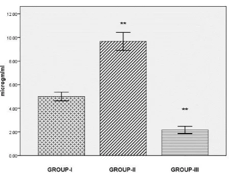
Significant increase(p<0.001) in serum lysozyme were found in Con A induced group compared to control group,where as in Con A plus Eupalitin-3-o-β-D-galactoside group lysozyme activity was reduced (p<0.001)markedly in comparison to induced group as illustrated in Figure-1.The suppression of Serum lysozyme levels suggest immunosuppressive effect of Eupalitin-3-o-β-D-galactoside
Figure-1: Serum lysozyme levels in control and treated fish. Comparisons are done between Group I
Vs Group II and Group II Vs Group III. Results are expressed as mean ± SE (n=6), ** (p<0.001) statistical significance difference between control and stimulant ConA treated group and within treated groups
Serum Myeloperoxidase activity
Serum Myeloperoxidase activity in Con A treated fish represented in Figure-2 were significantly (p=0.021) higher in comparison to the respective control fish.In Con A plus Eupalitin-3-o-β-D-galactoside administered fish myeloperoxidase levels (p=0.026) were reduced markedly.
Figure 2:Serum Myeloperoxidase levels in control and treated fish. Comparisons are done between Group I Vs Group IIand Group II Vs Group III. Results are expressed as mean ± SE (n=6),*(P=0.021) statistical significance difference between control and stimulant ConA treated group and*(P=0.026) within treated groups.
TNF-α mRNA expression
Quantitative RTPCR as represented in Figure-3 for TNF-α gene expression were analyzed in Head kidney for Conconavalin A - stimulated and Conconavalin A - stimulated Eupalitin-3-o-β-D-galactoside treated fish.
The Conconavalin A- stimulated Head kidney TNF-α mRNA levels represented in Figure-4 were elevated significantly (p<0.001). TNF-α cytokine mRNA expression) as illustrated in Figure-4 was significantly (p<0.001) lowered in Eupalitin-3-o-β-D-galactoside treated conconavalin A - stimulated fish.
Table 1: Primer sequence
Gene
name Primer sequence
Primer size (bases)
Gene bank number
TNF-α F:CAGAAACCCTGGACTGGAAA R:CATGTAGCGGCCATAGGAAT 20 AJ311800.2
Β-Actin R:CTTCTGCATACGGTCAGCAA F: CTCTTCCAGCCTTCCTTCCT 20 JQ619774.1
Figure 3: Effect of Eupalitin-3-o-β-D-galactoside on TNF-α mRNA expression in Conconavalin A stimulated head kidney tissue of koi carp. Lane 1, Lane 3 and Lane 5 represent the amplicons of TNF-α in Control (Lane 1), Conconavalin A (Lane 3) and Conconavalin A plus Eupalitin-3-o-β-D-galactoside treated groups (Lane5). Lane 2,Lane 4 and Lane 6 represents the amplicons of β-actin in Control(Lane 1), Conconavalin A(Lane 3) and Conconavalin A plus Eupalitin-3-o-β-D-galactoside treated groups(Lane5) Lane.Expression of TNF-α mRNA after 48 hrs for Conconavalin A - stimulated and Conconavalin A - stimulated Eupalitin-3-o-β-D-galactoside treated fish were analyzed byRT-PCR.
Figure 4: Densitometric scanning of TNF-α mRNA expression. Gene expression was normalized relative to the β-Actin as internal reference gene. Bars represent relative gene expression in each replicate experiment (mean ± SE,n =3)(p<0.001)for TNF-α-165bp β-actin-221bp
Conconavalin A – stimulated group compared with control group. Significant effect of Eupalitin-3-o-β-D-galactoside as determined by one-way ANOVA followed by Bonferroni post-hoc test (p<0.001) compared to Conconavalin A – stimulated group.
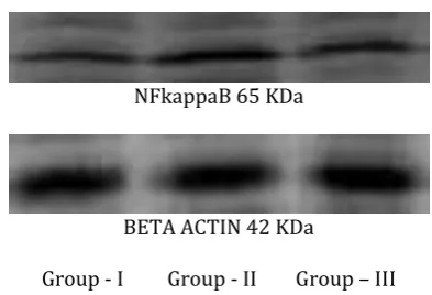
NFkB protein expression
Western blot analysis of NF-kB (Figure-5) was carried out to assess the immunosuppressive effect of Eupalitin-3-o-ß-D-galactoside in Conconavalin A induced fish. In the present study NF-kB protein
expression levels were increased
significantly(p=0.003)in Conconavalin A -stimulated Head kidney for 48 hrs as illustrated in Figure-6 . The Conconavalin A- stimulated Head kidney NF-kB protein levels were suppressed significantly (p=0.027) after Eupalitin-3-o-ß-D-galactoside treatment for 48 hrs.
NFkappaB 65 KDa
BETA ACTIN 42 KDa
Group - I Group - II Group – III
Figure 5: Immuno blot analysis for the effect of Eupalitin-3-o-β-D-galactoside on NFkB in Conconavalin A stimulated Head kidney.
Figure 6: Densitometric analysis of the expression of NFkB in Control, Conconavalin A and Conconavalin A plus Eupalitin-3-o-β-D-galactoside treated groups. Bars represent relative gene expression in each replicate experiment(mean ± SE,n =3) (p=0.003)for Conconavalin A – stimulated group compared to control group. Significant effect of Eupalitin-3-o-β-D-galactoside as a determined by one-way ANOVA
followed by Bonferroni post-hoc test (p=0.027) compared to Conconavalin A – stimulated group.
p38 MAPkinase protein expression.
The p38 MAPkinase takes part in inflammatory responses by regulating the tumor necrosis factor expression. Western blot analysis of p38MAPkinase represented in Figure-7 and densitometric scanning as represented in Figure-8, revealed p38MAPK protein expression was enhanced significantly (p=0.003) in Conconavalin A induced head kidney and reduced significantly (P=0.009) after treatment with Eupalitin-3-o-β-D-galactoside for 48hrs.
Down regulation of NFkB and p38MAPK protein expression in Eupalitin-3-o-β-D-galactoside treated head kidney tissue in the present work indicate the immunosuppressive effect of Eupalitin-3-o-β-D-galactoside in koi carp.
p38 38KDa
BETA ACTIN 42 KDa
Group-I Group-II Group-III
Figure 7: Immunoblot analysis for the effect of Eupalitin-3-o-β-D-galactoside on p38MAP kinase activation in Conconavalin A stimulated Head kidney. Conconavalin A strongly stimulated a rapid increase in activation of p38 mapkinase activity.The induced p38 mapkinase activity was significantly inhibited after treatment with Eupalitin-3-o-β-D-galactoside.
Eupalitin-3-o-β-D-galactoside as determined by one-way ANOVA followed by Bonferroni post-hoc test (p=0.009) compared to Conconavalin A – stimulated group.
DISCUSSION
Modulation of immune functions using medicinal plants and their products as a possible therapeutic measure has become fundamental principle of therapeutic approach. The present study was done to evaluate Eupalitin-3-o-β-D-galactoside for its possible immuno suppressive activity in koi carp.
Lysozyme is an important parameter in the immune defence of both invertebrates and vertebrates. Lysozyme is found in a wide range of vertebrates including fish and is one of the defensive factors against invasion by microorganisms.[9] In the present study significant increase in serum lysozyme were found in Con A induced group compared to control group, where as in Con A plus Eupalitin-3-o-β-D-galactoside lysozyme activity was reduced markedly in comparison to induced group.The suppression of serum Lysozyme levels suggest immunosuppressive effect of Eupalitin-3-o-β-D-galactoside.Significantly elevated levels of serum lysozyme were found on 28 days of an immunostimulant levamisole post exposure in Indian carp(CatlaCatla).[10] Significant increase in lysozyme were reported in koi fed with diets supplemented with a combination of the chitosan oligosaccharides and B. coagulans.[11]Exposure to UVB radiation (50-500mJcm-2)induced a decrease in plasma lysozyme activity in Rainbow trout indicating immunosuppression.[12] Lysozyme level in the kidney was significantly lowered than in the control group in common carp exposed to 5 mg/l cadmium for 96 hrs indicating the immuno-suppressive effect of the cadmium.[13]
In the present study serum myeloperoxidase activity in Con A treated fish were significantly higher in comparison to the respective control fish.In Con A plus Eupalitin-3-o-β-D-galactoside administered fish myeloperoxidase levels were reduced markedly. Enhanced myeloperoxidase activity was observed in
Indian carp (Catla Catla) treated with 1.25 and 2.5mg/l levamisole. [10] Significant (P < 0.05) decrease in MPO activities 72 hrs after cyclophosphamide (CYP) 200 mg kg−1 body weight) treatment when compared with control fish, support the immunosuppressive action of CYP in freshwater catfish, C. batrachus.[14]The results indicating a significant decrease in Lysozyme and serum myeloperoxidase activity could be attributed to the immunosuppressive effect of Eupalitin-3-o-β-D-galactoside.
Cytokine expression analysis allows us to perceive the immunologic status of fish.[15] TNF-α, a proinflammatory cytokine is elevated in inflammatory disease and plays an important role in immune and inflammatory response.Hence investigation on mRNAexpression of TNF-α in ConA stimulated and ConA induced Eupalitin-3-o-β-D-galactoside treated fish were done by RTqPCR. In this study TNF-α gene expression was downregulated in head kidney of Con A induced Eupalitin-3-o-β-D-galactoside treated group than the control group. Eupalitin-3-o-β-D-galactoside has been reported to inhibit LPS- stimulated TNF-α production in human PBMCs and it also blocked the activation of NFkB and AP-1,two major transcription factors involved in the expression of IL-2 and IL-2R gene ,which are necessary for T-cell activation and proliferation1. Kawada et al,[16]have reported the production of tumor necrosis factor alpha (TNF-alpha) by lipopolysaccharide-stimulated Kupffer cells was strongly inhibited by quercetin. TNF-α was downregulated by quercetin in a dose-dependent manner in LPS-stimulated DCs.[17] Wogonoside not only dose-dependently decreased the production of inflammatory mediators but also inhibited the release of pro-inflammatory cytokine TNF-α in LPS-induced RAW264.7 cells.[18] Kaempferol3-o-(3-o-acetyl-α-l rhamnopyranoside) isolated from flowers of
Nymphaea mexicana zucc was reported to have the
most prominent inhibitory effect on the LPS-stimulated tumor necrosis factor-alpha (TNF-α)
production in raw 264.7 macrophages.[19]
Suppression of the LPS-activated production of TNF-α in rat peritoneal cells and human peripheral blood mononuclear cells suggested the immunomodulatory effects of flavonoids casticin and chrysosplenol D of
ArtemisiavannuaL. (Qinghao). [20]
In the present study, it was investigated whether inhibition of p38 MAPK kinase and NF-kB by Eupalitin-3-o-β-D-galactoside alters the inducible expression of TNF-α in head kidney of koi carp. Down regulation of NFkB and p38MAPK protein expression in Eupalitin-3-o-β-D-galactoside treated head kidney tissue in the present work indicate the immunosuppressive effect of Eupalitin-3-o-β-D-galactoside in koi carp.
The p38 MAPkinase is involved in most of immunological responses. It plays an important role in innate as well as adaptive immune response. It is involved in signaling for the expression of certain NF-κB target genes which plays crucial role in the apoptosis pathways [21] mainly in the macrophages that are key cells involved in innate immune response. The p38 MAPkinase also takes part in inflammatory responses by regulating the interleukin and tumor necrosis factor expression. [22] Owing to these key properties this kinase can be an excellent target for the therapy of the immunological and inflammatory disorders.
Cho et al, (2002)[23] have described the suppression of TNF-α production through mapkinases and NFkB
pathway in LPS-stimulated RAW264.7
cells.SB202190,a p38 inhibitor has been reported to inhibit the expression of TNF-α expression in head kidney leucocytes isolated from Atlantic salmon(Salmo salar) fed with soybean oil or fish oil based diets.[24] Exposure of LPS-stimulated murine bone marrow neutrophils to sauchinone diminished production of tumor necrosis factor (TNF)-α and decreased the phosphorylation of p38 MAPK.Reduced levels of phosphorylation of p38 in Western blot analysis of p38 in Lung tissues were observed in LPS induced mice, injected intraperitoneally with
sauchinone suggesting attenuation of proinflammatory neutrophil activity of sauchinone.[25] Butanolic fraction from A. cochliacarpos (BFAC) and its major flavonoid, (+)-catechin, was reported to inhibit p38 in LPS-stimulated murine peritoneal macrophages.[26] Collart et al (1990) [27] have reported that NFkB, a transcription factor is necessary for the transcription of TNF-α in endotoxin (LPS) -stimulated macrophages. Inhibition of TNF-α gene expression was observed in LPS (20μg/ml) stimulatedhead kidney phagocytes of carp treated with NFkB inhibitor pyrollidine dithiocarbomate( 5μM).[28] Several reports have indicated that NF-kB is regulated by plant derived substances such as quercetin and green tea extracts,[29] that may potentially ameliorate disease states influenced by uncontrolled NF-kB activation. Human keratinocyte (HaCaT) cells exposed to 15 μM QGR for 20 min and 10 ng/ml TNF-α in combination with QGR for 15 minreduced the levels of NF-κB p65, NF-κB p50 and phospho-IκB-α indicating attenuation of NF-κBactivation.[30] Astragalin attenuated the activity of myeloperoxidase (MPO) and the expression of tumor necrosis factor-α (TNF-α) in a murine model of LPS-induced mastitis. It also decreased nuclear factor-kappaB (NF-κB) activation by inhibiting the degradation and phosphorylation of IκBα and the nuclear translocation of p65. Results suggested that astragalin exerts anti-inflammatory properties in LPS-mediated mastitis, possibly through inhibiting inhibition of the NF-κB signaling pathway that mediates the expression of pro-inflammatory cytokines. [31]
Quercetin inhibited TNF-α expression , NF-κβ1 gene expression and phosphorylation of Iκβα and Iκββ on cultured PBMCs indicating the modulation of immune response.It has been hypothesized that quercetin exerted anti-inflammatory effect on PBMCs inhibiting the endogenous production of the proinflammatory cytokine TNF-α and that these effects are mediated through the regulation of NF-κβ and Iκβ.[32] Propolis and caffeic acid was reported to suppress LPS-induced
p38 MAPK and NF-κB signaling pathways in Raw 264.7 cells.[33]
The present study on down regulation of TNF-α gene expression, NFkB and p38MAPK protein expression in Eupalitin-3-o-β-D-galactoside treated head kidney tissue demonstrates the possible mechanism of action of Eupalitin-3-o-β-D-galactoside in koi carp(Cyprinus carpio).
CONCLUSION
In this study, it is reported for the first time that Eupalitin-3-o-β-D-galactoside act as an immunosuppressor in a fish model and provided strong evidence that Eupalitin-3-o-β-D-galactoside may be a promising agent for the prevention and treatment of inflammatory and autoimmune diseases.However, further investigations are necessary to elucidate the exact mechanism underlying the immuno suppression of Eupalitin-3-o-β-D-galactoside and the potential usefulness of this compound as an immunosuppressant.
Source of support – Nil, Conflict of Interest – Nil.
REFERENCES
1. Pandeya R, Maurya R, Singh G,Sathiamoorthy B and Sita Naika S, Immunosuppressive Properties of Flavonoids isolated from Boerhaavia diffusa Linn.International Immunopharmacology. 2005; 5: 541–553.
2. Fathima K A and Parameswari C S, Immunomodulatory effect of Eupalitin-3-O-ß-D-galactopyranoside in Koi carp (Cyprinus carpio). Journal of Pharmacy Research. 2011; 4(10): 3396-3398.
3. Van Muiswinkel WB, Anderson D P, Lamers C H J, Egberts E , Van Loo J J A, and Ijessl J P, Fish immunology and fish health. In Fish immunology (eds. M.J. Manning and M.F. Tatner). Academic Press, London. 1985.
4. Lv-yun Zhu, Li Nie, Guan Zhu, Li-xin Xiangand Jian-zhong Shao, Advances in research of fish immune-relevant genes: A comparative overview of innate and adaptive immunity in teleosts Developmental & Comparative Immunology.2013; 39 (1–2): 39–62.
5. Dey M, Ripoll C, Pouleva R, Dorn R, Aranovich I, Zaurov D, Kurmukov A, Eliseyeva M, Belolipov I, Akimaliev A, Sodombekov I, Akimaliev D, Lila MA and Baskin I, Plant extracts from central asia showing anti-inflammatory activities in gene expression. Phytotherapy Research.2008; 22: 929-934.
6. Anderson DP, Siwicki AK, Basic Haematology and Serology for fish health programs, In Shariff M, Authur JR, Subasinge(eds),Diseases in Asian Aquaculture II, Fish Health Section, Asian Fisheries Society, Manila.1995; 185-202.
7. Quade M J and Roth J A, A rapid, direct assay to measure degranulation of bovine neutrophil primary granules. Vet. Immunol. Immunopathol. 1997; 58: 239–248.
8. Kain SR, Mai K, Sinai P. Human multiple tissue western blots: a new immunological tool for the analysis of tissue-specific protein expression. Bio-Techniques 1994;17:982-7.
9. Evelyn TPT, Finfish immunology and its use in preventing infectious diseases in cultured finfish. In
Lavilla-Pitogo, C.R. and Cruz-Lacierda, E.R. (eds.). Diseases in Asian Aquaculture IV, Fish Health Section, Asian Fisheries Society, Manila. 2002; 303-324.
10. Perera HACC and Asoka Pathiratne, Enhancement of Immune Responses in Indian Carp, Catla catla, Following Administration of Levamisole by Immersion. Diseases in Aquaculture VI, Fish health section, Asian fisheries society, Manila, Philippines: 2008;129-142.
11. Shimei Lin , Shuhong Mao , Yong Guan , Lin Luo, Li Luo and Yu Pan, Effects of dietary chitosan oligosaccharides and Bacillus coagulans on the growth,innate immunity and resistance of koi (Cyprinus carpio koi).Aquaculture. 2012; 342-343: 36-41.
12. Eveliina Markkula S,HarriM. Salo,Anna Kaisa Rikalainen and Ilmari Jokinen E,Different sensitivity of carp ((Cyprinus carpio ) and rainbow Trout
(Oncorrhynchus mykiss) to the immunomodulatory effects of UVB irradiation. Fish and Shellfish Immunology.2006:21:70-79.
13. Sovenyi J and Szakolczai J, Studies on the toxic and immunosuppressive effects of cadmium on the common carp. Acta Vet Hung. 1993;41(3-4):415-26.
14. Sahoo P.K and Jaya Kumari, Effects of cyclophosphamide on the immune system and disease resistance of Asian catfish Clarias batrachus,Fish & Shell fish Immunology, 2005; 19(4): 307–316.
15. Harms C A, Kennedy-Stoskopf S, Fuller F J, Horne W A, and Tompkins WAF, Cloning and sequencing hybrid striped bass (Morone saxatilis x M.chrysops) transforming growth factor-ß (TGF-ß) and development of a reverse transcription quantitative competitive polymerase chain reaction (RT-qc PCR) assay to measure TGF-ß mRNA of teleost fish. Fish and Shellfish Immunology.2000a: 10:61-85.
16. Kawada N, Seki S, Inoue M and Kuroki T, Effect of antioxidants, resversatol, quercetin, and n-actetylcysteine, on the functions of cultured rat hepatic stellate cells and Kupper cells. Hepatology. 1998; 27:1256-1274.
17. Huang RY, Yu YL, Cheng WC, OuYang CN, Fu E and Chu CL, Immunosuppressive Effect of Quercetin on Dendritic Cell Activation and Function. J Immunol. 2010; 184; 6815-6821.
18. Yang YZ, Tang YZ and Liu YH, Wogonoside displays anti-inflammatory effects through modulating inflammatory mediator expression using RAW264.7 cells. Journal of Ethnopharmacology 2013;148 (10): 271–276.
19. HSU CL, Fang SC and Yen GC, Anti-inflammatory effects of phenolic compounds isolated from the flowers of Nymphaea mexicana Zucc. Food Funct. 2013; 4: 1216-1222.
20. Zhu XX, Yang L, Li YJ, Zhang D, Chen Y, Kostecka P, Kmonickova E and Zídek Z.Effects of sesquiterpene, flavonoid and coumarintypes of compounds from
Artemisia annua L.on production of mediators of angiogenesis.Pharmacological Reports.2013; 65: 410-420.
21. Park JM, Greten FR, Zhi-Wei Li and Karin M. Macrophage Apoptosis by Anthrax Lethal Factor Through p38 MAP Kinase Inhibition. Science.2002; 297 Suppl 5589: 2048-51.
22. Ridley SH, Dean JL, Sarsceld SJ, Brook M, Clark AR and Saklatvala J, A p38 MAP kinase inhibitor regulates stability of interleukin-1-induced cyclooxygenase 2 mRNA.FEBS Letters. 1998; 439: 75-80.
23. Cho SY, Park SJ, Kwon MJ, Jeong TS, Bok SH, Choi WY, Jeong WI, Ryu SY, Do SH, Lee CS, Song JC and Jeong KS,Quercetin suppresses proinflammatory cytokines production through MAP kinases andNF-kappaB pathway in lipopolysaccharide-stimulated macrophage. Molecular and Cellular Biochemistry. 2003; 243: 153-160.
24. Holen E, Winterthun S, Du ZY and Krovel AV, Inhibition of p38 MAPK during cellular activation modulate gene expression of head kidney leukocytes isolated from Atlantic salmon (Salmo salar) fed soy bean oil or fish oil based diets Fish & Shellfish Immunology 2011;30: 397- 405.
25. Han HJ, Li M, Son JK, Seo CS, Song SW, Kwak SH and Bae HB, Sauchinone, a lignan from Saururus chinensis, attenuates neutrophil proinflammatory activity and acute lung injury.International Immunopharmacology. 2013 ; 17 (2): 471- 477. 26. Sanchez-Fidalgo S, da Silva MS, Cardeno A,
Aparicio-Soto M, Salvador MJ, Frankland Sawaya AC, Souza-Brito AR and de la Lastra C A, Abarema cochliacarpos reduces LPS-induced inflammatory response in murine peritoneal macrophages regulating ROS-MAPK signal pathway. Journal of Ethnopharmacology. 2013;149 ( 1): 140–147. 27. Collart MA, Baeuerle P and Vassalli P,Regulation of
tumor necrosis factor alpha transcription in macrophages: involvement of four kappa B-like motifs and of constitutive and inducible forms of NF-kappa B. Mol. Cell. Biol. 1990; 10:1498-1506. 28. Saeij, Jeroen PJ, Stet, René JM, de Vries, Beja J, van
Muiswinkel, Willem B and Wiegertjes, Geert F, Molecular and functional characterization of carp TNF: a link between TNF polymorphism and trypanotolerance?. Developmental and Comparative Immunology.2003; 27 (1): 29-41.
29. Muraoka, K., Shimizu, K., Sun, X., Tani, T., Izumi, R., Miwa, K and Yamamoto, K., Flavonoids exert diverse inhibitory effects on the activation of NF-kappaB. Transplant. Proc. 2002; 34:1335–1340.
30. Lee C S, l Jeong EJ, KimYJ, Lee MS, Seong Jun Seo SJ, Park KH and Lee MW, Quercetin-3-O-(2″ -galloyl)-α-l-rhamnopyranoside inhibits TNF-α-activated NF-κB-induced inflammatory mediator production by suppressing ERK activation.International Immunopharmacology. 2013;16(4): 481–487.
31. Li F, Liang D, Yang Z, Wang T, Wang W, Song X, Guo M, Zhou E, Li D, Cao Y and Zhang N,Astragalin suppresses inflammatory responses via down-regulation of NF-κB signaling pathway in lipopolysaccharide-induced mastitis in a murine model.International Immunopharmacology. 2013;17(2): 478-482.
32. Nair MS, Mahajan S, Reynolds JL, Aalinkeel R, Nair H, Schwartz SA, and Kandaswami C. The Flavonoid Quercetin Inhibits Proinflammatory Cytokine (Tumor Necrosis Factor Alpha) Gene Expression in Normal Peripheral Blood Mononuclear Cells via Modulation of the NF-κβ System. Clin Vaccine Immunol 2006;13 (3) :319-328.
33. Bufalo MC, Ferreira I, Costa G, Francisco V, Liberal J, Cruz MT, Lopes MC, Batista MT and Sforcin JM, Propolis and its constituent caffeic acid suppress LPS-stimulated pro-inflammatory response by blocking NF-κB and MAPK activation in macrophages. Journal of Ethnopharmacology. 2013;149(1): 84-92.
How to cite this article: K. A. Fathima*, C. S. Parameswari2;
OSDrC® OptiDose™: Immunosuppressive effect of Eupalitin-3-O-Β-D-Galactoside in Conconavalin-A induced KOI CARP (Cyprinus Carpio); J. Adv. Pharm. Edu. & Res. 2014: 4(3): 298-307.
Source of Support: Nil, Conflict of Interest: Nil