R E V I E W
Open Access
Interaction of host immunity with
HER2-targeted treatment and tumor
heterogeneity in HER2-positive breast
cancer
Gaia Griguolo
1,2,3,4, Tomás Pascual
1,2, Maria Vittoria Dieci
3,4, Valentina Guarneri
3,4and Aleix Prat
1,2*Abstract
Growing evidence suggests a clear role of the host immune system in HER2+ breast cancer. In addition, HER2+ breast cancer is generally considered more immunogenic than hormone receptor-positive (HR+)/HER2-, and specific molecular HER2+ subgroups (e.g. HER2-enriched disease) are more immunogenic than others (e.g. Luminal A or B). From a clinical perspective, the immune system plays a relevant prognostic role in HER2+ breast cancer and contributes to the therapeutic effects of trastuzumab. However, as more HER2-targeted agents become available, a better understanding of the role played by the immune system in modulating therapy response to different agents will be needed. Furthermore, the recent introduction in oncology of immune checkpoint inhibitors capable of unleashing anti-tumor immune response opens new possibilities for therapeutic combinations in HER2+ breast cancer. Here, we review the current pre-clinical and clinical data on the interplay between the immune system and HER2+ breast cancer, focusing on different HER2-targeted treatments and the biological heterogeneity that exists within HER2+ disease. Finally, we discuss new therapeutic approaches exploiting the immune system to increase activity or revert resistance to HER2-targeted agents.
Keywords:Breast cancer, HER2, Targeted treatment, Immunity, Tumor infiltrating lymphocytes, Immune checkpoints
Introduction
HER2 is overexpressed in 15–20% of breast cancers (BC) and is associated with clinically aggressive disease [1]. Targeting this oncogene has led to striking improve-ments in survival outcomes for HER2+ BC patients. To date, several HER2-targeted treatments are available, including monoclonal antibodies (trastuzumab, pertuzu-mab), tyrosine kinase inhibitors (lapatinib, neratinib), and antibody–drug conjugates (Ado-trastuzumab emtansine [T-DM1]) [2–6].
The role of the host immune system in HER2+ BC is becoming an important topic to study for several reasons. First, HER2+ BCs have higher stromal tumor-infiltrating lymphocytes (TILs) levels in general than hormone
receptor positive (HR+)/HER2- BCs, implying that HER2 + disease is usually more immunogenic [7,8]. Second, not all HER2+ tumors are immunogenic and specific molecu-lar HER2+ subgroups (e.g. HER2-enriched) are more immunogenic than others (e.g. Luminal A/B) [9]. Third, the percentage of TILs is clinically relevant due to its asso-ciation with better prognosis [10, 11]. Fourth, the recent introduction in oncology of therapeutic agents capable of unleashing anti-tumor immune response, such as check-point inhibitors, opens new treatment strategies [12]. Finally, the immune system not only plays a prognostic role but also seems to contribute substantially to the therapeutic effects of trastuzumab, originally credited to induce cell death by direct inhibition of HER2 intracellular signaling [13].
To date, several reviews have analyzed the prognostic role of immunity in HER2+ BC and its capability of modulating response to trastuzumab [13]. However, as more HER2-targeted agents have become available in
© The Author(s). 2019Open AccessThis article is distributed under the terms of the Creative Commons Attribution 4.0 International License (http://creativecommons.org/licenses/by/4.0/), which permits unrestricted use, distribution, and reproduction in any medium, provided you give appropriate credit to the original author(s) and the source, provide a link to the Creative Commons license, and indicate if changes were made. The Creative Commons Public Domain Dedication waiver (http://creativecommons.org/publicdomain/zero/1.0/) applies to the data made available in this article, unless otherwise stated. * Correspondence:alprat@clinic.cat
1Translational Genomics and Targeted Therapeutics in Solid Tumors, IDIBAPS, Barcelona, Spain
recent years, better understanding of the role played by the immune system in modulating response to these new treatments might help optimize or tailor treatment. Furthermore, a deeper understanding of the interaction between immunity and combination of anti-HER2 drugs with hormonotherapy and chemotherapy, in the context of the biological heterogeneity within HER2+ BC, will be required to design biologically meaningful therapy combinations.
HER2 as the antigen
HER2 overexpression in BC is often described as a typical case of oncogene addiction, thus defining tumors that are almost exclusively dependent on a single onco-genic pathway. As is the case for many oncogenes, its overexpression on the cell membrane and its essential role in tumor cell biology makes it a perfect antigen to guide immune response towards HER2+ cells [14].
In the attempt to induce host immune response towards HER2 for therapeutic uses, vaccines have been designed. A number of HER2-derived peptides have been investigated [15] and some have been shown capable of inducing immune response. However, despite some positive results in phase I-II trials, the develop-ment of BC vaccines has been a story of setbacks and efficacy has not been proved in phase III trials (Additional file1: Table S1) [16].
On the other hand, host immune response plays a key role in the activity of anti-HER2 monoclonal antibodies.
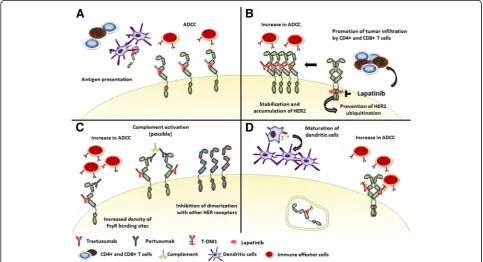
Indeed, trastuzumab has several mechanisms of action (Fig. 1a). By binding to the extracellular domain of HER2, it prevents receptor dimerization inhibiting downstream signaling. It also increases HER2 internal-ization and endocytic degradation, thus enhancing HER2 peptide presentation on major histocompatibility com-plex (MHC) receptors. In addition, while the antibody binds to HER2 on the cell surface, the crystalline fragment (Fc) of the immunoglobulin interacts with Fc-gamma-receptors (FcγR) on innate immune effector cells, like natural killer (NK) cells, neutrophils and
γδT-cells, activating antibody-dependent cellular cyto-toxicity (ADCC) [17,18]. This cytolytic activity increases availability of tumor antigens in the tumor microenvir-onment (TME), favoring antigen presentation. Antigen presentation is also enhanced by FcγR-mediated phago-cytosis of immune complexes by antigen-presenting cells. Hence, the interaction between trastuzumab and the innate immune system facilitates the development of tumor-specific T-cell immunity. On one hand, NK-cells prime dendritic cells, enhancing tumor antigen presenta-tion to cytotoxic CD8+ T-cells and polarizapresenta-tion of CD4+ T-cells towards an anti-tumor T-helper type 1 (Th1) phenotype. On the other hand, trastuzumab-dependent NK-cell activation leads to cytokine secretion contribut-ing to the recruitment and functional polarization of myeloid and T-cells [19].
Through these mechanisms, anti-HER2 antibodies exert a vaccine-like effect activating the adaptive as well
as the innate immune system. Consistently, activation of anti-HER2 CD4+ Th1 response correlates with patho-logic complete response (pCR) and disease-free survival (DFS) following anti-HER2 based neoadjuvant chemo-therapy [20,21].
Evaluating immunity in BC
Generally, TILs are typically believed to reflect immuno-logical response; however, they include different cell types usually dominated by T-cells, with variable propor-tions of B-cells, NK cells, macrophages and dendritic cells. While CD8+, CD4+ Th1 and NK cells are generally considered to favor a tumor-suppressive response, CD4+ T-helper 2 (Th2), FOXP3+ T-regulatory and dendritic cells might play a pro-tumorigenic role [22].
TILs are easily assessed on hematoxylin/eosin stained slides, both in intratumoral and stromal areas (sTILs). Current recommendation is to use sTILs as principal parameter.
However, all mononuclear cells are scored and semi-quantitative evaluation of TILs does not distinguish specific immune cells subtypes. Moreover, no clear cut-off exists to define what is a high infiltrate. Traditionally, the lymphocyte predominant BC (LPBC) definition (≥ 50–60% sTILs) has been used. More recently, TILs are measured as a continuous parameter to better represent the continuity of immune response [22,23].
Gene-expression analysis can also be used to infer proportions of infiltrating immune cell populations, pro-viding more information regarding different lymphocyte subpopulations, and to measure immune checkpoint gene expression [24]. Interestingly, checkpoint expres-sion significantly correlates with other immune markers and TILs [25,26].
Host immunity in HER2 + BC treated with chemotherapy and trastuzumab
Prognostic role of baseline immunity in early HER2+ BC To date, most data regarding the clinical validity of pre-existing immune response in HER2+ BC come from patients treated with trastuzumab-based chemotherapy for early BC (Additional file 1: Table S1 and Table S2). From a prognostic perspective, several studies in HER2+ BC patients receiving neoadjuvant (Table1) or adjuvant
[7, 24, 27–30] anti-HER2-based chemotherapy have
shown that expression of immune-associated gene signa-tures and infiltration by TILs in pre-treatment biopsies associated with longer DFS [10], independently of known prognostic clinical-pathological variables.
Role of immunity in residual disease after neoadjuvant treatment
Timing of TILs evaluation might be important. In residual disease after neoadjuvant therapy, TILs might have a
different prognostic meaning. In a retrospective study, in-cluding 175 HER2+ BC patients treated with neoadjuvant chemotherapy+/−trastuzumab, sTILs generally decreased during treatment (78% of patients). Presence of high TILs (> 25%) in patients with residual disease after neoadjuvant therapy was associated with worse DFS [31]. This pattern is opposite to that reported for triple-negative BC (TNBC), where high TILs in residual disease associated to better prognosis [32, 33]. These inconsistencies may be explained by differences in TILs composition across BC subtypes and by changes in TILs composition induced by neoadjuvant antiHER2-containing treatment. A decrease in FOXP3+ TILs has been described in HER2+ tumors achieving pCR, while an increase in FOXP3+ TILs has been described in HER2+ residual disease [34,35]. Indeed, another study, assessing post-neoadjuvant TILs in 111 HER2+ BC patients treated with chemotherapy+/− trastu-zumab, reported that low levels of CD8+ lymphocytes were associated with poor DFS, while low levels of FOXP3 + lymphocytes were associated with better DFS [36].
particularly from 1-year trastuzumab over 9-weeks, whereas high-TILs patients experienced an excellent out-come irrespectively of trastuzumab duration (interaction p= 0.015) [37].
One important aspect is that TILs might also predict sensitivity to the chemotherapy component. In the Geparsixto trial, 273 HER2+ BC patients received a combination of paclitaxel, anthracycline, trastuzumab, Table 1Neoadjuvant trials with trastuzumab-containing regimens which assessed the prognostic values of TILs and immune related gene signatures
Study Treatment N.
pts.a
Biomarker Tested Outcome Tested
Association
CALGB 40601 [100] NCT00770809
P-H P-L P-HL
265 Immune gene signatures [100]
pCR IgG signature independently associated with pCR at multivariate analysis
CherLOB [55] NCT00429299
P-H→FEC-H P-L→FEC-L P-HL→FEC-HL
105 TILs [55] pCR Associated with pCR at univariate analyses (no statistical significance beyond PAM50)
EFS Associated with EFS at univariate analyses
86 Immune gene signatures [55]
pCR 3 out of 4 signatures maintained association with pCR after correction for PAM50
GeparQuattro [57] NCT00288002
EC-H→D-H +/−X 178 TILs [57] pCR
Associated with pCR at multivariate analysis
GeparQuinto [57] NCT00567554
EC-H→D-H 162 TILs [57] pCR
EC-L→D-L 158 pCR Not associated with pCR
GeparSixto [38] NCT01426880
PM-HL +/−C 266 TILs [38] pCR Associated with pCR at multivariate analyses
226 mRNA expression of immunologic genes
pCR All 12 immune mRNA markers were associated with pCR (10/12 at multivariate analysis)
NeoALTTO [58] NCT00553358
P-H P-L P-HL
387 TILs [58] pCR Associated with pCR at multivariate analysis
EFS Associated with EFS at multivariate analysis
254 Immune gene signatures [59]
pCR two T-cell immune signatures were associated with pCR (only confirmed at multivariate analysis in P-HL arm)
NOAH [75] ISRCTN86043495
AP→P→CMF 51 Four immune metagenes [45]
pCR Not associated with pCR
AP-H→P-H→CMF-H→H 63 pCR 3/4 associated with pCR
NeoSphere [45] NCT00545688
DH DPrtz DHPrtz HPrtz
243 TILs [45] pCR Not significantly associated with pCR
305 PDL1 by IHC [45] pCR Not significantly associated with pCR
337 Immune genes and metagenes [45]
pCR 5 associated with pCR at multivariate analysis (different results in the DHPrtz arm)
Tryphaena [44] NCT00976989
FEC→DHPrtz FECHPrtz→DHPrtz CycloDHPrtz
213 TILs [43] pCR Not significantly associated with pCR
EFS Associated with EFS at multivariate analysis
173 Immune signatures and genes [43]
pCR 2 signatures and 4 genes associated with pCR at multivariate analysis
EFS Not associated with EFS at multivariate analysis
PAMELA [9] NCT01973660
HL 134 TILs at day15 pCR Associated with pCR at multivariate analysis
a
Number of patients included in the biomarker analysis
Adoxorubicin,Ccarboplatin,Cyclocyclophosphamide,CMFcyclophosphamide-methotrexate-fluorouracil,Ddocetaxel,ECepirubicin-cyclophosphamide,EFS
event-free survival,FECfluorouracil-epirubicin-cyclophosphamide,Htrastuzumab,IHCimmunohistochemistry,Llapatinib,Ppaclitaxel,pCRpathologic complete response,PMweekly paclitaxel + non pegylated liposomal doxorubicin,Prtzpertuzumab,TILtumor infiltrating lymphocytes,Xcapecitabine
lapatinib +/− carboplatin as neoadjuvant treatment. When 266 HER2+ baseline samples were analyzed [38], not only both sTILs as a continuous variable and LPBC were associated with pCR, but both significantly inter-acted with the addition of carboplatin (LPBCs showing higher pCR rates when receiving carboplatin). This suggests that TILs might also predict for sensitivity to chemotherapy.
Overall, the current data establishes the clinical valid-ity of pre-existing TILs as a prognostic biomarker. How-ever, more studies are needed to establish the clinical utility of TILs. In early HER2+ BC, several escalation (i.e. adding a second anti-HER2 agent) and de-escalation (i.e. shorter trastuzumab regimens, less-chemotherapy or non-chemotherapy) approaches have been or are being tested. In this context, TILs together with other prog-nostic clinicopathological variables might allow the con-struction of prognostic risk models that might help better treat our patients [39].
Host immunity and other HER2-targeted therapies
Due to their different nature and mechanisms of action, the interaction between immune system and new HER2-targeted treatments might be different from that described with trastuzumab alone (Fig.1).
Pertuzumab
Pertuzumab is a monoclonal antibody directed against the extracellular dimerization domain of HER2 (a differ-ent epitope than trastuzumab). Its binding inhibits dimerization of HER2 with other receptors of the HER family. As trastuzumab, pertuzumab can mediate ADCC and simultaneous binding of both antibodies to different HER2 epitopes increases the density of FcγR binding sites on HER2+ cells, possibly enhancing NK-mediated ADCC responses [40]. Consistently, studies on mouse models have reported that combining the two antibodies increases the total number of tumor infiltrating NK-cells and the proportion of them actively engaged in killing tumor cells [41]. In addition, only tumor cells treated with the combination are likely to have a sufficient number of cell-bound antibodies to induce efficient C3 opsonization, required to initiate complement-mediated-cytotoxicity and macrophage-mediated tumor cell killing [42]. However, if this might explain the mechanism of action of pertuzumab or if its improved efficacy in combination with trastuzumab only relies on a more profound pathway inhibition [43] is still unclear.
Pertuzumab in the neoadjuvant setting
As pertuzumab only shows significant activity when used in combination with trastuzumab, separating its immune effect in the clinical setting is almost impossible. In the neoadjuvant setting, pertuzumab has been tested in
several trials. In the TRYPHAENA trial, testing neoadju-vant pertuzumab and trastuzumab with multi-agent chemotherapy, TILs confirmed their prognostic role. Every 10% increase in baseline TILs was associated with a 25% reduction in DFS hazard, after adjusting for clinicopathological characteristics and pCR. Immune gene-expression signatures were also significantly as-sociated with pCR at multivariate analysis, but not with DFS [44].
The NEOSPHERE trial is a 4-arm study testing neoad-juvant docetaxel in association with trastuzumab, pertu-zumab, both or the combination of the two antibodies without chemotherapy. In this trial, baseline TILs as a continuous variable were not significantly associated with pCR, although this might be due to a non-linear ef-fect, as the low TILs group had, as expected, a signifi-cantly lower pCR rate. Interestingly, differences were observed across treatment arms. Patients treated with dual anti-HER2 blockade plus docetaxel showed higher rates of pCR, as compared to other treatment arms, in low and intermediate TILs groups, but not in the LPBC group. The impact of immune activation was also explored using gene expression analysis. In the trastuzumab-docetaxel, pertuzumab-docetaxel and in the chemotherapy-free arm, high expression of PDL1, MHC1, and IF-I metagenes associated with lower pCR rates, while high expression of PD1, STAT1, and MHC2 associated with higher pCR rates in multivari-able analyses. However, the impact of immune-related metagenes differed across treatment groups. In tumors with high activation of the immune system, the activity of all treatments tested appeared similar (including the chemotherapy-free arm and docetaxel-trastuzumab-pertu-zumab arm), while in tumors with low expression of PD1, CTLA4, and MHC1 the use of docetaxel-trastuzumab--pertuzumab was associated with a 2 to 20-fold higher like-lihood of pCR as compared to other arms. Indeed, the group in which chemotherapy-trastuzumab performed the least appeared to derive the most benefit from adding per-tuzumab [45].
Recently, results from the neoadjuvant PerElisa trial, testing pertuzumab, trastuzumab and letrozole in HR +/HER2+ BC patients selected using Ki67 response after short-course hormonotherapy, were reported. In this trial, baseline TIL levels did not show any impact on pCR [46]. Whether this might be due to the small num-ber of patients, to enrichment in luminal subtypes, to the combination with hormonotherapy or to the absence of chemotherapy remains unclear.
Pertuzumab in the metastatic setting
BC (mBC), confirmed the positive prognostic role of sTILs. Even if the association between sTILs and progression-free survival (PFS) was not significant, each 10% increase in sTILs significantly associated with lon-ger overall survival (OS). The prognostic effect of TILS appeared to be stronger for OS than for PFS, while no significant interaction with treatment was reported [11]. Two phase III trials assessing addition of pertuzumab to standard treatment in HER2+ mBC, the CLEOPATRA and PHEREXA trials [3, 47], consistently reported a higher magnitude of benefit in terms of OS than of PFS. Enhancement of anti-tumor immune activity by combination of pertuzumab and trastuzumab has been proposed as a possible mechanism for this delayed treat-ment benefit. Evocatively, a similar benefit in OS with limited benefit in PFS has been described in trials asses-sing immune checkpoint inhibitors [48,49].
However, in the CLEOPATRA trial only a small num-ber of patients was pretreated with trastuzumab (10.9%) and most samples analyzed came from primary tumors (93%). In fact, while in untreated BC TILs are associated with a T-effector phenotype, allegedly reflecting an ef-fective antitumour response, immunogenicity is sup-posed to decrease in the metastatic setting due to activation of immune-evasion mechanisms and to treatment-induced modifications of TME [35]. Consist-ently, a recent study which assessed sTILs in metastatic samples from 51 HER2+ BCs, mostly pretreated with HER2-targeted agents, did not observe any favorable impact of high sTILs on OS; indeed, a not statistically significant inverse relationship between TILs and prog-nosis was observed [50].
Lapatinib
Lapatinib is a reversible inhibitor of both HER2 and EGFR intracellular tyrosine kinase domains. Due to its intracellular activity, lapatinib might appear to lack the immune activity classically reported with trastuzumab. However, while trastuzumab mediates downregulation and degradation of HER2, lapatinib inhibits phosphoryl-ation of HER2 tyrosine domain, thus preventing ubiquitina-tion. This induces accumulation of HER2 on the cell membrane [51], increasing trastuzumab-dependent ADCC when administered in combination [52]. Lapatinib can also modulate TME. In animal models lapatinib promotes tumor infiltration by CD4 + CD8 + IFN-γ-producing T-cells through a Stat1 dependent pathway. Stat1-deficiency re-duces therapeutic activity of lapatinib, suggesting that immune activation can play a role in its antitumor activity [53].
Lapatinib in the neoadjuvant setting Lapatinib has been tested in various clinical settings, either alone or in combination with chemotherapy, trastuzumab or
hormonotherapy. Recently, a metanalysis of five neoad-juvant trials reported the impact of TILs in HER2+ BC treated with chemotherapy plus trastuzumab, lapatinib or their combination [54]. Four trials used a combined regimen of anthracyclines and taxanes (CherLOB, GeparQuattro, GeparQuinto and GeparSixto [55–57]), while paclitaxel alone was administered in the NeoALTTO trial [58]. In patients receiving anthracy-clines and taxanes, high baseline TILs were signifi-cantly associated with pCR, irrespective of the anti-HER2 treatment received (interaction p = 0.077). In the NeoALTTO trial, the relationship between TILs and pCR was nonlinear and rates of pCR in-creased sharply for TIL levels greater than 5% (p = 0.01), regardless of treatment group (interaction p = 0.519) [58]. However, the relationship between TILs and DFS was linear, regardless of treatment group, and patients with high TILs at baseline had better outcomes independently of whether they achieved pCR. Authors suggested anthracyclines given after surgery might explain the linear relationship with event-free survival.
Immune activation gene signatures were also tested in some of these trials. In the NeoALTTO trial, two T-cell– driven immune signatures significantly associated with pCR. However, this association was only confirmed in multivariable analysis in the combination arm and a significant interaction between these gene signatures and treatment (combination vs single arms) was reported [59]. In the CHERLOB trial, a T-cell gene signature and two immune-related gene signatures significantly corre-lated with pCR in a multivariate model adjusted by PAM50 [55]. FcγR polymorphisms (FcγRIIa-H131R, FcγRIIIa-V158F) were also tested: only FcγRIIIa V allele carriers showed significant improvement in pCR with dual HER2-blockade (trastuzumab-lapatinib-chemother-apy), and a significant interaction between FcγRIIIa V allele and combination treatment was observed [60]. This might hint to a relevant role for ADCC in deter-mining benefit of combining trastuzumab and lapatinib as compared to single agent, consistently with the sig-nificant interaction between immune signatures and combination treatment reported in the NeoALTTO trial [59].
Lapatinib in the metastatic setting In the metastatic setting, the CCTG MA.31 trial [62], a phase III trial randomizing 652 HER2+ mBC patients to receive either trastuzumab or lapatinib with taxane, casts an interest-ing perspective on the role of immunity in modulatinterest-ing activity of HER2-targeted treatment. In this trial, TILs assessed on primary tumor specimens were neither prognostic nor predictive. However, patients with low numbers of CD8+ TILs showed higher risk of progres-sing when treated with lapatinib compared with trastu-zumab (Hazard ratio 2.94;P = 0.003) than patients with high CD8+ TILs (Hazard ratio 1.36; P = 0.02). The dif-ferential effect and a significant interaction was con-firmed in a multivariable model [62].
Thus, low CD8+ sTILs in primary tumor predict infer-ior response to lapatinib vs trastuzumab in the meta-static setting. In fact, the immunogenic state of tumors before and after metastasis is possibly different. As immunogenic tumors present better response to neo/ad-juvant treatment, we could speculate that most of the metastatic population will be made up by not immuno-genic or immune evasive tumors (probably reflected by low CD8+ sTILs). In this group, trastuzumab can still be expected to function through enhancement of ADCC and priming of antitumor-adaptive T-cell responses. Indeed, previous treatment might modify the immune status of tumors and this should be taken into account in future clinical trials.
T-DM1
T-DM1 is an antibody-drug conjugate formed by trastu-zumab linked to the cytotoxic agent DM1. After binding HER2, T-DM1 is internalized, degraded in the endo-some, releasing DM1. In addition, T-DM1 blocks HER2 signaling pathway and mediates ADCC.
Antitumor immunity might contribute to T-DM1 therapeutic activity. Ansamitocin P3, the precursor of DM1, induces maturation of dendritic cells, facilitates antigen uptake and migration of tumor-resident dendritic cells to tumor-draining lymph nodes, thereby potentiating antitumor immunity [63, 64]. In a HER2+ mouse model, T-DM1 induced infiltration by effector T-cells, which was essential for its therapeutic activity. After T-DM1 treatment (but not with trastuzumab alone), tumors showed a shift towards a T-cell–inflamed phenotype with an increase in γδT-cells and NK-cells and expansion of CD45+. Consistently, the infiltrate showed a strong Th1 immune deviation. In addition, despite the tumor model presented primary resistance to anti–CTLA-4/PD-1 agents, combined use of these agents with T-DM1 resulted in strong antitumor efficacy and in development of immunologic memory [65]. Furthermore, analysis of paired samples from 28 HER2+ BC patients treated with preoperative T-DM1 showed an
increase in number and density of tumor-infiltrating T-cells after treatment [65].
Neratinib
Neratinib is a pan-HER tyrosine kinase inhibitor. It bonds covalently to a conserved cysteine residue, leading to irreversible inhibition of all four HER receptors, block of downstream pathways and in vitro inhibition of prolif-eration in tumor cells with trastuzumab resistance [66]. Neratinib recently received approval from FDA and EMA for the extended treatment of early-stage HER2+ BC, based on the phase III ExteNEt trial. This trial re-ported a small but statistically significant benefit in 5-year DFS for women receiving neratinib for one year after adjuvant trastuzumab versus placebo (90.2% vs 87.7%, p = 0.009) [6]. Discordantly from what is ob-served with pertuzumab and lapatinib, HR+ BC patients appeared to derive greater benefit than HR- patients. It has been suggested that this benefit might rely on intra-cellular irreversible inhibition of the downstream pathway, potentially limiting crosstalk between the HER2 and endocrine pathways, rendering cells more endocrine-responsive. Despite some evidence suggesting that neratinib can alter HER2 antigen levels, whether this might influence trastuzumab-mediated ADCC remains unknown [67]. Thus, limited available evidence exists regarding the role of immunity in modulating neratinib efficacy.
Anti-HER2 therapy in breast ductal carcinoma in situ (DCIS)
Anti-HER2 therapy is currently being investigated in DCIS. In a window-of-opportunity trial, patients with HER2-positive DCIS received a single dose of trastuzumab before definitive surgery. No evidence of response was ob-served. However, trastuzumab augmented NK-cell medi-ated ADCC, and in one case induced T-cell dependent humoral immunity [68]. A phase III trial of adjuvant trastuzumab in high-risk DCIS (NSABP-43) is currently ongoing to determine if adding trastuzumab to radiother-apy is beneficial in preventing recurrence. Moreover, the effect of tratuzumab on contralateral breast cancer will be evaluated. However, at this time there is no role for routine use of anti-HER2 therapy in DCIS patients.
Looking deeper: Heterogeneity of HER2+ BC and interaction with immune system
with immune activity. Estrogen can regulate the tran-scription of SerpinB9/proteinase inhibitor 9, a gran-zyme B inhibitor known to decrease susceptibility of HR + BC cells to NK and CD8+ T-cell cytotoxicity in vitro [70, 71]. Moreover, estrogens might modulate susceptibility of cells to NK-mediated ADCC by up-regulating MHC1 transcription [72, 73].
However, HR status does not fully recapitulate hetero-geneity in HER2+ BC. In fact, when HER2+ BCs are classified using PAM50 intrinsic subtypes, all subtypes are well represented [61, 74, 75]. The HER2-enriched subtype shows the highest number of mutations [76,77] and is enriched with high frequency of APOBEC3B-associ-ated mutations [78]. APOBEC-mediated mutagenesis is linked to the acquisition of subclonal mutations [79], gen-omic instability and potential neoantigens expression. This might explain differences observed in immune infiltrate: in neoadjuvant trials (PAMELA and CHERLOB), HER2-enriched tumors showed the highest levels of TILs as compared to other subtypes, especially Luminal A/ B [9, 55]. Luminal and HER2-enriched HER2 + BCs also show genetic differences. The 17q12 chromo-somal region, containing genes encoding chemokines and located proximal to the ERBB2 amplicon, is more frequently coamplified in luminal HER2+ BCs as com-pared to HER2-enriched HER2+ BCs [80]. Lack of co-amplification, typically observed in HER2-enriched tumors, is associated with higher expression of immune activation and exhaustion-related genes and higher levels of T-cells infiltration [80].
The exceptional sensitivity of HER2-enriched subtype to anti-HER2 treatment, with and without chemotherapy [61,75], might, at least in part, be due to high immune infiltrate. However, while baseline TILs provide add-itional independent value to intrinsic subtyping in predicting pCR after neoadjuvant chemotherapy plus HER2-targeted treatment, they have not shown inde-pendent predictive value when dual HER2-blockade is used without chemotherapy [9,46].
Looking deeper: Interaction with other cancer therapies
HER2-targeted agents are mostly administered in com-bination with other treatments, such as chemotherapy or endocrine therapy. Even though chemotherapy is often considered immunosuppressive, several cytotoxic agents used in BC, including anthracyclines and cyclo-phosphamide, induce immunogenic cell death, leading to activation of anti-tumor immune responses. More-over, cyclophosphamide can reduce the number of circu-lating T-regulatory cells [81]. In addition, it has been suggested that the synergistic effect of taxanes with tras-tuzumab might be partly explained by an improvement in NK effectiveness, by up-modulation of NK-activator ligands, and enhancement of trastuzumab-mediated
ADCC. Accordingly, NK-cells derived from HER2+ BC patients after taxane-containing therapy expressed higher levels of NKG2D receptor than before [82], sug-gesting that concomitant administration of taxanes with trastuzumab might maximize the immune effect of the antibody [63,64].
Less is known about immune-modulating effects of hormonotherapy. In preclinical studies, tamoxifen in-creased HER2 expression in non HER2-amplified BC cell lines, thus increasing NK cell-mediated ADCC. How-ever, in HR+/HER2-amplified cells, tamoxifen failed to improve NK-cell function, probably because the number of HER2 receptors exceeded the number of FcγRIIIa on NK-cells, already maximizing the potential for NK-cell mediated ADCC [83]. In preclinical trials, the aromatase inhibitor anastrozole has been shown to induce immune activation by inhibiting differentiation of naïve T-cells to T-regulatory, increasing pro-inflammatory and reducing anti-inflammatory cytokines levels [84]. Furthermore, immune activation can be enhanced by the combination of hormonotherapy and CDK4/6 inhibitors. In fact, pre-clinical data has shown that CdK4/6 inhibitors enhance antitumor immunity by increasing antigen presentation and suppressing proliferation of immunosuppressive T-regulatory cells. Consistently, in patients receiving neoadjuvant palbociclib, an enhanced expression of immune-related signatures was observed [85]. However, efficacy of CdK4/6 inhibitors in HER2+ BC is being tested in clinical trials (NCT02947685, NCT02448420) and these agents are not routinely used in the HER2+ subtype [86].
Most of the clinical information we have regarding the interplay between immune system and hormonotherapy is derived from HR+/HER2- BC [87]. In this setting, gene signatures associated with resistance to tamoxifen, both in the advanced and the adjuvant setting, include immune response genes (FCGBP, OTUD7B, WFDC2 in the adjuvant, FCGRT, PSME1, HLA-C, NFATC3 in the advanced setting) [87]. In addition, several inflammation related-genes were identified in gene signatures predict-ive for poor anti-proliferatpredict-ive response to neoadjuvant aromatase inhibitors [88]. TILs have also been assessed in this context, with discordant results [88,89].
and it cannot be excluded that differences in residual tumor biology might be implicated. For example, in the neoadjuvant chemotherapy GIOB trial, high baseline TILs were associated with a lower rate of Ki67 suppres-sion [89]. Whether a similar effect might be present at least in some subgroups of HR+/HER2+ BC remains unknown.
Finally, the hormonal asset of the patient might play, per se, a modulating role on the immune system. Estro-gen signaling has been shown to accelerate progression of various estrogen-insensitive tumor models through mobilization of myeloid-derived suppressor cells and en-hancement of their immunosuppressive activity, suggesting that anti-estrogenic agents might boost T-cell-dependent antitumor immunity [90]. Menopausal status of patients might therefore potentially play a modulating role, especially in patients not receiving hormonotherapy.
As interest is growing around the use of chemotherapy-free combinations in HER2+ BC, often containing endo-crine agents, an accurate assessment of the interplay between immune system and endocrine therapy in HR +/HER2 + BC is warranted.
Looking forward: Harnessing the immune system in HER2 + BC
As the contribution of immunity to the activity of HER2-targeted agents has become more apparent, sev-eral attempts to exploit it to increase activity or revert resistance to these agents have been made (Table2).
A first strategy is to optimize ADCC. The interaction between the IgG Fc and the FcγR on an effector cell is the first step leading to immune cell activation and some common single-nucleotide polymorphisms (SNPs) in FcγR genes have been associated with different antibody-binding affinities. Even if reported associations of SNPs with response to trastuzumab are discordant
[91–93], attempts have been made to enhance antitumor
activity through the design of anti-HER2 antibodies engineered for increased affinity for these SNPs. Marge-tuximab (MGAH22) is a monoclonal antibody which binds the same epitope of HER2 as trastuzumab, with similar affinities and the same anti-proliferative activity. It carries five aminoacid substitutions in the Fc domain to increase binding to low affinity isoforms of FcγR and reduce binding to CD32B, an inhibitory FccR, resulting in superior engagement of effector cells [94]. Phase I trial testing margetuximab single agent in HER2-overex-pressing solid tumors reported meaningful clinical activ-ity [94]. Recently, a press release reported that the phase III trial, comparing the addition to chemotherapy of margetuximab vs trastuzumab in HER2+ mBC patients with progression on prior HER2-targeted treatment, demonstrated a 24% risk reduction in PFS with
margetuximab as compared to trastuzumab. However, complete data is still awaited (NCT02492711).
Immune activation can also be enhanced using bispe-cific antibodies, capable of targeting both HER2 and T-cells, redirecting immune effector cells to the tumor site. Ertumaxomab is a trifunctional bispecific antibody which targets HER2 and CD3. Despite the strong immunologic responses and initial clinical responses ob-served in HER2+ mBC, the phase II trial was terminated prematurely due to changes in the company’s develop-ment plan [95]. Another CD3/HER2 bispecific antibody, GBR 1302, is currently under evaluation in a phase I trial (NCT02829372).
Immunity can also be targeted towards HER2+ cells through HER2 Bi-armed activated T-cells. These T-cells are activated through exposure to murine anti-CD3 monoclonal antibodies and interleukin-2 and then armed with an anti-CD3/anti-HER2 bispecific antibody. This agent was well tolerated in a phase I trial [96] and a phase I/II trial with pembrolizumab is currently on-going (NCT03272334).
In addition, two phase I/II trials are currently testing autologous HER2 chimeric antigen receptor (CAR)-ex-pressing T-cells in HER2+ mBC and solid tumor patients (NCT02713984, NCT02547961 completed without re-sults). CARs are genetically engineered hybrid T-cell receptors composed by a single-chain variable fragment (scFv) of the B-cell receptor linked to the T-cell receptor CD3ζtransmembrane and intracellular signaling domains plus one or more costimulatory domains, which can induce T-cell mediated cytotoxicity in vitro and regression of tumors in mice models [97]. Moreover, another phase I/II trial is ongoing, testing the combination of trastuzu-mab and NK immunotherapy in relapsed HER2 + BC (NCT02843126).
Table 2Active trials testing immune optimized anti-HER2 treatments for HER2+ BC
Strategy Tested Study Phase Setting Treatment N. Patients
Immune-optimized anti-HER2 antibodies
SOPHIA NCT02492711
III HER2+ mBC progressed on HER2-targeted treatment
Randomized:
-Chemotherapy+ Margetuximab -Chemotherapy+ Trastuzumab
530 (active, not recruiting)
Bispecific antibodies
NCT02829372 I Progressive HER2+ Solid Tumors
GBR1302 (CD3/HER2 bispecific mAb) 60 (recruiting)
Vaccines NCT03387553 I During neoadjuvant treatment (HER2+ BC)
HER-2 Pulsed Dendritic cell vaccine 24 (recruiting)
NCT02061423 I Post-neoadjuvant residual disease HER-2+ BC
HER-2 Pulsed Dendritic cell vaccine 7 (active, not recruiting)
NCT02063724 I Adjuvant (High Risk HER2+ BC)
HER-2 Pulsed Dendritic cell vaccine 15 (active, not recruiting)
NCT00436254 I Stage III-IV HER2+ BC or OC pNGVL3-hICD vaccine (plasmid-based DNA vaccine) + GM-CSF
66 (active, not recruiting)
NCT01730118 I Solid tumors with 1–3+ HER2/Neu Expression
Adenoviral Transduced Autologous HER2/Neu Dendritic Cell Vaccine
65 (recruiting)
NCT01376505 I Advanced solid tumors Synthetic peptides of HER-2 compris-ing B cell epitopes with a Promiscu-ous T cell epitope of Measles Virus
36 (recruiting)
NCT01355393 I/II Stage II-IV HER2+ BC HER-2/neu peptide vaccine + rintatolimod and/or GM-CSF
50 (active, not recruiting)
NCT00194714 I/II Stage IV HLA-A2+ HER2+ BC or OC receiving Trastuzumab
HER2 cytotoxic T-cell peptide-based vaccine
20 (enrolling by invitation)
NCT01922921 I/II Stage IV HER2+ BC receiving HER2-targeted mAb
Randomized:
-HER2 ICD peptide-based vaccine +polysaccharide-K
-HER2 ICD peptide-based vaccine +Placebo
31 (active, not recruiting)
NCT00343109 II HER2+ stage IIIB- IV BC receiving trastuzumab
HER-2/neu intracellular domain peptide-based vaccine mixed with GM-CSF
38(active, not recruiting)
NCT00266110 II HLA-A0201+ HER2+ mBC Dendritic cell Vaccine + GM-CSF + trastuzumab + vinorelbine
17(active, not recruiting)
NCT03384914 II Adjuvant HER2+ BC Randomized:
-Dendritic Cell (DC1) Vaccine -pUMVC3-IGFBP2-HER2-IGF1R (WOKVAC)
110 (recruiting)
NCT00640861 NA Treated Stage II/III MUC1+ HLA-A2+ BC
Randomized: combinations of MUC1/HER-2/Neu Peptide Based Immunotherapeutic Vaccines
45 (active, not recruitng)
NCT02297698 II Adjuvant (High Risk HER2+ BC)
Randomized:
Trastuzumab/GM-CSF +/− nelipepimut-S
100 (recruiting)
Immune-stimulating agents concomitantly with trastuzumab
NCT03571633 II Operable HER2+ BC Randomized:
Paclitaxel/trastuzumab +/− Pegfilgrastim
90 (not yet recruiting)
NCT03112590 I/II HER2+ BC IFN-γ+ Paclitaxel+Pertuzumab +Trastuzumab
48 (recruiting)
Cellular immunotherapy
NCT02843126 I/II Recurrent HER2 + BC Randomized: Trastuzumab +/−NK immunotherapy
30 (recruiting)
NCT02713984 I/II Relapsed or refractory HER2+ solid tumors
anti-HER2 CAR-modified T cells 60 (recruiting)
On the other hand, the KATE2 phase II trial failed to demonstrate an overall PFS benefit from adding atezoli-zumab to T-DM1 in HER2+ mBC. However, a PFS bene-fit for the combination was present in PD-L1+ and high CD8+ TILs tumors, although the magnitude of benefit was uncertain given the limited number of patients [99].
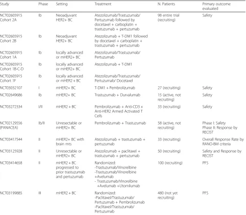
Several studies testing immune checkpoint inhibitors in combination with HER2-targeted therapies are cur-rently ongoing (Table 3). These will help us understand better the interaction between immune system and HER2-targeted agents and define successful combina-tions for future clinical trials.
Conclusions
The role of immunity in cancer treatment has recently moved into the spotlight, as mechanisms related to
immune surveillance, immune equilibrium, and immune escape have progressively been elucidated in several solid tumors and new drugs have entered clinical practice. Re-cent evidence from early phase trials supports the thera-peutic role of immunity in HER2 + BC and more data from ongoing trials will be available in the next few years.
In HER2+ BC, the interplay between immune system and tumor is complex and dynamic, involving the inter-action with different HER2-targeted treatments, chemo-therapy, hormonotherapy and the modulating action of HR status and tumor biology. A deeper understanding of these mechanisms might help optimize treatment personalization in HER2+ BC and design biologically meaningful trials that will eventually change the way we treat patients with HER2+ disease.
Table 3Clinical trials testing the association of checkpoint inhibitors and HER2-targeted treatment in HER2 + BC
Study Phase Setting Treatment N. Patients Primary outcome
evaluated
NCT02605915 Cohort 2A
Ib Neoadjuvant HER2+ BC
Atezolizumab/Trastuzumab/ Pertuzumab followed by docetaxel + carboplatin + trastuzumab + pertuzumab
98 entire trial (recruiting)
Safety
NCT02605915 Cohort 2B
Ib Neoadjuvant HER2+ BC
Atezolizumab + T-DM1 followed by docetaxel + carboplatin + trastuzumab + pertuzumab
NCT02605915 Cohort 1A
Ib locally advanced or mHER2+ BC
Atezolizumab/Trastuzumab/ Pertuzumab
NCT02605915 Cohort 1B-C-D
Ib locally advanced or mHER2+ BC
Atezolizumab + T-DM1
NCT02605915 Cohort 1F
Ib locally advanced or mHER2 + BC
Atezolizumab/Trastuzumab/ Pertuzumab/ Docetaxel
NCT03032107 I mHER2+ BC T-DM1 + Pembrolizumab 27 (recruiting) Safety
NCT02649686 Ib mHER2+ BC Trastuzumab + Durvalumab 15 (active, not recruiting)
Safety
NCT03272334 I/II mHER2 + BC Pembrolizumab + Anti-CD3 x Anti-HER2 Armed Activated T Cells
33 (recruiting) Safety
NCT02129556 (PANACEA)
Ib/II Unresectable or mHER2+ BC
Pembrolizumab + Trastuzumab 58 (active, not recruiting)
Phase I: Safety Phase II: Response by RECIST
NCT03417544 II mHER2+ BC with brain mts
Atezolizumab + trastuzumab + pertuzumab
33 (recruiting) Overall Response Rate by RANO-BM criteria
NCT03125928 II Unresectable or mHER2+ BC
Atezolizumab + paclitaxel + trastuzumab + pertuzumab
50 (recruiting) Safety and Response by RECIST
NCT03414658 II mHER2 + BC progressed to prior trastuzumab and pertuzumab
Randomized:
-Trastuzumab/Vinorelbine -Trastuzumab/Vinorelbine +Avelumab
- Trastuzumab/Vinorelbine +Avelumab +Utomilumab
100 (recruiting) PFS
NCT03199885 III mHER2 + BC Randomized:
-Paclitaxel/Trastuzumab/ Pertuzumab + Pembrolizumab -Paclitaxel/Trastuzumab/ Pertuzumab
480 (not yet recruiting)
PFS
Additional file
Additional file 1:Table S1.Summary of prognostic/predictive value of tumor infiltrating lymphocytes (TILs) in HER2+ breast cancer across prospective interventional clinical trials.Table S2.Summary of prognostic/predictive value of expression of immune genes and immune gene signatures in HER2+ breast cancer across prospective interventional clinical trials.Table S3.Peptide-based vaccine strategies targeting HER2 for the treatment of invasive HER2+ BC (early and advanced setting). (DOCX 48 kb)
Acknowledgements
No additional acknowledgements other than those listed as authors.
Funding
This work was partly supported by Pas a Pas, Save the Mama, Instituto de Salud Carlos III-PI16/00904 and Career Catalyst Grant CCR13261208 from the Susan Komen Foundation to A.P.
Availability of data and materials
Not applicable (review article).
Authors’contributions
GG collected the data and drafted the manuscript. TP, MV and AP revised the manuscript. VG provided additional revisions for the manuscript. All authors read and approved the final manuscript.
Ethics approval and consent to participate
Not applicable (review article).
Consent for publication
Not applicable (review article).
Competing interests
The authors declare that they have no competing interests.
Publisher’s Note
Springer Nature remains neutral with regard to jurisdictional claims in published maps and institutional affiliations.
Author details
1Translational Genomics and Targeted Therapeutics in Solid Tumors, IDIBAPS, Barcelona, Spain.2Department of Medical Oncology, Hospital Clínic, Barcelona, Spain.3Department of Surgery, Oncology and Gastroenterology, University of Padova, Padova, Italy.4Medical Oncology 2, Istituto Oncologico Veneto IRCCS, Padova, Italy.
Received: 26 September 2018 Accepted: 27 February 2019
References
1. Slamon D, Clark G, Wong S, Levin W, Ullrich A, McGuire W. Human breast cancer: correlation of relapse and survival with amplification of the HER-2/ neu oncogene. Science. 1987;235:177–82.
2. Balduzzi S, Mantarro S, Guarneri V, Tagliabue L, Pistotti V, Moja L, et al. Trastuzumab-containing regimens for metastatic breast cancer. Cochrane Database Syst Rev. 2014;6:CD006242.
3. Swain SM, Baselga J, Kim SB, Ro J, Semiglazov V, Campone M, et al. Pertuzumab, trastuzumab, and docetaxel in HER2-positive metastatic breast cancer. N Engl J Med. 2015;372:724–34.
4. Verma S, Miles D, Gianni L, Krop IE, Welslau M, Baselga J, et al. Trastuzumab emtansine for HER2-positive advanced breast cancer. N Engl J Med. 2012; 367:1783–91.
5. Geyer CE, Forster J, Lindquist D, Chan S, Romieu CG, Pienkowski T, et al. Lapatinib plus Capecitabine for HER2-positive advanced breast cancer. N Engl J Med. 2006;355:2733–43.
6. Martin M, Holmes FA, Ejlertsen B, Delaloge S, Moy B, Iwata H, et al. Neratinib after trastuzumab-based adjuvant therapy in HER2-positive breast cancer
(ExteNET): 5-year analysis of a randomised, double-blind, placebo-controlled, phase 3 trial. Lancet Oncol. 2017;18:1688–700.
7. Loi S, Sirtaine N, Piette F, Salgado R, Viale G, Van Eenoo F, et al. Prognostic and predictive value of tumor-infiltrating lymphocytes in a phase III randomized adjuvant breast cancer trial in node-positive breast cancer comparing the addition of docetaxel to doxorubicin with doxorubicin-based chemotherapy: BIG 02–98. J Clin Oncol. 2013;31:860–7.
8. Stanton SE, Adams S, Disis ML. Variation in the Incidence and Magnitude of Tumor-Infiltrating Lymphocytes in Breast Cancer Subtypes. JAMA Oncol. 2016;2:1354.
9. Nuciforo P, Pascual T, Cortés J, Llombart-Cussac A, Fasani R, Paré L, et al. A predictive model of pathologic response based on tumor cellularity and tumor-infiltrating lymphocytes (CelTIL) in HER2-positive breast cancer treated with chemo-free dual HER2 blockade. Ann Oncol. 2018;29:170–7. 10. Denkert C, von Minckwitz G, Darb-Esfahani S, Lederer B, Heppner BI, Weber
KE, et al. Tumour-infiltrating lymphocytes and prognosis in different subtypes of breast cancer: a pooled analysis of 3771 patients treated with neoadjuvant therapy. Lancet Oncol. 2018;19:40–50.
11. Luen SJ, Salgado R, Fox S, Savas P, Eng-Wong J, Clark E, et al. Tumour-infiltrating lymphocytes in advanced HER2-positive breast cancer treated with pertuzumab or placebo in addition to trastuzumab and docetaxel: a retrospective analysis of the CLEOPATRA study. Lancet Oncol. 2017;18:52–62. 12. Loi S, Giobbe-Hurder A, Gombos A, Bachelot T, Hui R, Curigliano G, et al.
Abstract GS2–06: Phase Ib/II study evaluating safety and efficacy of pembrolizumab and trastuzumab in patients with trastuzumab-resistant HER2-positive metastatic breast cancer: Results from the PANACEA (IBCSG 45–13/BIG 4–13/KEYNOTE-014) study. Cancer Res. 2018;78:GS2–06. 13. Bianchini G, Gianni L. The immune system and response to HER2-targeted
treatment in breast cancer. Lancet Oncol. 2014;15:e58–68.
14. Savas P, Caramia F, Teo ZL, Loi S. Oncogene addiction and immunity: clinical implications of tumour infiltrating lymphocytes in breast cancers overexpressing the HER2/neu oncogene. Curr Opin Oncol. 2014;26:562–7.
15. Ladjemi MZ, Jacot W, Chardès T, Pèlegrin A, Navarro-Teulon I. Anti-HER2 vaccines: new prospects for breast cancer therapy. Cancer Immunol Immunother. 2010;59:1295–312.
16. Huber CH, Wölfel T. Immunotherapy of cancer: from vision to standard clinical practice. J Cancer Res Clin Oncol. 2004;130:367–74.
17. Molina MA, Codony-Servat J, Albanell J, Rojo F, Arribas J, Baselga J. Trastuzumab (herceptin), a humanized anti-Her2 receptor monoclonal antibody, inhibits basal and activated Her2 ectodomain cleavage in breast cancer cells. Cancer Res. 2001;61:4744–9.
18. Baselga J, Albanell J. Mechanism of action of anti-HER2 monoclonal antibodies. Ann Oncol Off J Eur Soc Med Oncol. 2001;12(Suppl 1):S35–41. 19. Muntasell A, Cabo M, Servitja S, Tusquets I, Martínez-García M, Rovira A, et
al. Interplay between Natural killer cells and Anti-HER2 antibodies: Perspectives for breast cancer immunotherapy. Front Immunol. 2017;8:1544. 20. Datta J, Berk E, Xu S, Fitzpatrick E, Rosemblit C, Lowenfeld L, et al. Anti-HER2 CD4(+) T-helper type 1 response is a novel immune correlate to pathologic response following neoadjuvant therapy in HER2-positive breast cancer. Breast Cancer Res. 2015;17:71 BioMed Central.
21. Datta J, Fracol M, McMillan MT, Berk E, Xu S, Goodman N, et al. Association of Depressed Anti-HER2 T-Helper Type 1 Response With Recurrence in Patients With Completely Treated HER2-Positive Breast Cancer. JAMA Oncol. 2016;2:242. 22. Hendry S, Salgado R, Gevaert T, Russell PA, John T, Thapa B, et al. Assessing
Tumor-infiltrating Lymphocytes in Solid Tumors. Adv Anat Pathol. 2017;24: 235–51.
23. Salgado R, Denkert C, Demaria S, Sirtaine N, Klauschen F, Pruneri G, et al. The evaluation of tumor-infiltrating lymphocytes (TILs) in breast cancer: recommendations by an International TILs Working Group 2014. Ann Oncol. 2015;26:259–71.
24. Perez EA, Thompson EA, Ballman KV, Anderson SK, Asmann YW, Kalari KR, et al. Genomic analysis reveals that immune function genes are strongly linked to clinical outcome in the North Central Cancer Treatment Group n9831 Adjuvant Trastuzumab Trial. J Clin Oncol. 2015;33:701–8.
25. Schalper KA, Velcheti V, Carvajal D, Wimberly H, Brown J, Pusztai L, et al. In Situ Tumor PD-L1 mRNA Expression Is Associated with Increased TILs and Better Outcome in Breast Carcinomas. Clin Cancer Res. 2014;20:2773–82. 26. Paré L, Pascual T, Seguí E, Teixidó C, Gonzalez-Cao M, Galván P, et al.
27. Perez EA, Ballman KV, Tenner KS, Thompson EA, Badve SS, Bailey H. et al., Association of stromal tumor-infiltrating lymphocytes with recurrence-free survival in the N9831 adjuvant trial in patients with early-stage HER2-positive breast cancer. JAMA Oncol. 2016;(1):56–64.
28. Kim S-R, Gavin PG, Pogue-Geile KL, Song N, Finnigan M, Bandos H, et al. Abstract 2837: A surrogate gene expression signature of tumor infiltrating lymphocytes (TILs) predicts degree of benefit from trastuzumab added to standard adjuvant chemotherapy in NSABP (NRG) trial B-31 for HER2+ breast cancer. Cancer Res. 2015;75:2837.
29. Loi S, Michiels S, Salgado R, Sirtaine N, Jose V, Fumagalli D, et al. Tumor infiltrating lymphocytes are prognostic in triple negative breast cancer and predictive for trastuzumab benefit in early breast cancer: results from the FinHER trial. Ann Oncol. 2014;25:1544–50.
30. Kim S-R, Song N, Gavin PG, Salgado R, Bandos H, Kos Z. et al. NRG Oncology/NSABP B-31: Stromal tumor infiltrating lymphocytes (sTILs) and outcomes in early-stage HER2-positive breast cancer (BC). 2018 ASCO Annu Meet. J Clin Oncol. 2018;36(suppl; abstr 12010).
31. Hamy A-S, Pierga J-Y, Sabaila A, Laas E, Bonsang-Kitzis H, Laurent C, et al. Stromal lymphocyte infiltration after neoadjuvant chemotherapy is associated with aggressive residual disease and lower disease-free survival in HER2-positive breast cancer. Ann Oncol. 2017;28:2233–40.
32. Dieci MV, Mathieu MC, Guarneri V, Conte P, Delaloge S, Andre F, et al. Prognostic and predictive value of tumor-infiltrating lymphocytes in two phase III randomized adjuvant breast cancer trials. Ann Oncol. 2015;26:1698–704. 33. Loi S, Dushyanthen S, Beavis PA, Salgado R, Denkert C, Savas P, et al. RAS/
MAPK activation is associated with reduced tumor-infiltrating lymphocytes in triple-negative breast cancer: therapeutic cooperation between MEK and PD-1/PD-L1 immune checkpoint inhibitors. Clin Cancer Res. 2016;22(6): 1499–509.
34. Ladoire S, Arnould L, Apetoh L, Coudert B, Martin F, Chauffert B, et al. Pathologic complete response to neoadjuvant chemotherapy of breast carcinoma is associated with the disappearance of tumor-infiltrating foxp3+ regulatory T cells. Clin Cancer Res. 2008;14:2413–20.
35. Force J, Howie LJ, Abbott SE, Bentley R, Marcom PK, Kimmick G, et al. Early stage HER2-positive breast cancers not achieving a pCR From neoadjuvant trastuzumab- or pertuzumab-based regimens have an immunosuppressive phenotype. Clin Breast Cancer. 2018;18(5):410–7.
36. Ladoire S, Mignot G, Dabakuyo S, Arnould L, Apetoh L, Rébé C, et al. In situ immune response after neoadjuvant chemotherapy for breast cancer predicts survival. J Pathol. 2011;224:389–400.
37. Dieci MV, Conte P, Bisagni G, Brandes AA, Frassoldati A, Cavanna L, et al. Association of tumor-infiltrating lymphocytes with distant disease-free survival in the ShortHER randomized adjuvant trial for patients with early HER2+ breast cancer. Ann Oncol. 2019; Epub 17 Jan 2019.
38. Denkert C, von Minckwitz G, Brase JC, Sinn BV, Gade S, Kronenwett R, et al. Tumor-infiltrating lymphocytes and response to neoadjuvant chemotherapy with or without carboplatin in human epidermal growth factor receptor 2-positive and triple-negative primary breast cancers. J Clin Oncol. 2015;33: 983–91.
39. Savas P, Salgado R, Denkert C, Sotiriou C, Darcy PK, Smyth MJ, et al. Clinical relevance of host immunity in breast cancer: from TILs to the clinic. Nat Rev Clin Oncol. 2016;13:228–41.
40. Yamashita-Kashima Y, Iijima S, Yorozu K, Furugaki K, Kurasawa M, Ohta M, et al. Pertuzumab in combination with trastuzumab shows significantly enhanced antitumor activity in her2-positive human gastric cancer xenograft models. Clin Cancer Res. 2011;17:5060–70.
41. Tóth G, Szöőr Á, Simon L, Yarden Y, Szöllősi J, Vereb G. The
combination of trastuzumab and pertuzumab administered at approved doses may delay development of trastuzumab resistance by additively enhancing antibody-dependent cell-mediated cytotoxicity. MAbs. 2016;8: 1361–70.
42. Mamidi S, Cinci M, Hasmann M, Fehring V, Kirschfink M. Lipoplex mediated silencing of membrane regulators (CD46, CD55 and CD59) enhances complement-dependent anti-tumor activity of trastuzumab and pertuzumab. Mol Oncol. 2013;7:580–94.
43. Scheuer W, Friess T, Burtscher H, Bossenmaier B, Endl J, Hasmann M. Strongly Enhanced antitumor activity of trastuzumab and pertuzumab combination treatment on HER2-positive human xenograft tumor models. Cancer Res. 2009;69:9330–6.
44. Ignatiadis M, Van den Eynden G, Roberto S, Fornili M, Bareche Y, Desmedt C, et al. Tumor-infiltrating lymphocytes in patients receiving trastuzumab/
pertuzumab-based chemotherapy: A TRYPHAENA substudy. JNCI J Natl Cancer Inst. 2019;111(1):69–77.
45. Bianchini G, Pusztai L, Pienkowski T, Im Y-H, Bianchi GV, Tseng L-M, et al. Immune modulation of pathologic complete response after neoadjuvant HER2-directed therapies in the NeoSphere trial. Ann Oncol. 2015;26(12):2429–36.
46. Guarneri V, Dieci M, Bisagni G, Frassoldati A, Bianchi G, De Salvo G, et al. De-escalated treatment with trastuzumab-pertuzumab-letrozole in patients with HR+/HER2+ operable breast cancer with Ki67 response after 2 weeks letrozole: Final results of the PerELISA neoadjuvant study. J Clin Oncol. 2018; 36:abstr 507.
47. Urruticoechea A, Rizwanullah M, Im S-A, Ruiz ACS, Láng I, Tomasello G, et al. Randomized phase III trial of trastuzumab plus capecitabine with or without pertuzumab in patients with human epidermal growth factor receptor 2– positive metastatic breast cancer who experienced disease progression during or after trastuzumab-based therapy. J Clin Oncol. 2017;35:3030–8. 48. Motzer RJ, Escudier B, McDermott DF, George S, Hammers HJ, Srinivas S, et
al. Nivolumab versus everolimus in advanced renal-cell carcinoma. N Engl J Med. 2015;373:1803–13.
49. Reck M, Rodríguez-Abreu D, Robinson AG, Hui R, Csőszi T, Fülöp A, et al. Pembrolizumab versus chemotherapy for PD-L1–positive non–small-cell lung cancer. N Engl J Med. 2016;375:1823–33.
50. Dieci MV, Tsvetkova V, Orvieto E, Piacentini F, Ficarra G, Griguolo G, et al. Immune characterization of breast cancer metastases: prognostic implications. Breast Cancer Res. 2018;20:62.
51. Scaltriti M, Verma C, Guzman M, Jimenez J, Parra JL, Pedersen K, et al. Lapatinib, a HER2 tyrosine kinase inhibitor, induces stabilization and accumulation of HER2 and potentiates trastuzumab-dependent cell cytotoxicity. Oncogene. 2009;28:803–14.
52. Maruyama T, Mimura K, Izawa S, Inoue A, Shiba S, Watanabe M, et al. Lapatinib enhances herceptin-mediated antibody-dependent cellular cytotoxicity by up-regulation of cell surface HER2 expression. Anticancer Res. 2011;31:2999–3005.
53. Hannesdóttir L, Tymoszuk P, Parajuli N, Wasmer M-H, Philipp S, Daschil N, et al. Lapatinib and doxorubicin enhance the Stat1-dependent antitumor immune response. Eur J Immunol. 2013;43:2718–29.
54. Solinas C, Ceppi M, Lambertini M, Scartozzi M, Buisseret L, Garaud S, et al. Tumor-infiltrating lymphocytes in patients with HER2-positive breast cancer treated with neoadjuvant chemotherapy plus trastuzumab, lapatinib or their combination: A meta-analysis of randomized controlled trials. Cancer Treat Rev. 2017;57:8–15.
55. Dieci MV, Prat A, Tagliafico E, Paré L, Ficarra G, Bisagni G, et al. Integrated evaluation of PAM50 subtypes and immune modulation of pCR in positive breast cancer patients treated with chemotherapy and HER2-targeted agents in the CherLOB trial. Ann Oncol. 2016;27:1867–73. 56. Issa-Nummer Y, Darb-Esfahani S, Loibl S, Kunz G, Nekljudova V, Schrader I, et
al. Prospective validation of immunological infiltrate for prediction of response to neoadjuvant chemotherapy in HER2-negative breast cancer--a substudy of the neoadjuvant GeparQuinto trial. PLoS One. 2013;8:e79775. 57. Ingold Heppner B, Untch M, Denkert C, Pfitzner BM, Lederer B, Schmitt W,
et al. Tumor-infiltrating lymphocytes: a predictive and prognostic biomarker in neoadjuvant-treated HER2-positive breast cancer. Clin Cancer Res. 2016; 22:5747–54.
58. Salgado R, Denkert C, Campbell C, Savas P, Nuciforo P, Aura C, et al. Tumor-Infiltrating lymphocytes and associations with pathological complete response and event-free survival in HER2-positive early-stage breast cancer treated with lapatinib and trastuzumab. JAMA Oncol. 2015;1:448. 59. Fumagalli D, Venet D, Ignatiadis M, Azim HA, Maetens M, Rothé F, et al. RNA
Sequencing to predict response to neoadjuvant anti-HER2 therapy: A secondary analysis of the NeoALTTO randomized clinical trial. JAMA Oncol. 2016;3:227. 60. Musolino A, Naldi N, Dieci MV, Zanoni D, Rimanti A, Boggiani D, et al.
Immunoglobulin G fragment C receptor polymorphisms and efficacy of preoperative chemotherapy plus trastuzumab and lapatinib in HER2-positive breast cancer. Pharmacogenomics J. 2016;16:472–7.
61. Llombart-Cussac A, Cortés J, Paré L, Galván P, Bermejo B, Martínez N, et al. HER2-enriched subtype as a predictor of pathological complete response following trastuzumab and lapatinib without chemotherapy in early-stage HER2-positive breast cancer (PAMELA): an open-label, single-group, multicentre, phase 2 trial. Lancet Oncol. 2017;18:545–54.
63. Müller P, Martin K, Theurich S, Schreiner J, Savic S, Terszowski G, et al. Microtubule-depolymerizing agents used in antibody-drug conjugates induce antitumor immunity by stimulation of dendritic cells. Cancer Immunol Res. 2014;2:741–55.
64. Martin K, Müller P, Schreiner J, Prince SS, Lardinois D, Heinzelmann-Schwarz VA, et al. The microtubule-depolymerizing agent ansamitocin P3 programs dendritic cells toward enhanced anti-tumor immunity. Cancer Immunol Immunother. 2014;63:925–38.
65. Müller P, Kreuzaler M, Khan T, Thommen DS, Martin K, Glatz K, et al. Trastuzumab emtansine (T-DM1) renders HER2+breast cancer highly
susceptible to CTLA-4/PD-1 blockade. Sci Transl Med. 2015;7:315ra188. 66. Canonici A, Gijsen M, Mullooly M, Bennett R, Bouguern N, Pedersen K, et al.
Neratinib overcomes trastuzumab resistance in HER2 amplified breast cancer. Oncotarget. 2013;4:1592–605.
67. Collins DM, Gately K, Hughes C, Edwards C, Davies A, Madden SF, et al. Tyrosine kinase inhibitors as modulators of trastuzumab-mediated antibody-dependent cell-mediated cytotoxicity in breast cancer cell lines. Cell Immunol. 2017;319:35–42.
68. Kuerer HM, Buzdar AU, Mittendorf EA, Esteva FJ, Lucci A, Vence LM, et al. Biologic and immunologic effects of preoperative trastuzumab for ductal carcinoma in situ of the breast. Cancer. 2011;117:39–47.
69. Chung YR, Kim HJ, Jang MH, Park SY. Prognostic value of tumor infiltrating lymphocyte subsets in breast cancer depends on hormone receptor status. Breast Cancer Res Treat. 2017;161:409–20.
70. Kanamori H, Krieg S, Mao C, Di Pippo VA, Wang S, Zajchowski DA, et al. Proteinase Inhibitor 9, an Inhibitor of Granzyme B-mediated Apoptosis, Is a Primary Estrogen-inducible Gene in Human Liver Cells. J Biol Chem. 2000; 275:5867–73.
71. Jiang X, Ellison SJ, Alarid ET, Shapiro DJ. Interplay between the levels of estrogen and estrogen receptor controls the level of the granzyme inhibitor, proteinase inhibitor 9 and susceptibility to immune surveillance by natural killer cells. Oncogene. 2007;26:4106–14.
72. Hamada K, Gleason SL, Levi BZ, Hirschfeld S, Appella E, Ozato K. H-2RIIBP, a member of the nuclear hormone receptor superfamily that binds to both the regulatory element of major histocompatibility class I genes and the estrogen response element. Proc Natl Acad Sci. 1989;86:8289–93. 73. Sim BC, Hui KM. A HLA class Icis-regulatory element whose activity can be
modulated by hormones. Int J Cancer. 1994;59:646–54.
74. Cejalvo JM, Pascual T, Fernández-Martínez A, Brasó-Maristany F, Gomis RR, Perou CM, et al. Clinical implications of the non-luminal intrinsic subtypes in hormone receptor-positive breast cancer. Cancer Treat Rev. 2018;67:63–70. 75. Prat A, Bianchini G, Thomas M, Belousov A, Cheang MCU, Koehler A, et al.
Research-based PAM50 subtype predictor identifies higher responses and improved survival outcomes in HER2-positive breast cancer in the NOAH study. Clin Cancer Res. 2014;20:511–21.
76. Koboldt DC, Fulton RS, McLellan MD, Schmidt H, Kalicki-Veizer J, McMichael JF, et al. Comprehensive molecular portraits of human breast tumours. Nature. 2012;490:61–70.
77. Perou CM, Sørlie T, Eisen MB, van de Rijn M, Jeffrey SS, Rees CA, et al. Molecular portraits of human breast tumours. Nature. 2000;406:747–52. 78. Roberts SA, Lawrence MS, Klimczak LJ, Grimm SA, Fargo D, Stojanov P, et al.
An APOBEC cytidine deaminase mutagenesis pattern is widespread in human cancers. Nat Genet. 2013;45:970–6.
79. Venkatesan S, Rosenthal R, Kanu N, McGranahan N, Bartek J, Quezada SA, et al. Perspective: APOBEC mutagenesis in drug resistance and immune escape in HIV and cancer evolution. Ann Oncol. 2018;29:563–72.
80. Gil Del Alcazar CR, Huh SJ, Ekram MB, Trinh A, Liu LL, Beca F, et al. Immune escape in breast cancer during in situ to invasive carcinoma transition. Cancer Discov. 2017;7:1098–115.
81. Harris SJ, Brown J, Lopez J, Yap TA. Immuno-oncology combinations: raising the tail of the survival curve. Cancer Biol Med. 2016;13:171–93.
82. Di Modica M, Sfondrini L, Regondi V, Varchetta S, Oliviero B, Mariani G, et al. Taxanes enhance trastuzumab-mediated ADCC on tumor cells through NKG2D-mediated NK cell recognition. Oncotarget. 2016;7:255–65. 83. Richards JO, Albers AJ, Smith TS, Tjoe JA. NK cell-mediated
antibody-dependent cellular cytotoxicity is enhanced by tamoxifen in HER2/neu non-amplified, but not HER2/neu-non-amplified, breast cancer cells. Cancer Immunol Immunother. 2016;65:1325–35.
84. Wang J, Zhang Q, Jin S, Feng M, Kang X, Zhao S, et al. Immoderate inhibition of estrogen by anastrozole enhances the severity of experimental polyarthritis. Exp Gerontol. 2009;44(6–7):398–405.
85. Goel S, DeCristo MJ, Watt AC, BrinJones H, Sceneay J, Li BB, et al. CDK4/6 inhibition triggers anti-tumour immunity. Nature. 2017;548(7668):471–5. 86. Ciruelos E, Villagrasa P, Paré L, Oliveira M, de la Peña L, Pernas S, et al.
Abstract P5–20-19: PAM50 intrinsic subtype predicts survival outcome in HER2-positive/hormone receptor-positive metastatic breast cancer treated with palbociclib and trastuzumab: a correlative analysis of the PATRICIA (SOLTI 13–03) trial. AACR; 2018.
87. Dieci MVV, Griguolo G, Miglietta F, Guarneri V. The immune system and hormone-receptor positive breast cancer: Is it really a dead end? Cancer Treat Rev. 2016;46:9–19.
88. Dunbier AK, Ghazoui Z, Anderson H, Salter J, Nerurkar A, Osin P, et al. Molecular profiling of aromatase inhibitor-treated postmenopausal breast tumors identifies immune-related correlates of resistance. Clin Cancer Res. 2013;19:2775–86.
89. Dieci MV, Frassoldati A, Generali D, Bisagni G, Piacentini F, Cavanna L, et al. Tumor-infiltrating lymphocytes and molecular response after neoadjuvant therapy for HR+/HER2−breast cancer: results from two prospective trials. Breast Cancer Res Treat. 2017;163.
90. Svoronos N, Perales-Puchalt A, Allegrezza MJ, Rutkowski MR, Payne KK, Tesone AJ, et al. Tumor cell–independent estrogen signaling drives disease progression through mobilization of myeloid-derived suppressor cells. Cancer Discov. 2017;7:72–85.
91. Musolino A, Naldi N, Bortesi B, Pezzuolo D, Capelletti M, Missale G, et al. Immunoglobulin G fragment C receptor polymorphisms and clinical efficacy of trastuzumab-based therapy in patients with HER-2/neu–positive metastatic breast cancer. J Clin Oncol. 2008;26:1789–96.
92. Hurvitz SA, Betting DJ, Stern HM, Quinaux E, Stinson J, Seshagiri S, et al. Analysis of Fc receptor IIIa and IIa polymorphisms: lack of correlation with outcome in trastuzumab-treated breast cancer patients. Clin Cancer Res. 2012;18:3478–86.
93. Gavin PG, Song N, Kim SR, Lipchik C, Johnson NL, Bandos H, et al. Association of Polymorphisms inFCGR2AandFCGR3AWith Degree of Trastuzumab Benefit in the Adjuvant Treatment of ERBB2/HER2–Positive Breast Cancer. JAMA Oncol. 2017;3:335.
94. Bang YJ, Giaccone G, Im SA, Oh DY, Bauer TM, Nordstrom JL, et al. First-in-human Phase 1 study of margetuximab (MGAH22), an Fc-modified chimeric monoclonal antibody, in patients with HER2-positive advanced solid tumors. Ann Oncol. 2017;28:mdx002.
95. Kiewe P, Hasmüller S, Kahlert S, Heinrigs M, Rack B, Marmé A, et al. Phase I trial of the trifunctional anti-HER2 x Anti-CD3 antibody ertumaxomab in metastatic breast cancer. Clin Cancer Res. 2006;12:3085–91.
96. Lum LG, Thakur A, Al-Kadhimi Z, Colvin GA, Cummings FJ, Legare RD, et al. Targeted T-cell Therapy in Stage IV Breast Cancer: A Phase I Clinical Trial. Clin Cancer Res. 2015;21:2305–14.
97. Li J, Li W, Huang K, Zhang Y, Kupfer G, Zhao Q. Chimeric antigen receptor T cell (CAR-T) immunotherapy for solid tumors: lessons learned and strategies for moving forward. J Hematol Oncol. 2018;11:22.
98. Stagg J, Loi S, Divisekera U, Ngiow SF, Duret H, Yagita H, et al. Anti-ErbB-2 mAb therapy requires type I and II interferons and synergizes with anti-PD-1 or anti-CD137 mAb therapy. Proc Natl Acad Sci. 2011;108:7142–7. 99. Emens LA, Esteva F, Beresford M, Saura C, De Laurentiis M, Kim S-B, et al.
Abstract PD3–01: Results from KATE2, a randomized phase 2 study of atezolizumab (atezo)+trastuzumab emtansine (T-DM1) vs placebo (pbo)+T-DM1 in previously treated HER2+ advanced breast cancer (BC). SABCS; 2018. 100. Carey LA, Berry DA, Cirrincione CT, Barry WT, Pitcher BN, Harris LN, et al.