R E S E A R C H
Open Access
Differential expression of lysosome-associated
protein transmembrane-4 beta (LAPTM4B) in
granulosa cells of ovarian follicles and in other
bovine tissues
Kalidou Ndiaye
*, Paul D Carrière, Jean Sirois, David W Silversides and Jacques G Lussier
Abstract
Background:LAPTM4B is a member of the lysosome-associated transmembrane protein superfamily that is differentially expressed in normal human tissues and upregulated in various types of carcinomas. These proteins are thought to be involved in the regulation of cell proliferation and survival. The objective of this study was to investigate the expression of bovine LAPTM4B during ovarian follicular development and in various bovine tissues. Methods and results:Northern blot analysis revealed a 1.8 kb transcript, with highly variable steady state levels among tissues. RT-PCR analysis showed that LAPTM4B mRNA transcripts were low in granulosa cells of small antral follicles, increased in large dominant follicles, and decreased in ovulatory follicles following injection of human chorionic gonadotropin (hCG; P < 0.003). Ovulatory follicles collected at various times after hCG injection revealed a significant reduction of LAPTM4B mRNA starting at 18 h post-hCG (P < 0.029). Immunobloting analysis using antibodies generated against bovine LAPTM4B recognized proteins of 26.3 and 31.5 kDa in granulosa cells of developing follicles and corpus luteum. Further analyses of affinity-purified His-tag LAPTM4B overexpressed in HEK cells showed that the 31.5 kDa protein represented the ubiquinated isoform of the 26.3 kDa native protein. The 26.3 kDa protein was differentially expressed showing highest amounts in dominant follicles and lowest amounts in ovulatory follicles 24 h post-hCG. Immunohistochemical analyses of LAPTM4B showed marked heterogeneity of labeling signal among tissues, with LAPTM4B mainly localized to perinuclear vesicles, in keeping with its putative lysosomal membrane localization. Conclusion:This study reports for the first time that bovine LAPTM4B in granulosa cells is present in both unubiquinated and ubiquinated forms, and is differentially expressed in developing ovarian follicles, suggesting a possible role in terminal follicular growth.
Keywords:Ovary, Follicle, Granulosa cells, LAPTM4B, Bovine, Ubiquitination
Background
The lysosome-associated protein transmembrane 4 beta (LAPTM4B) belongs to the LAPTM gene family along with LAPTM4A and LAPTM5. These proteins are classi-fied according to the number of their transmembrane do-mains [1,2] and are thought to be involved in the regulation of cell proliferation and survival [3,4]. The members of the LAPTM family share similarities to
lysosomal proteins since they are membrane proteins that contain multiple tyrosine residues. These tyrosine-based motifs function as addressing signals, targeting the pro-teins toward lysosomes or late endosomes [3,5-7]. Specif-ically, LAPTM4B was localized to lysosomes as well as to the plasma membrane and internal organelles such as the Golgi apparatus and endosomes [8]. LAPTM4B mRNA was originally reported to be overexpressed in human he-patocellular carcinoma [9] and widely expressed in other normal human tissues, including total human ovarian ex-tract [10]. Furthermore, its mRNA level was increased in various types of carcinomas including breast, ovarian, * Correspondence:k.ndiaye@umontreal.ca
Centre de recherche en reproduction animale, Département de biomédecine vétérinaire, Faculté de médecine vétérinaire, Université de Montréal, P.O. Box 5000, St-Hyacinthe, Québec J2S 7C6, Canada
uterine, lung and gastric cancers [11-14]. On a functional basis, LAPTM4B promotes autophagy, a cell survival mechanism mediated by lysosomes that renders tumor cells resistant to metabolic and genotoxic stress, which promotes a faster tumor growth rate [15]. Moreover, knockdown of LAPTM4B mRNA using siRNA resulted in increased sensitivity of tumor cells to anthracyclines, an intercaling agent and a free radical inducer. Further ana-lyses indicated that LAPTM4B overexpression resulted in specific sequestration of anthracycline and delayed the entry of anthracycline into the nucleus [15].
LAPTM4B has been studied mainly in human and specifically in regard to its association with various car-cinomas, as referenced above. In the present study, we investigated the expression, localization, and regulation of the LAPTM4B mRNA and protein in the bovine spe-cies specifically during the final stages of ovarian follicu-lar development. In a preliminary expression study, we observed that LAPTM4B mRNA was strongly expressed in granulosa cells of dominant or preovulatory follicles. We also extended the analysis to other bovine tissues to allow for comparisons since no data is available in the bovine species.
The cyclic ovarian activity results in profound modifica-tions that require spatio-temporal coordination of prolifer-ation, apoptosis and differentiation of various cell types within the follicles resulting from changes in gene expres-sion. The growth of antral follicles is mainly under the in-fluence of the follicle-stimulating hormone (FSH) [16]. During follicular selection and dominance, granulosa cells acquire luteinizing hormone receptors (LHr) that allows the transfer of follicle dependency from FSH to LH and the increase in synthesis of oestradiol-17β [17,18]. The preovulatory surge of LH from the anterior pituitary gland results in ovulation and the release of the oocyte, and the remaining granulosa and theca cells of the follicle wall dif-ferentiate into luteal cells to form the corpus luteum and produce progesterone [19]. Granulosa cells thus play a critical role in reproductive functions as they contribute to steroid hormone synthesis [20], oocyte maturation [21], and corpus luteum formation after ovulation [22]. The transcription of genes in granulosa cells that control the growth of a bovine dominant or preovulatory follicle is rapidly downregulated or silenced as a result of LH-mediated increases in intracellular signaling [23]. Con-versely, LH upregulates or induces the expression of genes involved in ovulation and luteinization [24-26]. These studies demonstrate the critical importance of gene expression and regulation studies during the final stages of follicular development and ovulation. In this study, we report the differential regulation and localization of LAPTM4B in granulosa cells during the periovulatory period as well as in other bovine tissues that lead to a bet-ter understanding of its physiological function.
Methods
Experimental animal models
The regulation of LAPTM4B expression during follicular development and ovulation was studied using in vivo models as previously characterized [23]. Estrous cycles of normal cycling crossbred heifers were synchronized with one injection of PGF2α (25 mg, im; Lutalyse, Upjohn,
Kalamazoo, MI) given in the presence of a corpus luteum, and ovarian follicular development was monitored by daily transrectal ultrasonography. Following estrous synchro-nization, heifers were randomly assigned to the dominant follicle group (DF, n = 4), or the ovulatory hCG-induced follicle group (OF, n = 4). In the DF group, the ovary bear-ing the DF on the mornbear-ing of day 5 of the estrous cycle (day 0 = day of estrus) was obtained by ovariectomy (via colpotomy). The DF was defined as > 8 mm in diameter and growing while subordinate follicles were either static or regressing. The OF were obtained following an injec-tion of 25 mg of PGF2α (Lutalyse) on day 7 to induce
luteolysis, thereby promoting the development of the DF of the first follicular wave into a preovulatory follicle. An ovulatory dose of hCG (3000 IU, iv; APL, Ayerst Lab, Montréal, QC) was injected 36 h after the induction of luteolysis, and the ovary bearing the hCG-induced OF was collected by ovariectomy at 0, 6, 12, 18, and 24 h after hCG injection (n = 2–4 cows/time point). Immediately fol-lowing ovariectomy, follicles were dissected into prepara-tions of follicular wall (theca interna with attached granulosa cells) [27] or further dissected into separate iso-lates of granulosa cells [23], and stored at −70°C. Add-itionally, GC were collected from 2 to 4 mm small follicles (SF) obtained from slaughterhouse ovaries, and a total of three pools of 20 SF was prepared. Concentrations of progesterone (P4), and estradiol-17β (E2), and their ratio
(P4/E2) were validated by radioimmunoassay of follicular
fluid as previously described [23]. Corpora lutea (CL) at day 5 of the estrous cycle were obtained by ovariectomy and were dissected from the ovarian stroma, frozen in liquid nitrogen, and then stored at −70°C. The Animal Ethics Committee of the Faculty of Veterinary Medicine of the University of Montreal approved all animal procedures.
Cloning of bovine LAPTM4B
purified by mini-prep (Qiagen, Mississauga, ON). The LAPTM4B cDNA was entirely characterized by sequen-cing on an ABI Prism 310 (Applied BioSystem).
The 5′-end of the bovine LAPTM4B was verified by screening a genomic DNA library to eventually yield all the 5′-untranslated region and promoter sequences. The gen-omic DNA library was prepared in Lambda phages (BD Biosciences Clontech) and 1×10⁷pfu was screened using the bovine LAPTM4B cDNA (GenBank NM_205802) as a P32-radiolabelled probe. Hybridized clones were analyzed by PCR using specific probes designed in the 5′-UTR of the LAPTM4B cDNA (forward: GCGAGCTCTTCGCGG GGAGAG; reverse: CAAGTACCAGACGCCGAGCAG). A second round of screening was performed to isolate a positive clone. Recombinant DNA was purified as de-scribed [29], cloned into the pDrive vector (Qiagen Clon-ing kit) followClon-ingBamH1 digestion, and characterized by sequencing.
mRNA expression analysis
RNA was isolated from various bovine tissues obtained from slaughterhouse by extraction in lysis buffer (4 M guanidium isothiocyanate, 0.5% Na-N-laurylsarcosine, 25 mM Na-citrate, pH 7), sedimentation, and centrifuga-tion on a cesium chloride cushion as previously described [28]. The concentration of total RNA was quantified by measurement of optical density at 260 nm, and quality was evaluated by visualizing the 28S and 18S ribosomal bands following electrophoretic separation on 0.66 M for-maldehyde denaturing 1% agarose gel with ethidium bromide [29]. Expression of LAPTM4B mRNA in various tissues was compared by Northern analysis. Total RNA (20μg/tissue) was size-fractionned on a 0.66 M formalde-hyde 1% agarose gel, transfered by capillarity onto a nylon membrane (Hybond-N; GE Healthcare Life Sciences), and UV-treated (150 mJ) as previously described [28]. The amount of ribosomal RNA (18S) was estimated after methylene blue staining and the image was digitized (FotoDyne Inc., Hartland, WI) and analyzed using the NIH Image software. The bovine LAPTM4B cDNA was used to generate a radioactive probe incorporating [α32
P]-dCTP (NEN Life Sciences, Boston, MA) that was subse-quently used to hybridize Northern blots as described [28]. The film images were digitized and the intensity of bands expressed as ratios of 18S ribosomal RNA.
Expression and regulation ofLAPTM4BmRNA during follicular development and following hCG injection was analyzed by semi-quantitative RT-PCR. Total RNA was extracted from bovine GC collected from follicles at dif-ferent developmental stages (SF, DF, OF) and CL, and from follicular wall (granulosa and theca cells) collected at 0, 6, 12, 18, and 24 hours after an injection of hCG. The sample at 0 hour was represented by day 7 domin-ant follicle (DF). Specific LAPTM4B PCR primers were
used and the number of cycles was limited and opti-mized for analysis ofLAPTM4BmRNA expression. PCR reaction products were separated on a 2% TAE-agarose gel with ethidium bromide, visualized by UV light, digi-tized and analyzed by densitometry using ImageQuant software (GE Healthcare Life Sciences). GAPDH was used as a control gene, and specific signals ofLAPTM4B were normalized with correspondingGAPDHsignals.
Production of polyclonal anti-bovine LAPTM4B antibodies
A fragment corresponding to amino acids Lys169 to Ala225(LAPTM4B; Molecular weight = 7.5 kDa) located at the carboxy-terminal end of the bovine LAPTM4B was used to generate a glutathione S-transferase fusion protein (GST Gene Fusion System; GE Healthcare Life Sciences) to produce specific polyclonal antibodies. To increase the molecular weight of LAPTM4B recombin-ant protein fragment and thus facilitate down-stream purification procedures, a DNA construct that included a tandem repeat of Lys169 to Ala225 (LA)2-LAPTM4B
fragments cloned at the carboxy-terminal end of the GST was generated. A set of forward (5′-AAGGGT TACTTGATTAGCTGTGTTTGG-3′) and reverse (5′ -GGCAGACACGTACGGGGGC-3′) primers that in-corporated BamHI (forward primer) and EcoRI (reverse primer) restriction sites were designed from the LAPTM4B cDNA to generate the first LAPTM4B fragment. The same set of primers that incorporated anEcoRI (forward primer) and SalI (reverse primer) were used to generate the second LAPTM4B fragment. The LAPTM4B frag-ments were amplified by PCR using the Expand High Fidelity polymerase (Roche Molecular Biochemicals) ac-cording to the manufacturer’s protocol. The fragments were isolated after electrophoresis, digested with either BamHI and EcoRI (for the first fragment), or EcoRI and SalI (for the second fragment). The first fragment was subcloned into the pGEX-2 T vector (GE Healthcare Life Sciences) in frame with the GST coding region, as de-scribed previously [28]. The second fragment was then subcloned down-stream of the GST-LAPTM4B first fragment, and the resulting plasmid sequenced to con-firm its integrity. Protease-deficient E. coli BL-21 (GE Healthcare Life Sciences) were transformed with the (LA)2-LAPTM4B/pGEX-2 T construct, and expression of
recombinant (LA)2-LAPTM4B/GST fusion protein was
induced with 0.1 mM isopropyl-1-thio-β-D-galactopyra-noside (IPTG) for 6 hours. Proteins from bacterial ex-tracts were obtained after sonication as previously described [28]. The (LA)2-LAPTM4B/GST-fusion protein
was purified by affinity on glutathione-Sepharose beads (GE Healthcare Life Sciences), and digested with thrombin (10units/1 mg of fusion protein) to release the tandem (LA)2-LAPTM4B fragment. Proteins were resolved by
membrane (0.45 μm Hybond C; GE Healthcare Life Sci-ences), and stained with Ponceau S red. The tandem (LA)2
-LAPTM4B band (MW = 15.3 kDa) was cut and used to immunize a rabbit as previously described [28]. Preceeding immunization, the identity of the purified (LA)2-LAPTM4B
fragment was verified by liquid chromatography-tandem mass spectrometry (Eastern Quebec Proteomics Center, Laval University, Quebec, Canada). Polyclonal antibodies against bovine LAPTM4B were validated by immunoblot-ting using the GST-affinity purified recombinant (LA)2
-LAPTM4B.
Cell extracts and immunoblotting analysis
Granulosa cells and CL were obtained as described above. They were homogenized in M-PER buffer (Pierce, Rockford, IL, USA) supplemented with complete prote-ase inhibitors (Roche Diagnostics, Laval, QC, Canada) as described by the manufacter’s protocol, and centrifuged at 16,000 × g for 10 min at 4°C. The recovered super-natant was stored at−70°C until electrophoretic analyses were performed. Protein concentrations were deter-mined according to Bradford method [30] (Bio-Rad Pro-tein Assay, Bio-Rad Lab, Mississauga, ON, Canada). Immunoblotting was performed as described previously [28]. Samples (50 μg of proteins) were resolved by one-dimensional denaturing Novex Tris-glycine gels (Invitrogen, Burlington, ON, Canada) and electrophoretic-ally transferred to polyvinylidene difluoride membranes (PVDF; GE Healthcare Life Sciences). Membranes were incubated with the polyclonal bovine LAPTM4B anti-body (1:2000), and immunoreactive proteins were visual-ized by incubation with horseradish peroxidase-linked donkey anti-rabbit secondary antibody (1:20 000 dilution) and the enhanced chemiluminescence system, ECL plus (GE Healthcare Life Sciences) according to the manufac-turer’s protocol followed by revelation using the Chemi-Doc XRS+ system (Bio-Rad).
Overexpression of bovine LAPTM4B in mammalian cells
To confirm the detection of the two forms of LAPTM4B protein with our antibodies, overexpression experiments in mammalian cells were performed. The LAPTM4B open reading frame was amplified by PCR using the Expand High Fidelity polymerase (Roche Molecular Biochemicals) with specific LAPTM4B primers. The PCR fragment was purified and cloned into pQE-TriSystem His-Strep2 (Qiagen). The final construct pQE2-LAPTM4B was used to transfect HEK cells using the CalPhos mammalian transfection kit (Clontech Laboratories, Inc.) according to the manufacturer’s protocol. Seventy-two hours post-transfection, cells were harvested and protein extraction as well as immunoblotting was performed as described above using the anti-LAPTM4B antibody generated from the present study and a commercial anti-LAPTM4B
antibody (Abcam Inc.; cat. # ab82810, at a concentration of 1μg/ml). To verify for ubiquitination of the LAPTM4B protein, immunoblotting was also performed using a monoclonal anti-ubiquitin antibody (Sigma U0508 at 1:2,500 dilution). Immunoreactive proteins were visualized by incubation with horseradish peroxidase-linked sheep anti-mouse secondary antibody (1:20 000 dilution) and the enhanced chemiluminescence system, ECL plus (GE Healthcare Life Sciences) according to the manu-facturer’s protocol followed by revelation using the ChemiDoc XRS+ system (Bio-Rad).
Immunohistochemistry
Immunohistochemistry was performed on PBS buffered formaline-fixed follicles and CL that were generated as described above, as well as on bovine tissues obtained from slaughterhouse. Paraffin-embedded tissues were cut at 4μm thickness, mounted on SuperfrostPlus slides (Fischer Scientific, QC), deparaffined and rehydrated. Tissue sections were heat-treated as previously described [28], and were incubated for 14 h at 4°C with our anti-LAPTM4B antibody at a dilution of 1:500 in Tris-buffered saline (TBS; 150 mM NaCl, 0.1 M Tris pH 7.5) containing 1% bovine serum albumin, and 1% fat-free skim milk. Control tissue sections were incubated simi-larly with preimmune serum. The primary antibody and LAPTM4B antigen complexes were detected by incuba-tion with a monoclonal anti-rabbit IgG conjugated with alkaline phosphatase (Sigma Chemicals) at a dilution of 1:200 for 2 h at room temperature, followed by several washes in TBS, and incubation with the NBT/BCIP alka-line phosphatase substrate (Roche Diagnostics). Sections were mounted in 5% gelatin, 27% glycerol, and 0.1% so-dium azide. Photographs were taken under bright field illumination using a Nikon Eclipse E800 microscope equipped with a digital camera (Nikon DXM 1200). Digital images were processed by the Photoshop soft-ware (Adobe Systems Inc., San Jose, CA) and assembled by the Illustrator software (Adobe Systems Inc.).
Statistical analysis
Results
Isolation and expression analysis of bovine LAPTM4B mRNA
The of LAPTM4B cDNA was isolated from a lambda phage cDNA library constructed from bovine GC collected from dominant follicles at day 5 of the estrous cycle, and characterized by sequencing [GenBank: AF276819]. LAPTM4B expression was analyzed at the mRNA level via Northern blot analysis using several bo-vine tissues. A single transcript of LAPTM4B was de-tected at 1.8 kb, and the intensity of the signal was highly variable among tissues (Figure 1). The LAPTM4B steady state mRNA levels were strongest in fetal ovary, testis, adrenal gland, liver and uterus, moderately expressed in other tissues, and the weakest expression was observed in the spleen (Figure 1). The expression pattern of LAPTM4B during follicular development and ovulation was investigated by semi-quantitative RT-PCR using mRNA samples derived from GC at different fol-licular developmental stages and CL at D5 of the estrous cycle. LAPTM4B mRNA was differentially expressed with the strongest expression in DF compared to SF, OF and CL (P< 0.003; Figure 2A). Levels of LAPTM4B tran-script increased by nearly 2-fold in GC of DF compared to SF, and declined by 2.4-fold in OF as compared to DF. Since hCG treatment decreased LAPTM4B mRNA
in GC 24 h following its injection, we studied its expres-sion in follicular walls obtained from ovulatory follicles that were isolated at different time points between 0 and 24 hours after hCG treatment (Figure 2B). A significant decline in LAPTM4B mRNA expression was observed at 18 hours following hCG when compared to 0 h (P< 0.029) and reached the lowest level at 24 hours.
Production of an anti-bovine LAPTM4B antibody and immunoblotting analysis
The recombinant bovine LAPTM4B fragment corre-sponding to Lys169- Ala226, with a theoritical MW of 7.5 kDa, was produced in tandem (LA)2-LAPTM4B and
released from GST by thrombin digestion. The (LA)2
-LAPTM4B peptide fragment migrated at 15.3 kDa on a denaturing SDS-PAGE gel and was sequenced by mass spectrometry, which confirmed its identity. The (LA)2
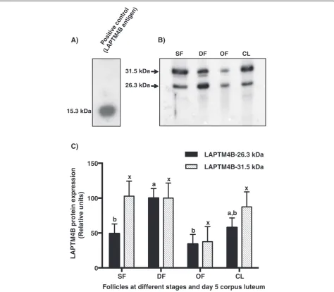
-LAPTM4B was used as immunizing antigen to generate anti-bovine LAPTM4B polyclonal antibodies. Immuno-blotting analyses showed that the antibody preparation recognized the affinity-purified and thrombin-cleaved (LA)2-LAPTM4B fragment that migrated at 15.3 kDa
(Figure 3A). The antibody recognized the native LAPTM4B migrating at a MW of 26.3 kDa (Figure 3B) in total protein extracts of GC and CL, which corre-sponds to its theoritical MW of 25.4 kDa. A higher
Fetal OvaryUterus PlacentomeAdult testisFetal testisEpidydimisSeminal glandBrain MuscleThyroidLiver StomachDuodenumSpleenHeart V.Lung KidneyAdrenal glandPituitaryCorpus luteum
LAPTM4B 1.8 kb
0 0.5 1 1.5 2
LAPTM4B / 18S RNA
(relative units)
A)
B)
Figure 1Analysis of bovine LAPTM4B mRNA expression in bovine tissues by Northern blot.Total RNA was extracted from various bovine
molecular size protein migrating at 31.5 kDa was also detected (Figure 3B). Comparison of the 26.3 kDa LAPTM4B protein expression showed greatest amounts in GC of DF whereas moderate expression was observed in CL, and significantly weaker expression was observed in OF and SF as compared to DF (P< 0.03; Figure 3C). The 31.5 kDa form showed no statistically significant variation among the different follicular stages and CL
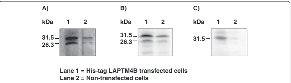
although its concentration was 2.6-fold weaker in OF than in DF. Overexpression experiments of the full-length LAPTM4B cDNA in HEK cells confirmed that two protein forms were produced from the single cDNA. These two forms were recognized at 26.3 and 31.5 kDa by the anti-LAPTM4B antibodies generated in this study (Figure 4A) and by the one acquired com-mercially (Figure 4B). The His-tag LAPTM4B protein
0 6 12 18 24
0 50 100 150
Hours post-hCG LAPTM4B/GAPDH (Relative units)
(2) (2)
a,b a
(2) a,b
(2) b
(2) b
B)
SF DF OF CL
0 50 100 150
Follicles at different stages and day 5 corpus luteum LAPTM4B/GAPDH (Relative units)
(4)
(4)
(3) (3)
a
b
b
b
A)
Figure 2Regulation of LAPTM4B mRNA expression during follicular development using semi-quantitative RT-PCR.RNA samples were
expressed in HEK cells was affinity purified and analyzed by immunoblotting using anti-bovine ubiquitin antibodies that detected a single protein band at 31.5 kDa indicating ubiquitination of LAPTM4B (Figure 4C).
Immunolocalization of LAPTM4B
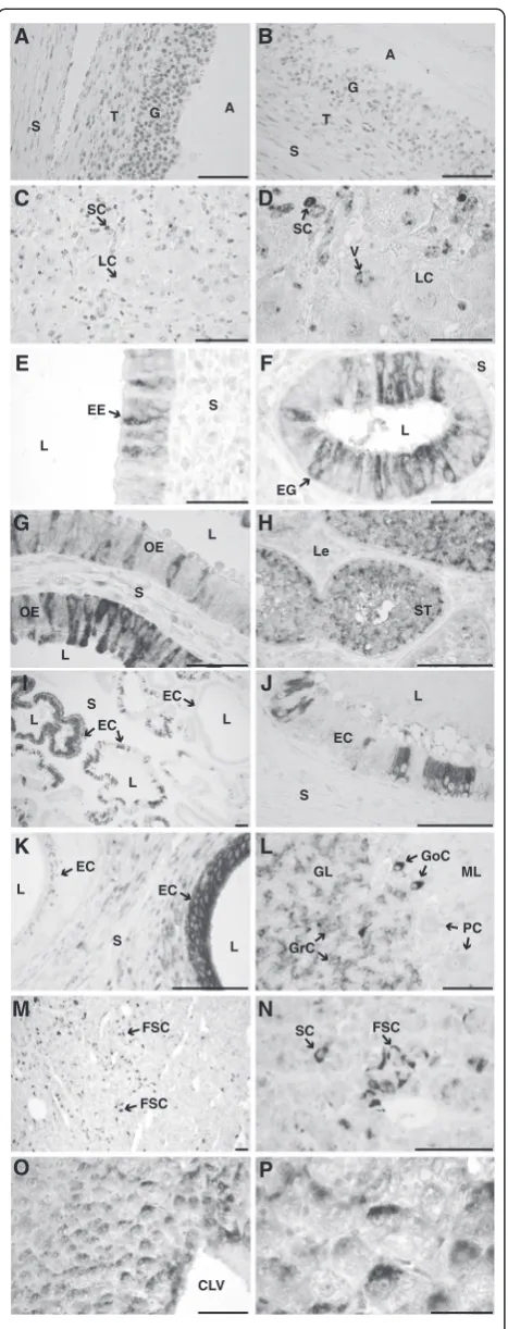
Immunohistochemical analyses provided evidence that LAPTM4B expression in bovine tissues is variable and spe-cific given the pattern of expression observed even among similar cell types within a tissue. In GC, a perinuclear
pattern of immunostaining was observed in all cells (Figure 5A). Dominant follicles obtained at day 5 showed a labeling signal in GC that was stronger compared to GC from follicles obtained 24 h following hCG injection (Figure 5B). Theca cells were also stained but the staining was weaker compared to GC (Figure 5A-B). For the CL, immunolabeling was variable among luteal cell types and within each cell type (Figure 5C,D). For instance, large luteal cells presented a weaker or no signal as compared to small luteal cells. Moreover, subcellular labeling was
SF DF OF CL
0 50 100 150
Follicles at different stages and day 5 corpus luteum
LAPTM4B protein expression
(Relative units)
LAPTM4B-26.3 kDa
LAPTM4B-31.5 kDa
a x
b a,b
b x
x
x
C)
Positive control (LAPTM4B antigen)
15.3 kDa
SF DF OF CL
31.5 kDa
26.3 kDa
A) B)
Figure 3Comparison of bovine LAPTM4B protein expression.Total protein extracts (50μg/well) from granulosa cells of 2–4 mm small
associated with perinuclear vesicles (Figure 5D). Since LAPTM4B mRNA expression in different tissues was vari-able among the tissues analyzed (Figure 1), we extended the immunohistochemical observations to other female and male reproductive tissues. Epithelial and glandular endometrial cells showed a variable pattern of immuno-staining even among adjacent cells (Figure 5E,F). Interest-ingly, endometrial glandular cells that were closer to the uterine lumen showed a stronger signal compared to the endometrial cells located deeper in the gland (not shown). In oviductal epithelial cells, heterogeneity of staining for LAPTM4B was also noticeable, with weak to strong signals associated with variable localization, being either peri-nuclear or at the baso-lateral or at apical side of cells (Figure 5G). In the testis, the intensity of immunostaining was variable between seminiferous tubules and was associ-ated with Sertoli cells (Figure 5H). Epithelial cells of the seminal gland showed a variable expression pattern associ-ated with a strong to weak signal even for adjacent groups of cells (Figure 5 I,J). Epithelial cells lining the epidydimis tail ducts showed immunolabeling signal from intense to weak that varied between ducts (Figure 5K). In the granu-lar layer of the cerebellum, granugranu-lar cells were weakly stained whereas strong perinuclear labeling was associated to Golgi cells. Purkinje cells were not labeled (Figure 5L). In the adenohypophysis, strong immunolabeling was asso-ciated to the folliculo-stellate cells (Figure 5M,N). In the liver, hepatocytes located near the centrolobular vein demonstrated a stronger perinuclear signal than hepato-cytes that were located eccentric to the centrolobular vein (Figure 5O,P).
Discussion
This study is the first to report that LAPTM4B expres-sion is differentially regulated at the mRNA and protein levels in granulosa cells at critical stages of follicular de-velopment, being stronger at the time of dominance
than at recruitment (2–4 mm diameter), at the time of ovulation and in luteal cells. LAPTM4B differential ex-pression extends to different bovine tissues. Moreover, immunohistochemical observations revealed that bovine LAPTM4B expression was highly variable among tissues and even among identical cell types of the same tissue.
The mRNA expression study of LAPTM4B in different bovine tissues revealed a single transcript of 1.8 kb, which corresponds well to the length of the cDNA char-acterized by sequencing from the granulosa cells cDNA library. Interestingly, two transcripts of 2.2 kb and 1.5 kb were reported in human and were named LAPTM4B variant 1 and variant 2, respectively [10,12,13,31]. The size of the human LAPTM4B variant 2 (GenBank acces-sion number: AF527412; [10]) corresponds to the bovine LAPTM4B cDNA characterized in this study. By im-munoblotting analysis of hepatocellular carcinoma and normal liver tissue, it was shown that human LAPTM4B was expressed as two isoforms that were translated from alternative ATG codons [12]. The apparent molecular weights of the isoforms were 35 and 24 kDa, corre-sponding to predicted 317- and 226-amino acid proteins, respectively. Expression of the 35-kDa isoform was greater in highly metastatic cell lines originating from liver, prostate, and pulmonary giant cell cancers than in syngenic low metastatic cell lines [12]. Immunoblotting analyses in the present study using antibodies raised against bovine LAPTM4B showed the presence of two proteins migrating at 26.3 kDa and 31.5 kDa, which corroborates the molecular weight of the protein ob-served in human [12]. However, contrary to the human LAPTM4B cDNA, the cDNA of the bovine LAPTM4B contains a single initiation codon of translation and the size of the bovine cDNA characterized herein corresponds to the size of the transcript estimated by Northern blot (1.8 kb). To further characterize and verify the 5′-UTR re-gion of bovine LAPTM4B, a genomic library was screened
31.5 31.5
26.3
31.5 26.3 B)
A) C)
Lane 1 = His-tag LAPTM4B transfected cells Lane 2 = Non-transfected cells
1 2 1 2 1 2
kDa kDa kDa
Figure 4Overexpression analysis of LAPTM4B in HEK cells.LAPTM4B was cloned into the his-tag pQE2 vector and used to transfect HEK cells.
to eventually yield the 5′-UTR and proximal promoter se-quences. The sequencing results showed that there was a single open reading frame coding for the 26.3 kDa form. Following the synthesis of the 26.3 kDa bovine protein, post-translational modifications may result in the 31.5 kDa protein. The anti-bovine as well as commercially available anti-human LAPTM4B antibodies recognized the two pro-tein isoforms when bovine LAPTM4B cDNA was overex-pressed in HEK cells. These results suggested a specific binding to the 26.3 and 31.5 kDa proteins rather than a cross reaction with a non-related protein. The affinity purified His-tag LAPTM4B protein overexpressed in HEK cells was analyzed by immunoblotting using anti-ubiquitin antibodies and demonstrated that the 26.3 kDa protein undergoes mono-ubiquitination thereby resulting in the 31.5 kDa isoform. The difference in molecular weight between the estimated 26.3 and 31.5 kDa corresponds well to the theoritical molecular weight of ubiquitin. This result is also supported by the recent observation that ubiquitination has a role in membrane sorting of LAPTM4 proteins [8].
Comparison of mRNA steady state levels in follicles at different developmental stages has shown that LAPTM4B was expressed in GC of SF, reached strongest expression in DF and declined in OF. Similarly, immunoblotting analysis revealed a variable expression of the 26.3 kDa LAPTM4B protein that paralled the mRNA results. Amounts of LAPTM4B protein were low in SF, highest in DF and lowest in OF. The comparison of LAPTM4B ex-pression by immunohistochemistry provided further sup-port that it is expressed in GC of DF, and this expression is reduced in OF 24 h following hCG treatment. Immuno-blotting analysis showed no significant variation in the
A G
S T
A
L
EE S
E
L
S
EG
F
Le
ST
H
LC SC
C
LC SC
V
D
A
G
T
S
B
S OE
L
L
G
OE
FSC
M
FSC
L
S EC
J
PC ML GL
L
SC L
S L
L EC EC
I
CLV
O P
S L
L EC EC
K
N
FSC GoC
GrC
Figure 5Immunohistochemical localization of LAPTM4B in bovine
tissues.Sections of paraffin-embedded tissues were incubated with the antibody directed against the recombinant bovine LAPTM4B (1: 500 dilution) and the complex was detected with a monoclonal anti-rabbit antibody coupled to alkaline phosphatase and NBT/BCIP as substrate. No counter staining was used.Bar= 0.1 mm if not otherwise stated. A. Follicular wall of a day 5 dominant follicle;Aantrum,Ggranulosa,
Sstroma,Ttheca interna.B. Follicular wall of an ovulatory follicle 24 h following hCG-injection.C. Corpus luteum at day 5 of the estrous cycle;
LClarge luteal cells,SCsmall luteal cells.D. Higher magnification of the corpus luteum section presented in C;Vperinuclear vesicles.Bar= 0.05 mm.E,F. Endometrium;EEendometrial epithelial cells,EGendometrial glandular cells, Llumen,Sstroma,bar= 0.05 mm.G. Oviduct;OE Oviductal epithelial cells,Sstroma,Llumen.H. Prepupertal testis;ST Seminiferous tubule,LeLeydig cells.I. Seminal vesicle;ECEpithelial cells,
Llumen,Sstroma.J. Higher magnification of seminal vesicle presented in J.K. Tail of epididymis;ECepithelial cells,Sstroma,L lumen.L. Cerebellum;GLgranular layer,MLmolecular layer,GrC granular cells,GoCGolgi cells,PCPurkinje cells.M. Adenohypophysis;
concentration of the 31.5 kDa protein, which could be related to the stability and accumulation of the 31.5 kDa isoform in lysosomes following ubiquitination of the 26.3 kDa protein. Thus, de novosynthesis of LAPTM4B protein may be better reflected by the 26.3 kDa isoform, which paralleled the variations observed at the mRNA level.
The immunohistochemistry results suggest that bovine LAPTM4B has a lysosomal localization as shown by the perinuclear staining in different cell types. Located at its carboxy terminal end, LAPTM4B contains a lysosomal tar-geting motif and was also shown to co-immunoprecipitate with ubiquitin ligase Nedd4 [3,8]. Ubiquitination of LAPTM4B by Nedd4 may contributes to its targeting towards late endosomes and lysosomes [8,32]. These ob-servations correlate well with a lysosomal localization as previously observed for human LAPTM4B [8] and for LAPTM4A [4,33]. Interestingly, folliculo-stellate cells in the adenohypophysis were strongly labeled and they are known as scavengers with high lysosomal activity [34]. In the ovarian follicle, granulosa and theca cells were labeled almost uniformly probably reflecting a synchrony in cell development necessary to insure active follicular develop-ment. In contrast, heterogeneity of labeling was observed in other tissues for similar and adjacent cell types. This was particularly striking in tissues such as the epithelial cells lining the oviduct, the uterus, the epididymis and the seminal gland. In these tissues, some epithelial cells did not stain while adjacent cells were strongly labeled. This could reflect a different physiological state between these epithelial cells within the tissue.
The results observed for the bovine LAPTM4B mRNA and protein regulation, which is predominant in DF while reduced in OF, suggest a potential role of LAPTM4B in the growth of ovarian follicles. It was proposed that human LAPTM4B promoted cell proliferation since it was over-expressed in liver tumors [11], and was differentially expressed with potential tumorigenic significance associated with pituitary tumorigenesis [14]. Moreover, knockdown and overexpression studies of LAPTM4B demonstrated its capacity in binding and sequestring chemotherapeutic agents towards lysosomes thereby inhibiting their action and conferring chemotherapeutic resistance [15,35-37]. Comparatively, another member of the LAPTM super-family, LAPTM4A, was involved in cellular detoxification processes through transport of toxic substances in late endosomes and toward lysosomes, thereby participating in cell survival [4,33]. Thus, expression of LAPTM4B in follicular and luteal cells could act as a survival factor through sequestration and detoxification of metabolites into lysosomes. Since granulosa cells of growing dominant follicle are metabolically and steroidogenically very active, LAPTM4B may confer protection against accumulation of metabolic byproducts and induction of apoptosis.
Competing interests
The authors declare that they have no competing interests.
Authors’contributions
KN contributed in the conception and design of the experiments, performed the experiments, performed analysis and interpretation of the results and wrote the paper; PDC contributed in the interpretation of results and revised the paper; JS participated in the experimental design of mRNA expression analysis and revised the paper; DWS contributed in the analysis and interpretation of results and revised the paper; JGL contributed in the conception and design of experiments, analysis and interpretation of results and revised the paper. All authors read and approved the final manuscript.
Acknowledgement
This work was supported by a Discovery Grant (to JGL) from the Natural Sciences and Engineering Research Council of Canada (NSERC).
Received: 28 October 2014 Accepted: 16 March 2015
References
1. Adra CN, Zhu S, Ko JL, Guillemot JC, Cuervo AM, Kobayashi H, et al. LAPTM5: a novel lysosomal-associated multispanning membrane protein preferentially expressed in hematopoietic cells. Genomics. 1996;35(2):328–37.
2. Cabrita MA, Hobman TC, Hogue DL, King KM, Cass CE. Mouse transporter protein, a membrane protein that regulates cellular multidrug resistance, is localized to lysosomes. Cancer Res. 1999;59(19):4890–7.
3. Hogue DL, Nash C, Ling V, Hobman TC. Lysosome-associated protein transmembrane 4 alpha (LAPTM4 alpha) requires two tandemly arranged tyrosine-based signals for sorting to lysosomes. Biochem J. 2002;365(Pt 3):721–30. 4. Hogue DL, Ellison MJ, Young JD, Cass CE. Identification of a novel
membrane transporter associated with intracellular membranes by phenotypic complementation in the yeast Saccharomyces cerevisiae. J Biol Chem. 1996;271(16):9801–8.
5. Marks MS, Woodruff L, Ohno H, Bonifacino JS. Protein targeting by tyrosine-and di-leucine-based signals: evidence for distinct saturable components. J Cell Biol. 1996;135(2):341–54.
6. Gough NR, Zweifel ME, Martinez-Augustin O, Aguilar RC, Bonifacino JS, Fambrough DM. Utilization of the indirect lysosome targeting pathway by lysosome-associated membrane proteins (LAMPs) is influenced largely by the C-terminal residue of their GYXXphi targeting signals. J Cell Sci. 1999;112(Pt 23):4257–69.
7. Cherqui S, Kalatzis V, Trugnan G, Antignac C. The targeting of cystinosin to the lysosomal membrane requires a tyrosine-based signal and a novel sorting motif. J Biol Chem. 2001;276(16):13314–21.
8. Milkereit R, Rotin D. A role for the ubiquitin ligase Nedd4 in membrane sorting of LAPTM4 proteins. PLoS One. 2011;6(11):e27478.
9. Liu J, Zhou R, Zhang N, Rui J, Jin C. Biological function of a novel gene overexpressed in human hepatocellular carcinoma. Chin Med J. 2000;113(10):881–5.
10. Shao GZ, Zhou RL, Zhang QY, Zhang Y, Liu JJ, Rui JA, et al. Molecular cloning and characterization of LAPTM4B, a novel gene upregulated in hepatocellular carcinoma. Oncogene. 2003;22(32):5060–9.
11. He J, Shao G, Zhou R. Effects of the novel gene, LAPTM4B, highly expression in hepatocellular carcinoma on cell proliferation and tumorigenesis of NIH3T3 cells. Beijing da xue xue bao Yi xue ban = Journal of Peking University Health sciences. 2003;35(4):348–52.
12. Liu XR, Zhou RL, Zhang QY, Zhang Y, Jin YY, Lin M, et al. Structure analysis and expressions of a novel tetratransmembrane protein, lysosoma-associated protein transmembrane 4 beta associated with hepatocellular carcinoma. World J Gastroenterol. 2004;10(11):1555–9.
13. Kasper G, Vogel A, Klaman I, Grone J, Petersen I, Weber B, et al. The human LAPTM4b transcript is upregulated in various types of solid tumours and seems to play a dual functional role during tumour progression. Cancer Lett. 2005;224(1):93–103.
15. Li Y, Zhang Q, Tian R, Wang Q, Zhao JJ, Iglehart JD, et al. Lysosomal transmembrane protein LAPTM4B promotes autophagy and tolerance to metabolic stress in cancer cells. Cancer Res. 2011;71(24):7481–9. 16. Mihm M, Bleach EC. Endocrine regulation of ovarian antral follicle
development in cattle. Anim Reprod Sci. 2003;78(3–4):217–37. 17. Ginther OJ, Beg MA, Bergfelt DR, Donadeu FX, Kot K. Follicle selection in
monovular species. Biol Reprod. 2001;65(3):638–47.
18. Webb R, Nicholas B, Gong JG, Campbell BK, Gutierrez CG, Garverick HA, et al. Mechanisms regulating follicular development and selection of the dominant follicle. Reprod Suppl. 2003;61:71–90.
19. Milvae RA, Hinckley ST, Carlson JC. Luteotropic and luteolytic mechanisms in the bovine corpus luteum. Theriogenology. 1996;45(7):1327–49. 20. Knight PG, Glister C. Potential local regulatory functions of inhibins, activins
and follistatin in the ovary. Reproduction. 2001;121(4):503–12.
21. Eppig JJ, Wigglesworth K, Pendola FL. The mammalian oocyte orchestrates the rate of ovarian follicular development. Proc Natl Acad Sci U S A. 2002;99(5):2890–4.
22. Rosenfeld CS, Wagner JS, Roberts RM, Lubahn DB. Intraovarian actions of oestrogen. Reproduction. 2001;122(2):215–26.
23. Ndiaye K, Fayad T, Silversides DW, Sirois J, Lussier JG. Identification of downregulated messenger RNAs in bovine granulosa cells of dominant follicles following stimulation with human chorionic gonadotropin. Biol Reprod. 2005;73(2):324–33.
24. Espey LL, Richards JS. Temporal and spatial patterns of ovarian gene transcription following an ovulatory dose of gonadotropin in the rat. Biol Reprod. 2002;67(6):1662–70.
25. Tesfaye D, Ghanem N, Carter F, Fair T, Sirard MA, Hoelker M, et al. Gene expression profile of cumulus cells derived from cumulus-oocyte complexes matured either in vivo or in vitro. Reprod Fertil Dev. 2009;21(3):451–61. 26. Li Q, Jimenez-Krassel F, Ireland JJ, Smith GW. Gene expression profiling of
bovine preovulatory follicles: gonadotropin surge and prostanoid-dependent up-regulation of genes potentially linked to the ovulatory process. Reproduction. 2009;137(2):297–307.
27. Filion F, Bouchard N, Goff AK, Lussier JG, Sirois J. Molecular cloning and induction of bovine prostaglandin E synthase by gonadotropins in ovarian follicles prior to ovulation in vivo. J Biol Chem. 2001;276(36):34323–30. 28. Bedard J, Brule S, Price CA, Silversides DW, Lussier JG. Serine protease
inhibitor-E2 (SERPINE2) is differentially expressed in granulosa cells of dominant follicle in cattle. Mol Reprod Dev. 2003;64(2):152–65. 29. Sambrook JF, Russell DW. Molecular cloning. A laboratory manual. vol. 3,
3rd ed. Cold Spring Harbor, NY: Cold Spring Harbor Laboratory Press; 2001. p. 2100.
30. Bradford MM. A rapid and sensitive method for the quantitation of microgram quantities of protein utilizing the principle of protein-dye binding. Anal Biochem. 1976;72:248–54.
31. Liu X, Zhou R, Zhang Q, Zhang Y, Shao G, Jin Y, et al. Identification and characterization of LAPTM4B encoded by a human hepatocellular carcinoma-associated novel gene. Beijing da xue xue bao Yi xue ban = Journal of Peking University Health sciences. 2003;35(4):340–7. 32. Dikic I, Wakatsuki S, Walters KJ. Ubiquitin-binding domains - from structures
to functions. Nat Rev Mol Cell Biol. 2009;10(10):659–71.
33. Hogue DL, Ellison MJ, Vickers M, Cass CE. Functional complementation of a membrane transport deficiency in Saccharomyces cerevisiae by recombinant ND4 fusion protein. Biochem Biophys Res Commun. 1997;238(3):811–6.
34. Devnath S, Inoue K. An insight to pituitary folliculo-stellate cells. J Neuroendocrinol. 2008;20(6):687–91.
35. Hogue DL, Kerby L, Ling V. A mammalian lysosomal membrane protein confers multidrug resistance upon expression in Saccharomyces cerevisiae. J Biol Chem. 1999;274(18):12877–82.
36. Li L, Wei XH, Pan YP, Li HC, Yang H, He QH, et al. LAPTM4B: a novel cancer-associated gene motivates multidrug resistance through efflux and activating PI3K/AKT signaling. Oncogene. 2010;29(43):5785–95.
37. Li Y, Zou L, Li Q, Haibe-Kains B, Tian R, Li Y, et al. Amplification of LAPTM4B and YWHAZ contributes to chemotherapy resistance and recurrence of breast cancer. Nat Med. 2010;16(2):214–8.
Submit your next manuscript to BioMed Central and take full advantage of:
• Convenient online submission
• Thorough peer review
• No space constraints or color figure charges
• Immediate publication on acceptance
• Inclusion in PubMed, CAS, Scopus and Google Scholar
• Research which is freely available for redistribution