Abstract
A 27-year old woman presented with primary amenorrhoea and infertility. On examination, she was found to have palpable inguinal gonads, normal female external genitalia, a blind-ending vagina with no cervix, almost complete absence of axillary and pubic hair, and good breast development. Further investigations confirmed the absence of internal female genitalia, the presence of a 46,XY karyotype and testosterone levels in the high normal male reference range. A diagnosis of complete androgen insensitivity syndrome was made, based on this clinical picture and laboratory findings. Two of her younger siblings were subsequently also diagnosed with this condition. She underwent an orchidectomy and is currently on female hormone replacement therapy. At the time of writing up this case study, her two younger sisters were still awaiting surgery.
Peer reviewed. (Submitted: 2013-01-13. Accepted: 2013-05-02.) © SEMDSA JEMDSA 2013;18(3):159-163
Case study
A 27-year old female African patient from Mpumalanga was referred to the Endocrinology Clinic at Steve Biko Academic Hospital with primary amenorrhoea and infertility. She was referred from the cardiology department after admission for a supraventricular tachycardia. A diagnosis of Wolff-Parkinson-White syndrome was made. She had sought help, four years prior to this referral, at the gynaecology clinic, but was lost to follow-up before the workup was completed.
She was one of six siblings, with a 26-year old brother and four younger sisters. There was no history of consanguinity in the family. Of her younger sisters, the youngest, aged 11, was prepubertal and premenarchal, and the 16-year old sibling had normal menses. Both the 19-16-year old and the 21-year old siblings experienced primary amenorrhoea. The patient reported a usual female gender role and female gender identity, and had strong maternal feelings. She had already adopted a child, but wanted biological children with her husband.
On examination, it was found that she had a normal female phenotype, but was relatively tall at 1.71 m. She reported that she was much taller than her mother, and was the same height as her father. Her body mass index was 26. She was found to have palpable inguinal gonads which caused some discomfort, and normal
female external genitalia, with no cliteromegaly or ambiguous genitalia present. Her vagina was short and blind-ending, with no palpable cervix. The patient reported having experienced severe dyspareunia for the first two years after becoming sexually active. She had good breast development, but an almost complete absence of axillary and pubic hair.
A transvaginal sonar confirmed the absence of a uterus and intraabdominal ovaries. The inguinal sonar detected a right gonad measuring 2.9 x 2.8 x 1.2 cm,
Three siblings with complete
androgen insensitivity syndrome
Kemp T, MBChB, MMed(Int Med), CertEndocrinologyandMetabolism(SA)Phys,
Senior Specialist Endocrinology Unit, Department of Internal Medicine, Steve Biko Academic Hospital; University of Pretoria
Correspondence to: Tanja Kemp; e-mail: kemp.tanja@gmail.com Keywords: siblings,completeandrogen insensitivity syndrome, disorders of sex development
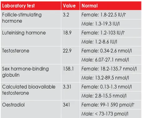
Table I: Laboratory findings in the index case
Laboratory test Value Normal
Follicle-stimulating hormone
3.2 Female: 1.8-22.5 IU/l*
Male: 1.3-19.3 IU/l Luteinising hormone 18.9 Female: 1.2-103 IU/l*
Male: 1.2-8.6 IU/l Testosterone 22.9 Female: 0.34-2.6 nmol/l
Male: 6.07-27.1 nmol/l Sex hormone-binding
globulin
158.1 Female: 18.2-135.7 nmol/l Male: 13.2-89.5 nmol/l Calculated bioavailable
testosterone
3.31 Female: 0.13-1.3 nmol/l Male: 2.8-15.5 nmol/l Oestradiol 341 Female: 99-1 590 pmol/l*
and a left one measuring 2.5 x 1.1 x 2.2 cm (Figure 1 a-c). The patient’s laboratory findings are summarised in Table II.
The serum testosterone values were in the adult male range, with an elevated luteinising hormone (LH). Chromosomal studies detected a 46,XY karyotype. This confirmed the diagnosis of a form of 46,XY disorder of sex development. Taking into account the clinical picture, especially the lack ambiguous genitalia, the
lack of secondary sexual hair, her female phenotype and the sonar findings, in combination with testosterone levels in the high normal male range, the diagnosis was most likely to be that of complete androgen insensitivity syndrome (CAIS). Ovotesticular disorder of sex development (especially in view of her high oestradiol level), and 5-alpha-reductase type 2 deficiency were also considered, but were much less likely in view of the clinical findings and special investigations.
The masses are annotated by arrows
Figure 1 a: Sonar of the left inguinal mass
The masses are annotated by arrows
Figure 1 b: Sonar of the right inguinal mass
The masses are annotated by arrows
Figure 1 c: Sonar of the right inguinal mass
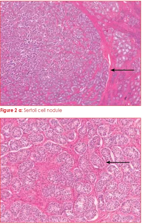
Figure 2 a: Sertoli cell nodule
Figure 2 b: Tubuli seminiferi without any spermatogenesis
The patient underwent a bilateral gonadectomy, and the left gonad measured 4 x 2 x 2 cm. Some of the histological findings are depicted in Figure 2 a-c. On histology, it was confirmed to be testicular tissue, and contained a small nodule measuring 0.5 cm. The right testis also measured 4 x 2 x 2 cm, and contained two nodules, as well as a cyst that was one centimetre in diameter. Neither testis demonstrated spermatogenesis, and the nodules were of Sertoli cell origin. Leydig cell hyperplasia was prominent in both testes. No ovarian tissue was present. The patient’s testosterone level dropped postoperatively to a level below 0.34 nmol/l, her oestradiol level fell to 139 pmol/l, the LH went up to 47.9 IU/l, and the follicle-stimulating hormone (FSH) increased to 99.6 IU/l. She was started on hormone replacement therapy in the form of Premarin®.
The patient was extensively counselled before surgery regarding her diagnosis and the implications of this condition with regard to fertility. She demonstrated very good insight into the condition. Unfortunately, her husband left her when he found out about her infertility. She did not disclose her karyotype to him.
Her two younger sisters (cases 2 and 3) were subsequently also evaluated and demonstrated almost identical clinical phenotypes, ultrasonographic findings and findings on special investigations. Their laboratory findings are summarised in Table II. Their karyotypes were also found to be 46,XY. They are both still awaiting gonadectomies. The youngest sister will soon be evaluated by the paediatric endocrinology clinic. Unfortunately, we were not able to obtain sequencing of the androgen receptor gene for the mutation.
Discussion
CAIS was previously called complete testicular feminisation. The reported incidence in the literature varies from between approximately one in 20 000 genetic males in Denmark, to one in 99 000 in the Netherlands.1-3 No data are available on how common this condition is in South Africa. Only one large case series has been described.4 It forms part of a wide spectrum of disorders caused by mutations in the gene coding for the androgen receptor, leading to resistance to androgens.1 Patients can present as phenotypically normal females, or with ambiguous genitalia in the case of partial androgen insensitivity syndrome, or in milder forms, as normal men with undervirilisation or fertility problems. These conditions are classified under the 46,XY disorders of sex development.5 The rest of this discussion will focus on CAIS.
Aetiology
Sentinel work was performed by Marcelli, Tilley and Griffin, and McPhaul, Marcelli and Zoppi, who identified the different mutations that caused CAIS.6,7Since the androgen receptor gene is on the X chromosome, the inheritance of this condition is X-linked recessive. It is caused by numerous mutations in the gene coding for the androgen receptor, leading to severe impairment in androgen receptor function. More than a thousand mutations have been identified so far.8 Unfortunately, we could not obtain sequencing of the gene mutation in this family.
Reported abnormalities include missense mutations, premature stop codons, deletions and splicing abnormalities. Loss-of-function mutations in the coding sequence of the androgen receptor can be found in most patients with CAIS. Several other defects have been identified in recent years.9,10 In a large kindred from Soweto, the genetic cause was found to be the missense mutation, D732Y, in exon 5 of the androgen receptor ligand-binding domain.4 Whereas mutations in the androgen receptor are natural, environmental endocrine disruptors have been implicated in some cases of PAIS.11
Clinical features
Occasionally, babies are diagnosed with CAIS, even prenatally or at birth, because of a discrepancy between the karyotype determined through amniocentesis and the anatomical gender.1,5 Babies with CAIS have phenotypically normal female external genitalia which are not ambiguous. A small percentage of these children present with inguinal hernias. In retrospective studies, the incidence of CAIS was estimated to be approximately 0.8-2.4% in girls with inguinal hernias. This Table II: Laboratory findings in cases 2 and 3
Laboratory test Case 2 Case 3 Normal
Follicle-stimulating hormone
1.7 2.1 Female: 1.8-22.5 IU/l*
Male: 1.3-19.3 IU/l
Luteinising hormone
16.3 13.1 Female: 1.2-103 IU/l*
Male: 1.2-8.6 IU/l
Testosterone 20.65 22.71 Female: 0.34-2.6 nmol/l
Male: 6.07-27.1 nmol/l
Sex hormone-binding globulin
68.6 70.9 Female: 18.2-135.7 nmol/l
Male: 13.2-89.5 nmol/l
Calculated bioavailable testosterone
6.23 6.79 Female: 0.13-1.3 nmol/l
Male: 2.8-15.5 nmol/l
Oestradiol 140 97 Female: 99-1 590 pmol/l*
Male: < 73-173 pmol/l
was confirmed in a prospective study which found an incidence of 1.1%.12 Some patients are also diagnosed at a young age because of the presence of palpable gonads in the inguinal area or in the labia majora.
The majority present later during puberty with primary amenorrhoea.1,5 It is probably not unusual for patients to present much later in developing countries, as occurred in our index case. The hallmark of this condition on examination is normal breast development, normal external female genitalia, with the absence of or scanty pubic and axillary hair. Very few other disorders of sex development have this combination of clinical findings. Other confirmatory findings are an absent, short or blind-ending vagina, an absent cervix and often palpable gonads in the inguinal area or in the labia majora. Testes can also be intra-abdominal. Special investigations confirm the absence of a uterus, a 46,XY karyotype, and demonstrate serum testosterone levels in the normal adult male range.1 Body habitus and breast development is similar to that of normal females. These patients have a usual female gender identity with normal maternal instincts.1,5
They tend to be taller and larger than would be expected following a calculation of mid-parental height. The mean height is 171.5 cm, as was also observed in these three sisters.13 Usually, the growth pattern follows that of girls, but the final achieved height is between that of average males and females.14 It is postulated that the Y chromosome may have a direct effect on growth and body size which is not mediated through the androgen receptor. The bone mineral density is decreased in the lumbar spine, even in subjects where gonadectomy, followed by oestrogen replacement, is performed early. This suggests an important role for androgens in bone mineral density that cannot be replaced by oestrogen.14
Laboratory findings
Our three patients had typical laboratory findings of CAIS. The serum testosterone level was in the normal adult male range, or slightly higher.15,16 Serum sex hormone-binding globulin concentrations can be increased. The testosterone production rate in the testes is increased because of an increase in serum LH. This is because of resistance to the negative feedback of androgens on LH secretion at hypothalamic-pituitary level.15 Serum FSH levels are usually normal. Oestrogen production is increased by up to 70% higher than that of normal men. This oestrogen originates from excess testosterone which is peripherally aromatised to oestrogen, as well as from the testicular secretion of oestrogen.1,17
Diagnosis
Diagnosis is confirmed by finding a 46,XY karyotype in the presence of the above clinical findings, with no female internal genitalia detected on imaging, and a serum testosterone level in the adult male range.1,5 The coding region of the gene that encodes the androgen receptor can be sequenced to find the specific mutation.5,7
Management
Gender assignment is not an issue in CAIS, even if diagnosed early. These patients should be raised as females. Full disclosure now forms part of the management plan.18,19 In the case of a minor, the parents should be fully informed and counselled, preferably by a psychologist who is familiar with this condition as disclosure of the diagnosis can lead to strong emotional reactions in the parents.20
The child may receive information based on age and understanding, but full disclosure should occur before adulthood.18,21 Extensive counselling and psychological support may also be required. The damage caused by non-disclosure and finding out the diagnosis inadvertently, much later, can cause more psychological distress.18-20 Female family members should also be offered testing. However, there are ethical and legal implications, especially with regard to patient confidentiality.22
Dilator therapy to increase vaginal depth and size can be started after puberty before sexual activity is contemplated.23 This may be sufficient, but occasionally vaginoplasty may be necessary.18,19,24 In a recent study, the majority of patients reported significant problems with desire, arousal and dyspareunia.25 Long-term sexual problems can occur, especially if surgical results are suboptimal. Sexual problems can occur if the vagina remains small after dilator therapy or vaginoplasty.26,27
The risk of malignancy is increased in patients with cryptorchid testes, which is why these three sisters were referred for gonadectomies. Germ cell tumours or gonadoblastomas can occur in the testes, especially if located intra-abdominally, and can become malignant. A recent review of adults with CAIS estimated the risk of a gonadal malignancy developing of approximately 14% (0-22%).28 Older figures have estimated the risk as being greater than 30% by 50 years of age, but these included patients with PAIS who, in general, have a much higher risk than patients with CAIS.29
safe to defer gonadectomy until after puberty.18,19,28,30 This will optimise normal breast development and may reaffirm the patient’s gender identity. Surgery may be deemed necessary earlier in the case of an inguinal hernia requiring surgical repair, if the gonads are uncomfortable or painful, or if there is doubt about the completeness of the androgen resistance. In the last scenario, incomplete resistance may lead to virilisation during puberty.24,30
Lifelong hormone replacement therapy is necessary post-gonadectomy in adults, or from the time of puberty if surgery was performed earlier, especially to optimise the already lower-than-normal bone mineral density.14,18,19 Whereas biological offspring has been impossible in the past, fertility is potentially becoming more feasible. If the testes can be preserved after gonadectomy, it may possible for these patients to have biological children of their own in the future, with intracytoplasmic injection of primitive germ cells or spermatocytes (which are often present) into a donor ovum, and with the assistance of a surrogate.29,31
Histology
The testes of these patients are of a similar appearance to those of cryptorchid testes in boys. The number of Leydig cells can be normal or increased. Usually, there is no spermatogenesis.29,32 Sertoli cell nodules, as found in our index case, or Sertoli cell adenomas, are common, but are not premalignant. The epididymis
and vas deferens are usually absent, but can be
histologically identified adjacent to the testes in up to 36% of patients.32 Carcinoma in situ or germ cell malignancies are rarely identified, but usually only occur in postpubertal patients.32
Acknowledgements
Special thanks are given to Dr Gerhard Davel and the Department of Anatomical Pathology for the use of their histology slides.
References
1. Hughes IA, Davies JD, Bunch TI, et al. Androgen insensitivity syndrome. Lancet. 2012;380(9851):1419-1428.
2. Boehmer AL, Brinkmann O, Brüggenwirth H, van Assendelft C, et al. Genotype versus phenotype in families with androgen insensitivity syndrome. J Clin Endocrinol Metab. 2001;86(9):4151-4160.
3. Banksboll S, Qvist I, Lebech PE, et al. Testicular feminization syndrome and associated gonadal tumors in Denmark. Acta Obstet Gynecol Scand. 1992;71(1):63-66.
4. Scott EC, Greenberg TS, Arndt S, et al. Complete androgen insensitivity syndrome in a black South African family: a clinical and molecular investigation. Endocr Pract. 2006;12(6):664-669.
5. Mendonca BB, Domenice S, Arnhold IJ, Costa EM. 46,XY disorders of sex development. Clin Endocrinol. 2009;70(2):173-187.
6. Marcelli M, Tilley WD, Wilson CM, et al. Definition of the human androgen receptor gene structure permits the identification of mutations that cause androgen resistance: premature termination of the receptor protein at amino acid residue 588 causes complete androgen resistance. Mol Endocrinol. 1990;4(8):1105-1116. 7. McPhaul MJ, Marcelli M, Zoppi S, et al. Genetic basis of endocrine disease. 4. The spectrum of mutations in the androgen receptor gene that causes androgen resistance. J Clin Endocrinol Metab. 1993;76(1):17-23.
8. Gottlieb B, Beitel LK, Nadarajah A, et al. The androgen receptor gene mutations database: 2012 update. Hum. Mutat. 2012;33(5):887-894.
9. Philibert P, Audran F, Pienkowski C, et al. Complete androgen insensitivity syndrome is frequently due to premature stop codons in exon 1 of the androgen receptor gene: an international collaborative report of 13 new mutations. Fertil Steril. 2010;94(2):472-476.
10. Ahmed SF, Cheng A, Dovey L, et al. Phenotypic features, androgen receptor binding and mutational analysis in 278 clinical cases reported as androgen insensitivity syndrome. J Clin Endocrinol Metab. 2000;85(2):658-665.
11. Sultan C, Paris F, Terouanne B, et al. Disorders linked to insufficient androgen action in male children. Hum Reprod Update. 2001;7(3):314-322.
12. Sarpel U, Palmer SK, Dolgin SE. The incidence of complete androgen insensitivity in girls with inguinal hernias and assessment of screening by vaginal length measurement. J Pediatr Surg. 2005;40(1):133-137.
13. Varrela J, Alvesalo L, Vinkka H. Body size and shape in 46,XY females with complete testicular feminization. Ann Hum Biol. 1984;11(4):291-301.
14. Danilovic DL, Correa PH, Costa EM, et al. Height and bone mineral density in androgen insensitivity syndrome with mutations in the androgen receptor gene. Osteoporos Int. 2007;18(3):369-374.
15. Faiman C, Winter JSD. The control of gonadotropin secretion in complete testicular feminization. J Clin Endocrinol Metab. 1974:39(4):631-638.
16. Boyar RM, Moore RJ, Rosner W, et al. Studies of gonadotropin-gonadal dynamics in patients with androgen insensitivity. J Clin Endocrinol Metab. 1978;47(5):1116-1122.
17. MacDonald PC, Madden JD, Brenner PF, et al. Origin of estrogen in normal men and in women with testicular feminization. J Clin Endocrinol Metab. 1979;49(6):905-916.
18. Hughes IA, Houk C, Ahmed SF, et al. Consensus statement on management of intersex disorders. Arch Dis Child. 2006;91(7):554-563.
19. Jorgensen PB, Kjartansdóttir KR, Fedder J. Care of women with XY karyotype: a clinical practice guideline. Fertil Steril. 2010;94(1):105-113.
20. Slijper FM, Frets PG, Boehmer AL, et al. Androgen insensitivity syndrome: emotional reactions of parents and adult patients to the clinical diagnosis of AIS and its confirmation by androgen receptor gene mutation analysis. Horm Res. 2000;53(1):9-15.
21. Goodall J. Helping a child to understand her own testicular feminization. Lancet. 1991;337(8732):33-35.
22. Berg JS, French SL, McCullough LB, et al. Ethical and legal implications of genetic testing in androgen insensitivity syndrome. J Pediatr. 2007;150(4):434-438. 23. Ismail-Pratt IS, Bikoo M, Liao LM, et al. Normalization of the vagina by dilator
treatment alone in complete androgen insensitivity syndrome and Mayer-Rokitansky-Kuster-Hauser syndrome. Hum Reprod. 2007;22(7):2020-2024. 24. Purves JT, Miles-Thomas J, Migeon C, Gearhart JP. Complete androgen
insensitivity: the role of the surgeon. J Urol. 2008;180(4 Suppl):1716-1719. 25. Köhler B, Kleinemeier E, Lux A, et al. Satisfaction with genital surgery and sexual
life of adults with XY disorders of sex development: results from the German clinical evaluation study. J Clin Endocrinol Metab. 2012;97(2):577-588. 26. Wisniewski AB, Migeon CJ, Meyer-Bahlburg HFL, et al. Complete androgen
insensitivity syndrome: long-term medical, surgical and psychosexual outcome. J Clin Endocrinol Metab. 2000;85(8):2664-2669.
27. Minto CL, Liao KL, Conway GS, Creighton SM. Sexual function in women with complete androgen insensitivity syndrome. Fertil Steril. 2003;80(1):157-164. 28. Deans R, Creighton SM, Liao L-M, Conway GS. Timing of gonadectomy in adult
women with complete androgen insensitivity syndrome: patient preferences and clinical evidence. Clin Endocrinol 2012;76(6):894-898.
29. Levin HS. Tumors of the testis in intersex syndromes. Urol Clin North Am. 2000;27(3):543-551.
30. Cheikhelard A, Morel Y, Thibaud E, et al. Long-term followup and comparison between genotype and phenotype in 29 cases of complete androgen insensitivity syndrome. J Urol. 2008;180(4):1496-1501.
31. Schlegel PN, Girardi SK. Clinical review 87: in vitro fertilization for male factor infertility. J Clin Endocrinol Metab. 1997;82(3):709-716.