IBBJ
Summer 2017, Vol 3, No 3
Effect of the Ethanolic Extract of Catha edulis Leaves on
the Electrical Activity of Some Brain Centers of Male
Rabbits
Mohammed Sadeg Abdullah Al-Awar*, Mohammed Abdullah Yahya Al-Eryan
Department of Biology, Faculty of Education, Amran University, Yemen .
Submitted 15 Apr 2017; Accepted 3 May 2017; Published 17 Jun 2017
Catha edulis is an evergreen tree with psychotropic effects.The present work was conducted to evaluate the
possible effects of the ethanolic extracts of Catha edulis leaves (ECE) on the electrical activity of 4 areas in the
brain of male rabbits. Forty male rabbits weighting 2200-2500 g were divided into 4 groups. The electrical activity
of motor, somato–sensory, occipital, and auditory areas were measured after 60, 120 and 180 min of
intraperitoneal administration of doses of 0, 5, 10, and 15 g of Catha edulis leaves in 10 % ethanolic extract, using
electroencephalography. Results showed that ECE had a significant (P< 0.001) pathological effects on the
electrical activity of the studied areas of the brain, except the auditory area. Further cellular and molecular studies
should reveal the mechanism of action of Catha edulis in the brain.
Keywords: Catha edulis, brain, electrical activity
hat is a common name given to the plant
(Catha edulis Forssk.); it is an evergreen tree
or large shrub with glabrous leaves. Peter Forsskal, (1736-1763) was the first to notice Khat.This
Swedish scientific botanist who died in Yemen,
described the plant and classified it Catha edulis, family Celastraceae (1).
Khat is widely cultivated in East Africa and
Southern Arabia (Yemen); it grows wild at altitudes of 1500-2000 meters above sea level. Khat is more
than a psychotropic plant, as it is the basis of a life
style and plays a dominant role in celebrations, especially at birth, marriage, funeral services and
political meetings (2).
Several pharmacological and chemical
investigations have been carried out on the isolation
and identification of the active constituents of the
Khat. Many authors isolated some alkaloids
identified as cathine, cathinine and cathidine
components (3). Wolfes isolated cathine in a
crystalline form and identified cathine as being
norpseudoephedrine (4). This alkaloid was
considered to be responsible for the effects of Khat (5-6). Latter studies revealed the presence of another
alkaloid in the leaves of Khat, identified as
cathinone and was suspected to be a labile precursor of cathine, and converted into cathine during the
drying process (6-7). The pharmacological aspects
of Khat are now well understood. Khat effects are mainly due to the cathinone, which is more potent
than cathine (8-10). In addition to alkaloids, the
leaves of Khat contain a variety of substances, including tannins, inorganic ions (K+, Mg++, Ca++,
Na+) and amino acids (6, 11). The study of United
Nations narcotics laboratory (UNNL), found that the
leaves of Khat contained not only cathine and its
diastereomer norephedrine [R, S (-)
phenylpro-panolamine], but also revealed the presence of
important amount of a further alkaloid named
cathinone [S (-) α – aminopropiophenone], with a
K
Original Article
structure closely related to that of cathine
(norpseudoephedrine) as well as to that of amphe-tamine (7, 12).
Since 1956 the United Nation commission of narcotics and drugs prohibited Khat’s sale and cultivation due to its addictive effects and danger on man’s life. World health organization (WHO), classified Khat as a drug of abuse, which can produce mild to moderate psychic dependence but
not physical dependence and tolerances (13-14). The
present work is the first study designed to determine the possible effects of the ethanolic extract of catha
edulis leaves on the electrical activity of 4 areas in
the brain of male rabbits.
Materials and methods
Plant extracts preparation
Catha adulis leaves were obtained from city of
Dalih, Dalih governorate, Yemen. Under shade –
dried powdered leaves were Soxhlet extracted with
10% Ethanol, to afford a gummy residue (ECE 10
%). Three doses were prepared: 10 % of ethanolic
extract of 5, 10, and 15 g of Catha edulis leaves. For
physiological testing the extracts, were dissolved in
5 ml propylene glycol (PG).
Animals
Forty male rabbits (2200-2500 g) were used.
They were adapted to condition of experiment for 5
days in a standard environmental condition and kept
under standard commercial diet with water ad
libitum according to the institutional guide for the
care and use of laboratory animals.
The animals were divided into 4 groups of 10
animals each and received the extracts intra
peritoneally (ip). Group 1 (control) were given
propylene glycol (PG) 5 ml/kg. Group 2 (ECE1 were
treated with 5 g ethanolic extract/. Group 3 (ECE2)
were treated with 10 g/kg extract.
Group 4 (ECE3) were treated with 15 g/kg of
extract.
Electroencephalography (EEG)
The electrical activity of rabbits brain was
recorded using electroencephalograph (EEG),
(Siemens ES1200, Germany), after 60, 120, and 180
minutes of administration of the ethanolic extracts of Catha edulis. The fur of the animals above the
skull was shaved; four punctures in the skull above
the studied areas were cautiously made, and the connecting to the EEG electrodes was inserted in the
punctures. The electrical activity of motor,
somato-sensory, occipital visual, and auditory areas, were recorded.
Statistical analysis
The statistical analysis was performed by statistical analysis system (SAS) software.
Continuous data are expressed as mean ± SE. Data
were compared using one– way ANOVA. P value <0.001 was considered to be statistically significant.
Results
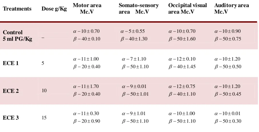
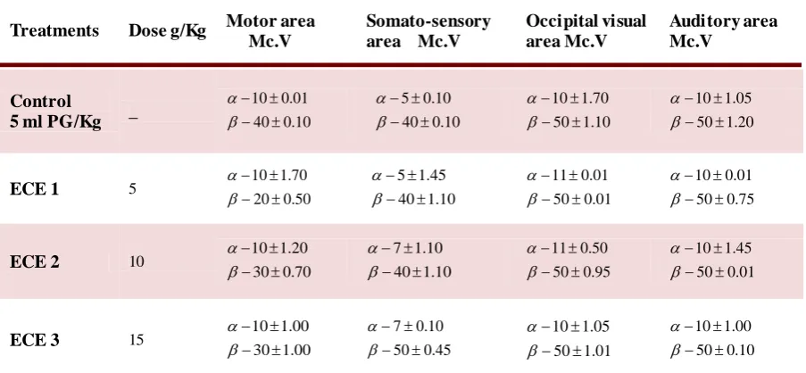
Tables 1-3 show the electrical activity of
motor, somato-sensory, occipital visual, and
auditory areas,in the brain after 60, 120, and 180
min, respectively, of ip administration of 10%
ethanolic extracts of Catha edulis.
Results showed that in the motor area, the
alpha wave values were significantly (P< 0.001)
increased in all treated animals after 60 min of ECE
administration (Table1). The increase of alpha wave
values continued after 120 min (Table 2), but was
less than after 60 min. The alpha wave values
returned to the normal after 180 min of ECE
administration (Table 3).
The beta wave values significantly (P< 0.001)
decreased in the motor area in all treated animals.
This decrease continued even after 180 min of ECE
administration (Table 3).
Regarding the somato-sensory area, the alpha
and beta waves values significantly increased after
60 min in ECE2 and ECE3 groups (P< 0.001) (Table
1). Both alpha and beta waves values significantly
increased after 120 min of ECE administration in
all treated animals (P< 0.001) (Table 2).
The increase in alpha wave values continued in animals in ECE2 and ECE3 after 180 min, whereas
the beta waves values returned to the normal except
in ECE3 animals (Table 3).
In occipital area, after 60 min the alpha wave values increased in ECE2 and ECE3 animals, the
values of beta wave also increased in ECE1 and
ECE2 groups (Table 1). The increase of alpha wave values continued (Table 2) after 120 min in ECE1
and ECE2, whereas the beta wave values of ECE1
and ECE2 groups were decreased. After 180 min the
alpha wave values continuedto be high but to a lesser extent than after 60 and 120 min, whereas the beta
wave values returned to the normal (Table 3).
No electrical activity change was recorded in the auditory region.
Treatments Dose g/Kg Motor area
Mc.V Somato- sensory area Mc.V Occipital visual area Mc.V Auditory area Mc.V Control
5 ml PG/Kg _
ECE 1 5
ECE 2 10
ECE 3 15
52 . 1 40 70 . 0 10 90 . 1 40 95 . 0 5 25 . 1 50 00 . 1 10 0 . 1 50 20 . 1 10 45 . 0 30 70 . 0 13 10 . 0 40 70 . 0 5 10 . 1 60 90 . 0 10 40 . 0 50 10 . 0 10 01 . 1 30 20 . 0 13 10 . 1 60 2 . 1 7 55 . 1 60 75 . 0 12 70 . 0 50 01 . 1 10 20 . 1 20 75 . 0 12 01 . 1 60 01 . 0 7 75 . 0 50 45 . 0 12 95 . 0 50 00 . 1 10
Table 1.The electrical activity of 4 areas in brain after 60 min of Catha edulis extracts administration
Values are: mean of 10 animals ±SE; PG: Propylene glycol, Mc.V: microvolt; α: alpha wave; β: beta wave.
Treatments Dose g/Kg Motor area
Mc.V
Somato-sensory area Mc.V
Occipital visual area Mc.V
Auditory area Mc.V
Control
5 ml PG/Kg _
ECE 1 5
ECE 2 10
ECE 3 15
10 . 0 40 70 . 0 10 30 . 1 40 55 . 0 5 60 . 1 50 70 . 0 10 75 . 0 50 90 . 0 10 40 . 0 20 00 . 1 11 10 . 1 50 10 . 1 7 45 . 1 40 10 . 0 12 50 . 0 50 20 . 1 10 40 . 0 20 70 . 1 11 01 . 1 50 01 . 0 9 10 . 1 40 75 . 0 12 45 . 0 50 20 . 1 10 90 . 0 20 30 . 0 11 10 . 1 50 01 . 1 9 10 . 1 50 00 . 1 10 30 . 0 50 01 . 0 10
Table 2.The electrical activity of 4 areas in brain after 120 min of Catha edulis extracts administration
Values are: mean of 10 animals ±SE; PG: Propylene glycol, Mc.V: microvolt; α: alpha wave; β: beta wave.
Discussion
The electroencephalography is not only a
method of determining the functional state of the
nervous cells, but in some cases it helps to establish
the character of the diseases and the localization of
the pathological process in the brain (15).
The above mentioned changes in the electrical
activity of nervous cells can occur only if the Na+
pumps in the cells membrane are temporary blocked
and/or if the enzymatic system of brain is damaged
(16). Therefore, it can be assumed that one or more compounds in Catha edulis have the ability to
temporary block the Na+ pumps in cells membrane,
and /or, the ability to affect the enzymatic system of the brain. Farag and Al-Qirbi reported that Catha
edulis affects the enzymatic system of rabbit's brain
(17).
The frequency changes in EEG values could
be due also to the acute change in the level of gamma
amino butyric acid (GABA) in the brain (16). The ability of the constituents of Catha edulis to
decrease the levels of 5- hydroxytryptophane
(5-HTP) and GABA in the brain of rabbits, was previously reported (Kennedy et al., 1983).
However, the change in the electrical activity of
nervous cells may lead to many pathological disorders in peoples with long– term Khat chewing,
and at the end, to the death of nervous cells. The
frequency changes in the values of EEG recorded in
our experiment, (i.e. abnormal (P< 0.001) increase
and decrease in the electrical activity of brain in the
treated animals compared with the control animals),
are similar to that recorded in patients with
psycho-neurological disorders dependent on narcotics (18).
The frequency change in the electrical activity of the
brain can lead to a withdrawal phenomenon (e.g.
restlessness, agitation, tremor, rarely convulsions)
(18). Some of Khat constituents were reported to cause similar symptoms (6). The changes in EEG
values in our experiments were similar to the EEG
changes recorded in patients with paradoxical reactions such as restlessness, hallucinations,
aggressiveness, agitation, unusual behavior,
psychosis, anxiety, nightmares, irritability and delusions (18). Jeger and Sireling, also reported that
Khat caused some of the above mentioned disorders
(19).
Other adverse effects such as confusion,
depression, increase or decrease in libido may also
occur in the case of frequency change in brain electrical activity (16). Some of khat contents cause
similar effects (6).
Tiredness (drowsiness, lethargy, dopiness, prolonged reaction time) tremor, dizziness, head-
Treatments Dose g/Kg Motor area
Mc.V
Somato-sensory area Mc.V
Occipital visual area Mc.V
Auditory area Mc.V
Control
5 ml PG/Kg _
ECE 1 5
ECE 2 10
ECE 3 15
10 . 0 40
01 . 0 10
10 . 0 40
10 . 0 5
10 . 1 50
70 . 1 10
20 . 1 50
05 . 1 10
50 . 0 20
70 . 1 10
10 . 1 40
45 . 1 5
01 . 0 50
01 . 0 11
75 . 0 50
01 . 0 10
70 . 0 30
20 . 1 10
10 . 1 40
10 . 1 7
95 . 0 50
50 . 0 11
01 . 0 50
45 . 1 10
00 . 1 30
00 . 1 10
45 . 0 50
10 . 0 7
01 . 1 50
05 . 1 10
10 . 0 50
00 . 1 10
Table 3.The electrical activity of 4 areas in brain after 180 min of Catha edulis extracts administration
Values are: mean of 10 animals ±SE; PG: Propylene glycol, Mc.V: microvolt; α: alpha wave; β: beta wave.
ache, ataxia, clouding of consciousness, and reduced
alertness, dysarthria, anterograade, amnesia, and tension can develop in case of frequency change in
electrical activity of brain (18). Similar disorders
were also reported as the effects of Khat consumption (20).
Further deep studies are needed to better
understand the mechanism of action of Khat in the brain.
Acknowledgments
Authors are grateful to the staff of unit of Neurology in Sheik Hamad hospital in Al-Manama,
Kingdom of Bahrain, for their technical assistance.
Conflict of interest
The authors declared no conflict of interest.
References
1. Numan N. The green leaf: khat. World Journal of Medical Sciences. 2012;7:210-23.
2. Luqman W, Danowski T S. The use of khat (Catha edulis) in Yemen. Social and medical observations. Ann Intern Med. 1976;85:246-9.
3. Cais M, Ginsburg D, Mandelbaum A, et al. Constituents of Catha edulis. Tetrahedron. 1975;31:2727-31.
4. Wolfes O. Über das Vorkommen von d-nor-iso-Ephedrin in Catha edulis. Archiv Pharm. 1930;268:81-3. [Article in Dutch] 5. Hofmann H, Opitz K, Schnelle H. Die Wirkung des nor-psi-Ephedrins. Arzneimittelforschung-Drug Research. 1955;5:36 7-70.
6. Alles G A, Fairchild M D, Jensen M, et al. Chemic a l pharmacology of Catha edulis. J Med Chem. 1960;3:323-52. 7. Szendrei K. The chemistry of khat. Bull Narc. 1980;32:5-35.
8. Kalix P. Khat: a plant with amphetamine effects. J Subst Abuse Treat. 1988;5:163-9.
9. Kalix P. Pharmacological properties of the stimulant khat. Pharmacol Ther. 1990;48:397-416.
10. Kalix P. Catha edulis, a plant that has amphetamine effects. Pharm World Sci. 1996;18:69-73.
11. Winterfeld K, Bernsmann G. Zur kenntnis der Inhaltsstoffe von Catha edulis Forskal. Archiv der Pharmazie. 1960;293:9 9 1-1000.
12. Schorno X, Steinegger E. CNS-active phenylpropylamines of Catha edulis FORSK. (Celastraceae) of Kenyan origin. Experientia. 1979;35:572-4.
13. Eddy N B, Halbach H, Isbell H, et al. Drug dependence: its significance and characteristics. Bull World Health Organ. 1965;32:721-33.
14. Al-Meshal I, Ageel A, Tariq M, et al. The anti gastric ulcer activity of Khat (Catha edulis). J Subst Abuse Treat. 1983;4:14 3 -50.
15. Tatarinov V G. Human anatomy and physiology;: Textbook for nurses' training schools. Moscow: Mir Publishers 1971. 16. Haines, D.E. The principles of Neurology. New-Yo rk Churchill Living-stone pub; 2007:729-740.
17. Farag R, Qirbi A. Effect of khat on some enzymatic levels in liver and brain of rabbits. Egyptian J Biochem. 1991;9:299-307. 18. Kandel E R, Schwartz J H, Jessell T M, et al. Principles of neural science: McGraw-hill New York; 2000.
19. Jager A D, Sireling L. Natural history of khat psychosis. Aust N Z J Psychiatry. 1994;28:331-2.
20. Geisshüsler S, Brenneisen R. The content of psychoactive phenylpropyl and phenylpentenyl khatamines in Catha edulis Forsk. of different origin. J Ethnopharmacol. 1987;19:269-77.