IRANIAN JOURNAL OF VETERINARY SURGERY
(IJVS) WWW.IVSA.IR
Enhancing Ectopic Bone Formation in Canine Masseter Muscle by Loading
Mesenchymal Stem Cells onto Natural Bovine Bone Minerals
Mohamadreza Baghaban Eslaminejad1∗, PhD
Mohammad Jafarian2, DMD
Arash Khojasteh2, DMD
Fatemeh Mashhadi Abbas2, DDS
Mohammad Mehdi Dehghan3, DVSc
Bahar Houshmand4, DDS
1
Stem Cell Department, Cell Science Research Center, Royan Institute, ACECR, Tehran, Iran.
2
Department of Oral and Maxillofacial Surgery, Taleghani University Hospital, Shahid Beheshti University of Medical Sciences, Tehran, Iran.
3
Department of Clinical Science, Faculty of Veterinary Medicine, University of Tehran, Tehran, Iran.
4
Iranian Center for Dental Research,
Shahid Beheshti University of Medical Sciences, Tehran, Iran.
Abstract
Objectives- To assess the ectopic bone formation in canine masseter muscle following the
implantation of the natural bovine bone minerals (NBM) loaded with canine mesenchymal stem cells (MSCs).
Design- Experimental study Animals- four mongrel dogs
Procedures- Tripotent MSCs isolated from the canine bone marrowwere loaded onto the NBM
sponges and allowed to adhere. The cell-loaded scaffolds were then implanted in parallel with cell-free control scaffolds in masseter muscles of four mongrel dogs. Eight weeks after, the animals were sacrificed and the ectopic bone formation in implantation site was studied using the sections prepared from the parts of the muscle containing the implants. Furthermore, the amount of bone formation in two studied groups was quantified using Image-Pro Plust software.
Results- The implants from the both groups were appeared to be encapsulated by fibrous tissue
in implantation site which included some trabecular bone containing osteocyte and osteoblast. There were no indications of inflammation and foreign body reaction, nor were there any indications of cartilage tissue formation.In contrast to control, in MSCs group, lamellar bone
∗
Corresponding Author:
Mohamadreza Baghaban Eslaminejad, PhD
was observed in some area. More importantly, in cell loaded scaffolds more amount of bone was formed compared to that of control cell free scaffolds (P<0.05).
Conclusion and Clinical Relevance- Taken together it seems that in vivo bone forming
capacity of the NBM sponges would be improved by loading it with MSCs.
Keywords- Mesenchymal Stem Cells, Bovine Bone Minerals, Canine.
Introduction
Large bone defects represent major clinical problems in the practice of reconstructive orthopedics and craniofacial surgery. As current treatment for these applications, such as autogenous or allogenous bone grafting have limitations, new approaches for bone tissue repair are essential 1-5. In this regards bone tissue engineering approach using mesenchymal stem cells and appropriate scaffold has been gained considerable attention 6,7.
Mesenchymal stem cells are multipotential cells capable of differentiating into osteoblasts, chondrocytes, adipocytes, tenocytes, and myoblasts 8-10. From a small bone marrow aspirate (~10 to 20 mL), mesenchymalstem cells can be isolated and expanded in culture into a large number because of their extensive proliferative capacity 11,12.
Calcium and phosphate ceramics are being increasingly used as bone substitutes in orthopaedic, oral and maxillo-facial surgery 13-15. Yoshikawa et al 16 have reported that hydroxyapatite (HA) loaded with mesenchymal stem cells (MSCs) has osteogenic potential comparable with autogenous particulate cancellous bone and marrow (PCBM). Furthermore, Yamada et al. have confirmed similar osteogenic potential in ß-TCP (tricalcium phosphate), which is a biodegradable material 17.
BioOss®, a natural bovine bone mineral is being largely used in reconstructive craniofacial
surgery as a bone fill. This scaffold is indeed a bovine deproteinized bone sterilized by gamma radiation and possesses osteoconductive property. A few in vitro studies have confirmed that this scaffold could support fibroblastic and mesenchymal stem cell culture, but little is known regarding their in vivo bone formation capacity when being loaded with bone forming cells 18-21. In present study, canine bone-marrow derived mesenchymal stem cells were loaded onto BioOss sponges and engrafted autologously in the canine masseter muscle to evaluate the ectopic bone formation of the cell/scaffold construct compared to that of cell-free materials. Study like this could help to understand in vivo bone formation capacity of commonly-used bone substitute (BioOss) when being enriched by MSCs.
Materials and Methods
Mesenchymal Stem Cell Isolation and Cultivation
Royan Institute. In the cell culture lab, canine MSCs were isolated according to the method by Kadiyala et al18 with some modification. In brief, the nucleated cell fraction of the marrow was enriched by gradient centrifugation and cultured in 150-cm2 flask at 5×10⁴ cells /ml in 15 ml low-glucose DMEM (Dulbecco Modified Eagle Medium, Gibco, UL) containing 15% FCS, 100 U/ml penicillin G and 100 U/ml streptomycin. The cells were incubated at 37 °C in a humidified 5% CO2 atmosphere and on day 7, the non-adherent cells were removed along with the culture media. The cultures were feed twice a week, passaged on days 17-21 by lifting the cells with 0.05% Trypsin 0.53 mM EDTA exposure for 5 minutes and split in a 1:3 ratio into new 150-cm2 culture flasks. Additional passages were performed to obtain adequate number of the cells and this was achieved upon passage 3.
Differentiation Potential
To evaluate the mesenchymal stem cell nature, the isolated cells were differentiated into osteogenic, chondrogenic and adipogenic cell lineages.
To induce osteogenic differentiation, confluenced passaged-3 cells were cultured in the DMEM medium supplemented with 50 mg/ml ascorbic2- phosphate (Sigma, USA), 10 nM dexamethazone( Sigma, USA) and 10 mM b gicerole phosphate ( Sigma, USA) for 3 weeks. At the end of this period, alizarin red staining was used to observe the matrix mineralization. For staining, the cultures were first fixed by methanol for 10 minutes and then subjected to alizarine red solution for 2 minutes.
To induce the cartilage differentiation, micro mass culture system was used. For this purpose, 2.5×10⁵ passaged-3 cells were pelleted under 1200 g for 5 minute and cultured in a DMEM medium supplemented by 10 ng/ml transforming growth factor-b3, 10 ng/ml bone morphogenetic protein-6, 50mg/ml insulin transferin selenium+ premix and 1.25 mg bovine serum albumin and 1% fetal bovine serum. Three weeks later, the pellets were subjected to the following: fixing in 10% formalin; dehydrating in an ascending ethanol; clearing in xylene; embedding in paraffin wax and sectioning in 5µ by microtome. The sections were then stained by toluidin blue for 30 second at room temperature.
For adipogenesis, DMEM medium containing 100 nM dexamethazone (Sigma, USA) and 50 mg/ml indomethasine (Sigma, USA) was used to induce the differentiation in the confluenced culture of the cells. Three weeks later, the culture was fixed with 4% formalin at room temperature, washed by 70% ethanol and stained by oil red solution in 99% isopropanol for 15 minute.
RNA Extraction and RT-PCR Analysis of Gene Expression
under linear conditions. The products were analyzed on 2% agarose gel and visualized by ethidium bromide staining.
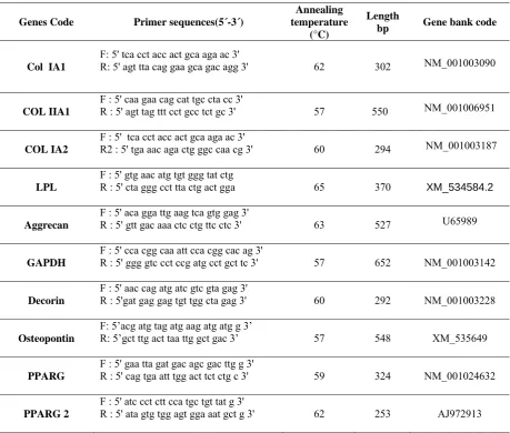
Table 1: The primers used in RT-PCR analysis
Genes Code Primer sequences(5´-3´)
Annealing temperature
(°C)
Length
bp Gene bank code
Col IA1
F: 5' tca cct acc act gca aga ac 3'
R: 5' agt tta cag gaa gca gac agg 3' 62 302 NM_001003090
COL IIA1
F : 5' caa gaa cag cat tgc cta cc 3'
R : 5' agt tag ttt cct gcc tct gc 3' 57 550 NM_001006951
COL IA2
F : 5' tca cct acc act gca aga ac 3'
R2 : 5' tga aac aga ctg ggc caa cg 3' 60 294 NM_001003187
LPL
F : 5' gtg aac atg tgt ggg tat ctg
R : 5' cta ggg cct tta ctg act gga 65 370 XM_534584.2
Aggrecan
F : 5' aca gga ttg aag tca gtg gag 3'
R : 5' gtt gac aaa ctc ctg ttc ctc 3' 63 527 U65989
GAPDH
F : 5' cca cgg caa att cca cgg cac ag 3'
R : 5' ggg gtc cct ccg atg cct gct tc 3' 57 652 NM_001003142
Decorin
F : 5' aac cag atg atc gtc gta gag 3'
R : 5'gat gag gag tgt tgg cta gag 3' 60 292 NM_001003228
Osteopontin
F: 5’acg atg tag atg aag atg atg g 3’
R: 5’gct ttg act taa ttg gct gac 3’ 57 548 XM_535649
PPARG
F : 5' gaa tta gat gac agc gac ttg g 3'
R : 5' cag tga att tgg act tct ctg c 3' 59 324 NM_001024632
PPARG2
F : 5' atc cct ctt cca tgc tgt tat g 3'
R : 5' ata gtg tgg agt gga aat gct g 3' 62 253 AJ972913
Implant Preparation
embedded in paraffin wax (Leica, Bensheim, Germany). Five micrometer sections were then cut, stained with H & E and observed by light microscope.
In-vivo Bone Differentiation
Four adult mongrel dogs with healthy teeth, weighing between 20-30 kg, were used in this study. The dogs were premedicated with Xylazine- HCl (1 mg/ kg) (Xylazine 2%, Alfasan, Woerden-Holland) intramuscularly and atropine sulphate (0.05 mg/kg) (Atropin 0.5, Daroupakhsh Pharmaceutical Mfg, Co, Tehran, Iran) subcutaneously. This was followed by general anaesthesia with sodium thiopental (10 mg/kg) (Nesdonal, Specia, France) intravenously and oroendotracheal intubation. After induction of general anaesthesia, infiltration anaesthesia was applied to the submandibular body area. Submandibular incision was made in the bilateral mandibular angle area and layered dissection was performed through the mandibular bone. Layered dissection was performed until investing fascia of the masseter muscle reached. Blunt dissection with curved hemostat performed to creat tunnel pouch measured 5×5 millimeter. In the right side of the mandible 4 granules of BioOss loaded with cMSCs were embedded with microforceps and in the other side the same amount of control cell free scaffolds were embedded. The pouch was closed in the layered fashion with resorbable sutures (Vhicril, Ethicon). Each dog was sacrificed 8 weeks after insertion of the implant with an overdose of sodium thiopental and subsequent perfusion through the carotid arteries with a fixative consisting of a mixture of glutaraldehyde (5%) and formaldehyde (4%) buffered to pH 7.2 .The implant site of the muscle were removed and placed in 10% formalin for an additional 10 days and decalcified in formic acid for 24 days. The specimens were washed with tap water, dehydrated with ascending concentrations of ethyl alcohol, cleared in xylene, infiltrated with paraffin and processed for histologic evaluation. Decalcified coronal 5 µm serial sections which incorporated total implant area were prepared and stained using H & E.
The stained sections were examined under light microscope in terms of the bone formation, the presence of scaffold piece, the presence of inflammation cells. Furthermore, to quantify the amount of bone formation, slides were photographed at 6.5 magnification using stereomicroscope equipped with digital camera (Nikon E8400, Japan). The percent area of bone formation was then calculated for either scaffold/cell or cell free scaffold implants using Image-Pro Plust software (Media Cybernetic, Silver Springs, MD, USA).
Statistical Analysis
All the data are presented as means and standard deviation. The data were subjected to statistical analysis using one-way ANOVA and Post-Hoc Tukey. Differences at P<0.05 were considered significant. Calculation were performed using the SPSS statistical package (SPSS 11, SPSS Inc., Chicago, IL, USA)
Results
Cell Culture
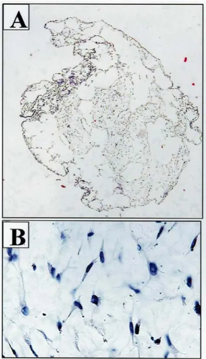
Figure 2: Decalcified scaffolds contained the
loaded MSCs A: magnification 10× and B) magnification 200×
Figure 1: Canine MSCs isolation, expansion and
differentitian. A) Primary culture, B) Passage 3 , C) Alizarin red staining of osteogenic culture, D) Bone gene expression of the differentiated cells, E) Oil red staining of adipogenic culture, F) Adipocyte specific genes expressed in differentiated culture, G) Toluidin blue staining of cartilage differentiation, H) Chondrocyte specific genes expressed in differentiating cultures.
Differentiation
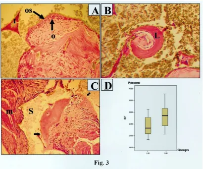
Figure 3: Representative histomicrograph of the bone formation within the muscle tissue. A) Bone trabecula (t) was
visible in implantation site in both groups. In some area, osteoblastic rim(o) and the newly formed osteoid was evident( os), Magnification 100×, H&E staining. B) Only in test group the laminar bone(L) was observed, Magnification 200×, H&E staining. C) The trabecula (Arrow) included many osteocytes and was lined with many osteoblasts. m: muscle fiber, s: scaffold, Magnification 100×, H&E staining D) The percentages of the newly formed bone were significantly high in test compared to control group, BF: Bone formation , 1.00: Cell free scaffold, 2.00: Cell/scaffold composite.
Decalcification
According to the sections prepared from cell-loaded scaffolds, the spaces within BioOss granules appeared to be occupied by canine mesenchymal stem cells. The cells were observed to be morphologically spindle-shape and adherent to scaffold internal surfaces (Fig. 2A and B).
Histologic Study
formation was not found at any time, and thus the process was the so-called intramembranous bone formation. There was a generalized absence of inflammation or foreign-body reaction in the sections.
The percentage of newly formed bone was 28.18%± 5.20 in the BioOss/MSCs implants while in
the control cell free matrices it was 24.16%± 4.22 (Fig. 3D). This demonstrated that there were
differences in the amount of new bone formed in response to the MSCs and it was statistically significant (p<0.05).
Discussion
In present study bone marrow-derived canine MSCs were loaded onto sponges of natural bovine bone minerals (BioOss) and implanted autologously in masseter muscle for a period of 8 weeks. Histological observation indicated that ectopic bone was formed in both MSCs loaded-scaffolds as well as the control cell-free implants. The main differences between two studied groups were in terms of the amount of bone being formed in implantation site as well as its histologic structure. Our quantitative results suggested that in cell-loaded scaffolds the amount of bone formation was significantly higher than of the control cell free implants. Furthermore, in contrast to control, in MSCs group, lamellar bone was observed in some area.
Implantation of culture-expanded autologous MSCs offers the advantage of directly delivering the cellular machinery responsible for synthesizing new bone and circumventing the otherwise slow steps leading to natural or enhanced bone repair. By incorporating living cells with specifically designed matrices, the shortcomings of osteoconductive factors alone to affect permanent bone repair may be overcome. A distinct advantage of using MSCs is that they are adult stem cells, isolated from donors capable of informed consent.
The observations made by Cooper et al (2004) indicated that BioOss loaded with human mesenchymal stem cells formed no bone when implanted in subcutaneous tissue of nude rats20. Our results stand in opposition with the report of Cooper et al in that we observed indications of lamellar bone formation in implant site. Such discrepancy in results could be attributable to differences in the kind of animal model (rat versus dog), implantation site (subcutaneous versus muscle tissue) and the period of implant maintenance (6 weeks versus 8 weeks) in each study. The findings by Mauney et al (2005) who implanted MSCs loaded BioOss subcutaneously in nude rats revealed that the histologic kind of the bone being formed in implantation site was embryonic 21. According to our results the bone being formed in implantation site seemed to be structurally mature bone. Again these differences could be explainable with considering the differences in each study setup including the kind of animal model and implantation site.
One limitation of MSCs study is that no distinct specific markers were introduced for them and for this reason, their identification among the others; hence their isolation would be a difficult task. In the lack of specific marker it was proposed that the golden standard to identify the MSCs is to differentiate them into two or more cell lineages 22. In present study, evaluation of the isolated cells indicated that they were able to produce differentiated progenies including osteoblastic, chondrocytic and adipocytic lineages, therefore; their mesenchymal stem cell nature was confirmed.
highly porous (70%-75%) with large pore size of 150-300 µm. To overcome this problem, the medium that was used for cell loading was rendered a little more viscous by adding a few µl of collagen I gel and the cell suspended in this mixture was placed on top surface of the scaffold. This procedure provides the cell with a chance of slow penetration and enough time of interacting with internal surfaces of the scaffolds.
Taken together it seems that in vivo bone forming capacity of the natural bovine bone mineral sponges would be improved by loading it with MSCs.
References
1. Gazdag AR, Lane JM, Glaser D, et al. Alternatives to autogenous bone graft: efficacy and indications. J Am Acad Orthop Surg 1995; 3: 1-8.
2. Li XQ, Stevenson S, Klein L, et al. Differential patterns of incorporation and remodeling among various types of bone grafts. Acta Anat 1991; 140: 236-44.
3. Rah Dk. Art of replacing craniofacial bone defects: Yonsei Med J 2000; 41:756-765.
4. Hannouche D, Raould A, Nizard RS, et al. Embedding of Bone Samples in methylmethacrylate: A Suitable Method for Tracking LacZ mesenchymal stem cells in skeletal tissues: J Histochem Cytochem 2006;55:255-262.
5. Shors EC. Coralline bone graft substitutes. Orthop Clin North Am 1999; 30:599-613.
6. Persidis A. Tissue engineering. Nat Biotechnol 1999; 177:508-510.
7. Hollinger JO, Winn S, Bonadio J. Options for tissue engineering to address challenges of the aging skeleton. Tissue Engineering 2000; 6: 341 -350.
8. Pittenger MF, Mackay AM, Beck SC, et al. Multilineage potential of adult human mesenchymal stem cells. Science 1999; 284: 143-147.
9. Caplan AI, Fink DJ, Goto T, Linton AE, Young RG, Wakitani S, Goldberg VM, Haynesworth SE: Mesenchymal stem cells and tissue repair. In: Jackson DW, Arnoczky SP, Woo SL, Frank CB, Simon TM, editors. The anterior cruciate ligament: current and future concepts. New York: Raven Press; 1993. p 405-17.
10.Jaiswal N, Haynesworth SE, Caplan AI, et al. Osteogenic differentiation of purified, culture-expanded human mesenchymal stem cells in vitro. J Cell Biochem 1997; 64:
295-312.
11.Lennon DP, Haynesworth SE, Bruder SP, et al. Human and animal mesenchymal progenitor cells from bone marrow. identification of serum for optimal selection and proliferation. In Vitro Cell Dev Biol 1996; 32: 602-11.
12.Beresford JN. Osteogenic stem cells and the stromal system of bone and marrow. Clin Orthop 1989; 240: 270-280.
13.Vrouwenvelder WCA, De Groot CC, De Groot K. Histological and biochemical evaluation of osteoblasts cultured on bioactive glass, hydroxyapatite, titanium alloy and stainless steel. J Biomed Mater Res 1993; 27:465-475.
14.Hench LL. Bioceramics: from concept to clinic. J Am Ceram Soc 1991; 74:1487-1510. 15.Oonish H. Orthopaedic applications of hydroxyapatite. Biomaterials 1991; 12: 171-178.
17. Yamada Y, Boo JS, Ozawa R, et al. Bone regeneration following injection of mesenchymal stem cells and fibrin glue with a biodegradable scaffold. J Craniomaxillofac Surg 2003; 31 :27-33.
18.Ignjatovic N, Ninkov P, Kojic V et al. Ctotoxicity and fibroblast properties during in vitro test of biphasic calcium phosphate/poly-dllactide-co-glycolide biocomposites and different phosphate materials. Mic Res Tech 2006; 69:976-982.
19.Turhani D, Weibenbock M, Watzinger E et al. In vitro study of adherent mandibualr osteoblast-like cells on carrier materials. Int J Oral Maxillfac Surg 2004;34:543-550. 20.Cooper LF, Harris CT, Bruder SP, et al. Incipient analysis of mesenchymal
stem-cell-derived osteogenesis. J Dent Res 2001; 80: 314 –320.
21.Mauney JR, Jaquiery C, Volloch V, et al. In vitro and in vivo evaluation of differentially demineralized cancellous bone scaffolds combined with human bone marrow stromal cells for tissue engineering, Biomaterials 2005; 26: 3173-3185.
22.Dominci M, Le Blanc K, Mueller I, et al. Minimal criteria for defining multipotent mesenchymal stromal cells. The international societry for cellular therapy position statement. Cytotherapy 2006; 4: 315-317.
هﺪﻴﻜﭼ
ﺖﻳﻮﻘﺗ
ﮓﺳ
ﻪﻐﺿﺎﻣ
ﻪﻠﻀﻋ
ﻞﺧاد
رد
ﻚﻴﭘﻮﺘﻛا
يزﺎﺳ
ناﻮﺨﺘﺳا
يﺮﻴﮔرﺎﺑ
ﻖﻳﺮﻃ
زا
لﻮﻠﺳ
ﻲﻤﻴﺸﻧاﺰﻣ
يدﺎﻴﻨﺑ
يﺎﻫ
وﺎﮔ
ﻲﻌﻴﺒﻃ
ﻲﻧﺪﻌﻣ
داﻮﻣ
زا
ﻞﻜﺸﺘﻣ
ﺖﺴﺑراد
رد
داﮋﻧﻲﻣﻼﺳانﺎﺒﻏﺎﺑﺎﺿرﺪﻤﺤﻣ
1 ، نﺎﻳﺮﻔﻌﺟﺪﻤﺤﻣ 2 ، ﻪﺘﺴﺠﺧشرآ 2 ،
سﺎﺒﻋيﺪﻬﺸﻣﻪﻤﻃﺎﻓ
2
،
نﺎﻘﻫديﺪﻬﻣﺪﻤﺤﻣ
3 ، ﺪﻨﻤﺷﻮﻫرﺎﻬﺑ 4 1
،يدﺎﻴﻨﺑيﺎﻫلﻮﻠﺳهوﺮﮔ ،ﻲﻫﺎﮕﺸﻧاددﺎﻬﺟﻲﻟﻮﻠﺳمﻮﻠﻋتﺎﻘﻴﻘﺤﺗﺰﻛﺮﻣ
نﺎﻳورهﺪﻜﺸﻫوﮋﭘ ناﺮﻳا،ناﺮﻬﺗ،
.
2
ﺶﺨﺑ ﻲﺘﺸﻬﺑﺪﻴﻬﺷهﺎﮕﺸﻧاد،ﻲﻜﺷﺰﭙﻧاﺪﻧدهﺪﻜﺸﻧاد،ترﻮﺻوﻚﻓ،نﺎﻫدﻲﺣاﺮﺟ ناﺮﻳاناﺮﻬﺗ،
.
3
ناﺮﻬﺗهﺎﮕﺸﻧاد،ﻲﻜﺷﺮﭙﻣادهﺪﻜﺸﻧاد،ﻲﻫﺎﮕﻧﺎﻣردمﻮﻠﻋهوﺮﮔ،ﻲﺣاﺮﺟﺶﺨﺑ ،ناﺮﻬﺗ،
ناﺮﻳا
.
4
،ناﺮﻬﺗ،ﻲﺘﺸﻬﺑﺪﻴﻬﺷهﺎﮕﺸﻧاد،ناﺮﻳاﻲﻜﺷﺰﭙﻧاﺪﻧدتﺎﻘﻴﻘﺤﺗﺰﻛﺮﻣ
ناﺮﻳا . فﺪﻫ
-زاﻞﻜﺸﺘﻣيﺎﻫﺖﺴﺑرادﺪﻧﻮﻴﭘلﺎﺒﻧد ﻪﺑﮓﺳﻪﻐﺿﺎﻣﻪﻠﻀﻋﻞﺧادردﻚﻴﭘﻮﺘﻛاناﻮﺨﺘﺳا ﻞﻴﻜﺸﺗﻲﺳرﺮﺑ ﻲﻌﻴﺒﻃﻲﻧﺪﻌﻣداﻮﻣ
ﻲﻤﻴﺸﻧاﺰﻣيدﺎﻴﻨﺑيﺎﻫلﻮﻠﺳﺎﺑهﺪﺷيﺮﻴﮔرﺎﺑ
.. ﺮﻃ ﻪﻌﻟﺎﻄﻣح -ﻲﺑﺮﺠﺗﻪﻌﻟﺎﻄﻣ اﻮﻴﺣ تﺎﻧ
-ﮓﺳساررﺎﻬﭼ
رﺎﻛشور
-ﻞﻜﺸﺘﻣيﺎﻫﺞﻨﻔﺳاﻞﺧادﻪﺑ،ﻲﻟﻮﻠﺳهدرﻪﺳﻪﺑﺰﻳﺎﻤﺗناﻮﺗﺎﺑ،ﮓﺳناﻮﺨﺘﺳاﺰﻐﻣزاﻖﺘﺸﻣﻲﻤﻴﺸﻧاﺰﻣيدﺎﻴﻨﺑيﺎﻫلﻮﻠﺳ
ﺪﺷيراﺬﮔرﺎﺑيوﺎﮔﻲﻧﺪﻌﻣداﻮﻣزا ﺪﻨﺒﺴﭽﺑنآحﻮﻄﺳﻪﺑﺎﻫلﻮﻠﺳﺎﺗﺪﻳدﺮﮔﺖﺸﻛزورﺪﻨﭼتﺪﻣﻪﺑو
.
يوﺎﺣ يﺎﻫﺞﻨﻔﺳاﺲﭙﺳ
،گﻮﻟﻮﺗارﻮﻄﺑلﻮﻠﺳ ﺪﺷﻪﺘﺷﺎﻛﮓﺳﻪﻐﺿﺎﻣﻪﻠﻀﻋﺖﻓﺎﺑﻞﺧادﻪﺑ
.
ﺮﺘﻨﻛهوﺮﮔناﻮﻨﻋﻪﺑﺰﻴﻧلﻮﻠﺳنوﺪﺑيﺎﻫﺞﻨﻔﺳا ﻪﻐﺿﺎﻣﻪﻠﻀﻋردل
ﺪﺷﺪﻧﻮﻴﭘﻞﺑﺎﻘﻣفﺮﻃ
..
يوﺎﺣﻪﻠﻀﻋزاﻲﺘﻓﺎﺑيﺎﻫشﺮﺑﻪﻴﻬﺗﺎﺑﻚﻴﭘﻮﺘﻛاناﻮﺨﺘﺳاﻞﻴﻜﺸﺗوﺪﻧﺪﺷﻲﻧﺎﺑﺮﻗﺎﻫﮓﺳ ،ﺪﻌﺑﻪﺘﻔﻫﺖﺸﻫ
ﺪﺷﻲﺳرﺮﺑ،ﺖﻨﻠﭙﻤﻳا
.
لﻮﻠﺳنوﺪﺑﺞﻨﻔﺳاولﻮﻠﺳيوﺎﺣﺞﻨﻔﺳاهوﺮﮔودردهﺪﺷﻞﻴﻜﺸﺗناﻮﺨﺘﺳاناﺰﻴﻣ،ﺮﺿﺎﺣﻪﻌﻟﺎﻄﻣردﻦﻴﻨﭽﻤﻫ
ﺮﻧزاهدﺎﻔﺘﺳاﺎﺑ راﺰﻓام Image-Pro Plust ﺪﻳدﺮﮔﻲﺳرﺮﺑ . ﺞﻳﺎﺘﻧ
-دﻮﺑهﺪﺷﻪﻃﺎﺣا يزوﺮﺒﻴﻓﺖﻓﺎﺑ ﻂﺳﻮﺗفاﺮﻃازا ﺎﻫﺖﻨﻠﭙﻤﻳا ،ﺎﻣتاﺪﻫﺎﺸﻣسﺎﺳاﺮﺑ
.
هوﺮﮔ ود ﺮﻫرد ﻲﻧاﻮﺨﺘﺳايﺎﻫ لﻮﻜﺑاﺮﺗ
دﻮﺑهﺪﻫﺎﺸﻣ ﻞﺑﺎﻗ
.
دﻮﺑﺖﺳﻼﺑﻮﺌﺘﺳاو ﺖﻴﺳﻮﺌﺘﺳايوﺎﺣ ،ﺎﻫلﻮﻜﺑاﺮﺗ ﻦﻳا
.
ﻓﺎﺑ ﻞﻴﻜﺸﺗ زا ﻲﻧﺎﺸﻧﭻﻴﻫ ،هوﺮﮔ ود ﺮﻫرد ، ﻲﻓوﺮﻀﻏﺖ
ﺖﺷاﺪﻧدﻮﺟوﻲﺟرﺎﺧﻢﺴﺟﺶﻨﻛاووبﺎﻬﺘﻟا
.
ﻎﻟﺎﺑرﻼﻣﻻناﻮﺨﺘﺳاﻲﻘﻃﺎﻨﻣرد،لﻮﻠﺳيوﺎﺣﺪﻟﻮﻓﺎﻜﺳاهوﺮﮔرد،لﺮﺘﻨﻛهوﺮﮔفﻼﺧﺮﺑ
ﺪﺷهﺪﻫﺎﺸﻣ
.
دﻮﺑلﺮﺘﻨﻛهوﺮﮔزاﺶﻴﺑيرادﻲﻨﻌﻣرﻮﻄﺑ،هﺪﺷﻪﺘﺧﺎﺳناﻮﺨﺘﺳاناﺰﻴﻣﺶﻳﺎﻣزآهوﺮﮔردﻪﻛدﻮﺑﻦﻳاﺮﺘﻤﻬﻣﻪﺘﻜﻧ
. يﺮﻴﮔﻪﺠﻴﺘﻧ دﺮﺑرﺎﻛو ﻲﻨﻴﻟﺎﺑ
-لﻮﻠﺳيﺮﻴﮔرﺎﺑﺎﺑ،وﺎﮔﻲﻌﻴﺒﻃ ﻲﻧﺪﻌﻣداﻮﻣيزﺎﺳناﻮﺨﺘﺳاﺖﻴﻓﺮﻇﻪﻛﺪﺳرﻲﻣﺮﻈﻧﻪﺑﻪﺘﻓرﻢﻫيور
ﺪﺸﺨﺑﻲﻣدﻮﺒﻬﺑﻲﻤﻴﺸﻧاﺰﻣيدﺎﻴﻨﺑ
.
نﺎﮔژاوﺪﻴﻠﻛ
ﻲﻤﻴﺸﻧاﺰﻣيدﺎﻴﻨﺑيﺎﻫلﻮﻠﺳ ناﻮﺨﺘﺳاﻲﻧﺪﻌﻣداﻮﻣ،