Mariet Allen, PhD* Sarah J. Lincoln, BS* Morgane Corda, BS* Jens O. Watzlawik, PhD Minerva M. Carrasquillo,
PhD
Joseph S. Reddy, PhD Jeremy D. Burgess, BS Thuy Nguyen, BS Kimberly Malphrus, BS Ronald C. Petersen, MD,
PhD
Neill R. Graff-Radford, MD
Dennis W. Dickson, MD Nilüfer Ertekin-Taner,
MD, PhD
Correspondence to Dr. Ertekin-Taner:
Supplemental data at Neurology.org/ng
ABCA7
loss-of-function variants,
expression, and neurologic disease risk
ABSTRACT
Objective: To investigate and characterize putative “loss-of-function” (LOF) adenosine triphosphate–binding cassette, subfamily A member 7 (ABCA7) mutations reported to associ-ate with Alzheimer disease (AD) risk.
Methods:We genotyped 6 previously reportedABCA7putative LOF variants in 1,465 partici-pants with AD, 381 participartici-pants with other neuropathologies (non-AD), and 1,043 controls and assessed the overall mutational burden for association with different diagnosis groups. We mea-sured brain ABCA7 protein and messenger RNA (mRNA) levels using Western blot and quantita-tive PCR, respecquantita-tively, in 11 carriers of the 3 most common variants, and sequenced all 47 ABCA7 exons in these participants to screen for other coding variants.
Results:At least one of the investigated variants was identified in 45 participants with late-onset Alzheimer disease, 12 participants with other neuropathologies, and 11 elderly controls. Associ-ation analysis revealed a significantly higher burden of these variants in participants with AD (p 5 5.00E-04) and those with other neuropathologies (p 5 8.60E-03) when compared with controls. Concurrent analysis of brain ABCA7 mRNA and protein revealed lower protein but not mRNA in p.L1403fs carriers, lower mRNA but not protein in p.E709fs carriers, and additional deleterious mutations in some c.557015G.C carriers.
Conclusions:Our results suggest that LOF may not be a common mechanism for theseABCA7
variants and expand the list of neurologic diseases enriched for them.Neurol Genet2017;3:e126; doi: 10.1212/NXG.0000000000000126
GLOSSARY
AD5Alzheimer disease;AGD5argyophilic grain disease;CADD5combined annotation dependent depletion;cDNA5 com-plimentary DNA;DLBD5diffuse Lewy body disease;GAPDH5glyceraldehyde 3-phosphate dehydrogenase;LOAD5late-onset Alzheimer disease;LOF5loss of function;mRNA5messenger RNA;NMD5nonsense-mediated decay;OR5odds ratio;PA5
pathologic aging;PSP5progressive supranuclear palsy;RNAseq5RNA sequencing;VaD5vascular dementia.
Enrichment of
ABCA7
nonsense, frameshift, or splice-site variants was identified in Icelandic
patients with Alzheimer disease (AD), and this association replicated in non-Icelandic cohorts.
1These AD risk variants were predicted to be loss-of-function (LOF) mutations that cause
pre-mature protein termination. In a Belgian cohort, 8
ABCA7
putative LOF mutations were
identified,
22 of which (E709fs, p.Trp1214X) showed reduced brain
ABCA7
messenger RNA
(mRNA). Additional putative LOF mutations were identified in North American Caucasian AD
cases of which p.E1679X
3was statistically significant. These initial studies were followed by
replications and/or identification of additional rare
ABCA7
putative LOF mutations in other
AD cohorts,
4–6in addition to one study in Parkinson disease.
7Although predicted to result in reduced
ABCA7
protein, brain
ABCA7
protein levels in
putative LOF mutation carriers are unknown. Correlative investigations of ABCA7 brain
*These authors contributed equally to this work.
Form the Department of Neuroscience (M.A., S.J.L., M.C., J.O.W., M.M.C., J.S.R., J.D.B., T.N., K.M., D.W.D., N.E.-T.), Department of Neurology (N.R.G.-R., N.E.-T.), Mayo Clinic, Jacksonville, FL; and Department of Neurology (R.C.P.), Mayo Clinic, Rochester, MN. Funding information and disclosures are provided at the end of the article. Go to Neurology.org/ng for full disclosure forms. The Article Processing Charge was paid by the authors.
protein and mRNA levels in putative LOF
mutation carriers and controls are necessary
to determine whether these variants lead to
reduced protein levels via nonsense-mediated
decay (NMD) of mRNA, as expected. Herein,
we investigated the frequency of 6 previously
reported
ABCA7
putative LOF variants
1–3in
our cohort of 1,465 participants with
late-onset Alzheimer disease (LOAD) and 381
par-ticipants with other non-AD neuropathologies,
in comparison with 1,043 clinically normal
controls. We evaluated the corresponding
brain ABCA7 protein and mRNA in 15
par-ticipants composed of carriers for 3 of the
most common mutations (E709fs, L1403fs,
and c.5570
1
5G
.
C) and noncarriers who
lacked any predicted deleterious
ABCA7
mutations. Our study provides a systematic
investigation of downstream effects of
ABCA7
predicted LOF mutations and has implications
for potential mechanism of action of these
variants.
METHODS Standard protocol approvals, registrations, and patient consents.This work was approved by the Mayo Clinic Institutional Review Board, and all participants and/or their proxies provided consent.
Participants and samples. We screened 2,889 Caucasian American participants recruited at Mayo Clinic Rochester (RS), Jacksonville (JS), or Brain Bank (AUT) (table 1) for previously reported1–3ABCA7rare putative LOF variants. Of the 1,465 participants with AD, 1,208 were diagnosed at autopsy as definite AD and 257 as probable or possible AD clinically, according to NINCDS-ADRDA criteria.8Autopsied participants who did not meet criteria for AD neuropathology, but who were enriched for other neurologic pathologies, such as vascular dementia (VaD), frontotemporal dementia, dementia with Lewy bodies, and progressive supranuclear palsy (PSP) were also screened (non-AD, n 5 381), in addition to 1,043 elderly clinical control participants without cognitive impairment. The clinical control
participants did not meet criteria for mild cognitive impairment, AD, or other dementias.
Genotyping analysis.Custom TaqMan SNP assays (Applied Biosystems, Foster City, CA) were designed and used to genotype
ABCA7 mutations p.Trp1214X (rs201060968),2 L1403fs, c.441612T.G (rs113809142),1,2p.E1679X,3and c.557015G.C (rs200538373).1,2Data were analyzed on ABI Prism 7900 Detection System using SDS version 2.2.2 software (Applied Biosystems). For genotyping E709fs 7-bp deletion mutation,1,2fluorescently labeled PCR products were generated and run on an ABI3730xl Genetic Analyzer (Applied Biosystems). Allele sizes were determined using GeneMapper Software 5.0 (Applied Biosystems).
Sequencing analysis.AllABCA7mutations identified in our cohort were validated by sequence analysis. PCR primers were designed to amplify and sequence the genomic regions flanking the mutations. Primer sequences are provided in table e-1 at Neurology.org/ng. PCR products were purified using the Agencourt AMPure system (Beckman Coulter, Brea, CA) and then sequenced in both directions using a Big Dye Terminator v3.1 Cycle Sequencing kit (Applied Biosystems). Sequencing reactions were purified using Agencourt CleanSEQ (Beckman Coulter) and run on an ABI3730xl Genetic Analyzer (Applied Biosystems). Sequence analysis was performed using Sequencher 4.8 software (Gene Codes Corporation, Ann Arbor, MI).
Quantitative PCR.BrainABCA7mRNA levels were measured from the temporal cortex of autopsied participants with L1403fs (n53), E709fs (n54), and c.557015G.C (n54) mutations and 4 participants who lacked these mutations. These 15 partic-ipants were also sequenced for additional mutations in the 47
ABCA7exons using Sanger sequencing as described above. Total RNA was extracted using TRIzol Reagent (Ambion Life Tech-nology) followed by DNase RNA cleanup step using RNeasy (Qiagen, Germantown, MD). The quantity and quality of RNA samples were determined by the Agilent 2100 Bioanalyzer using an Agilent RNA 6000 Nano Chip. Complementary DNA (cDNA) synthesized from 1mg of RNA with Applied Biosystems High-Capacity cDNA Archive Kit was used as a template for relative quantitative PCR using ABI Taqman chemistry (Applied Biosystems).ABCA7mRNA expression was quantified using assay Hs01105117_m1 (exons 45–46). Hs00951083_m1 (TFRC), Hs02800695_m1 (HPRT), and Hs00355488_m1 (TXNL1) assays were used as endogenous controls for global normalization. Each sample was run in replicates of 4 on the QuantStudio 7 Real-Time PCR System, and analysis was performed using the applied biosystems App 3.0.0-PRC-build8 (Thermo Fisher Cloud).
Allele-specific expression in Glu709fs mutation carriers.
To further investigate allele-specific expression, semiquantitative PCR was performed on cDNA from the temporal cortex of Glu709fs carriers. Fluorescently labeled wild-type and/or Glu709fs mutant PCR products were generated with sizes expected at 281 and 274 bp, respectively. PCR amplification was stopped in the log phase after 25 cycles. PCR products were run on an ABI3730xl Genetic Analyzer (Applied Biosystems).
RNA sequencing. We had RNA sequencing (RNAseq) data from temporal cortex tissue of a participant with c.557015G.C and another participant with c.441612T.G mutation. RNA was isolated as described above, and the TruSeq RNA Sample Prep Kit (Illumina, San Diego, CA) was used for library preparation. The library concentration and size distribution were determined on an Agilent Bioanalyzer DNA 1000 chip. Samples underwent 101-bp paired-end sequencing on Illumina HiSeq2000 sequencers. RNAseq measures were processed through the Mayo Clinic MAP-RSeq Table 1 Sample demographics
Series N Mean age, y (SD) Female, n (%) APOEe41, n (%)
AUT-AD 1,208 81.4 (8.7) 706 (58) 741 (61)
RS-AD 236 72.1 (5.4) 140 (59) 149 (63)
JS-AD 21 67.1 (5.1) 15 (71) 11 (53)
RS-CON 1,043 76.5 (7.2) 551 (53) 280 (27)
AUT–non-AD 381 76.6 (9.0) 165 (43) 83 (22)
Abbreviations: AD5participants with a diagnosis of Alzheimer disease; AUT5
autopsy-confirmed participants; CON5cognitively normal control participants; JS5clinical
partic-ipants from Mayo Clinic Jacksonville; non-AD5participants with other neurodegenerative
diseases; RS5clinical participants from Mayo Clinic Rochester.
pipeline.9Indexed bam files were used for visualizing alignments in Integrative Genomics Viewer.10
Western blot analysis.Protein lysate was extracted from tem-poral cortex human brain tissue using a lysis buffer (150 mM NaCl, 50 mM Tris pH 7.5, 0.1% Triton). Protein concentration was determined using NanoDrop 2000 (ThermoFisher Scientific, Waltham, MA), and equal amounts of protein were separated on an NuPAGE 4%–12% Bis-Tris gel and transferred to an Immobilon P membrane (Millipore, Billerica, MA). Membranes were blocked in 5% milk in tris-buffered saline with 0.1% Tween 20 (TBST) and labeled overnight at 4°C with ABCA7 primary antibodies designed to epitope aa 2096-2146 (LS-C291064, LifeSpan BioSciences, Seattle, WA). We validated this antibody for specificity to ABCA7 as described in the supplemental results. Blots were incubated with rabbit IgG horseradish peroxidase–linked secondary antibody (Pierce, SA1-9510) for 1 hour, and proteins were detected using Western Lightning Plus-ECL (Perkin Elmer, Waltham, MA). Equivalent sample loading was confirmed by probing with anti–glyceraldehyde 3-phosphate dehydrogenase (GAPDH) antibody. Protein bands were quantified using ImageJ, and relative amounts of ABCA7 protein were determined. In the Western blots from human brain lysates, both the full-length ABCA7 protein and the shorter 218 KDa ABCA7 product from its known isoform (ENSP00000465322) were observed. For Western blot quantifications, the sum of intensity of these 2 bands divided by the intensity of GAPDH was used.
Statistical analysis.ABCA7protein or mRNA levels in muta-tion carriers were compared with noncarriers using the nonpara-metric 1-sided Mann-WhitneyUtest. Combined counts for the putative LOFABCA7alleles were compared between different diagnostic groups using the Fisher exact 2-sided test. StatsDirect v2.7.8 was used for these analyses.
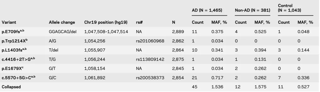
RESULTS We evaluated 6 previously reported1–3 ABCA7rare putative LOF variants predicted to lead to truncated protein due to the premature insertion of a stop codon into the transcript. Forty-five of 1,465 participants with LOAD vs 11 of 1,043 elderly cognitively normal controls (table 2) harbored one of these variants, resulting in AD risk association (odds ratio [OR]52.97,p55.00E-04) (table 3).
In 381 autopsied non-AD participants who did not meet criteria for AD neuropathology, but who were enriched for other neurologic pathologies, 12 had one of these ABCA7 variants, leading to association (OR5 3.05,p5 8.60E-03) when compared with controls. These non-AD participants with ABCA7
variants consisted of 7 with PSP, 3 with VaD, 1 with diffuse Lewy body disease (DLBD), and 1 with pathologic aging (PA) and argyophilic grain disease (AGD). Three PSP participants had the secondary diagnoses of VaD, PA, or AGD. PSP participants had p.E709fs, p.E1679X, p.L1403fs, or c.557015G.C; and VaD participants had p.E1679X, c.441612T.G, or p.L1403fs variants. The p.E709fs variant was also observed in 2 participants with DLBD and PA/AGD. There was no difference in the frequency of putativeABCA7LOF variants between participants with AD and non-AD neuropathologies (p 5
8.70E-01).
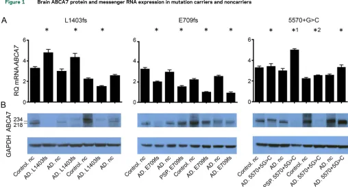
Of the 6 variants evaluated, p.E709fs, p.L1403fs, and c.557015G.C had the highest frequencies, which enabled the evaluation of several mutation car-riers for brain mRNA expression and protein levels (figure 1, A and B, table e-2). There was considerable variance in brainABCA7protein and mRNA levels, both in mutation carriers and noncarriers. Despite this, brain ABCA7 full-length protein (;234 kD) levels were consistently lower in p.L1403fs carriers compared with noncarriers (p5 0.029) (figure 1A). However, p.L1403fs carriers did not have corresponding lower brainABCA7mRNA expression (figure 1B). In contrast, p.E709fs carriers had consistently lower brainABCA7
mRNA expression (p50.029) (figure 1B), which was not reflected in their brain protein levels (figure 1A). Lower brainABCA7 mRNA expression was observed in both the LOAD participants and a PSP participant who were p.E709fs carriers. Semiquantitative assessment
Table 2 ABCA7loss-of-function mutations’frequency in patients with AD, controls, and participants with other non-AD neuropathologies
Variant Allele change Chr19 position (hg19) rs# N
AD (N51,465) Non-AD (N5381)
Control (N51,043)
Count MAF, % Count MAF, % Count MAF, %
p.E709fsa,b GGAGCAG/del 1,047,508–1,047,514 NA 2,889 11 0.375 4 0.525 1 0.048
p.Trp1214Xb A/G 1,054,256 rs201060968 2,862 1 0.034 0 0 0 0
p.L1403fsa,b T/del 1,055,907 NA 2,864 10 0.341 3 0.394 3 0.144
c.441612T>Ga,b T/G 1,056,244 rs113809142 2,875 1 0.034 1 0.131 0 0
p.E1679Xc G/T 1,058,154 NA 2,845 1 0.034 2 0.262 0 0
c.557015G>Ca,b G/C 1,061,892 rs200538373 2,854 21 0.717 2 0.262 7 0.336
Collapsed 45 1.536 12 1.575 11 0.527
Abbreviations: AD5Alzheimer disease; MAF5minor allele frequency; NA5not available.
aVariant reported by Steinberg et al., 2015.1
bVariant reported by Cuyvers et al., 2015.2
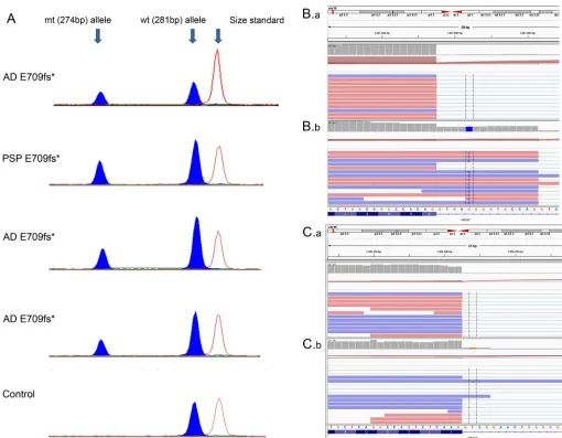
of 4 p.E709fs carriers revealed lower brain levels of the shorter mutant allele (274 bp) in comparison with the wild-type allele PCR product (281 bp) (figure 2A). Similar assessment of p.L1403fs carriers did not support lower brain levels of the 1-bp deletion mutant vs wild-type allele (data not shown).
Investigations of brainABCA7protein and mRNA expression levels in 4 c.557015G.C carriers vs 4 noncarriers did not reveal consistent differences (figure 1, A and B). RNAseq data from carriers of both splice-site mutations, c.557015G.C and 441612T.G, demonstrate that reads containing the mutant allele
extend into the intronic sequence (figure 2, B and C). The inclusion of the mutant intronic sequence within mRNA indicates that these mutations lead to altered splicing. RNAseq data from a c.557015G.C carrier (figure 2B.b) and a noncarrier (figure 2B.a) illustrate the difference in reads mapped to the intron when compared with the exon. The gray bar chart at the top of each panel displays the depth of reads at the locus. In the c.557015G.C carrier, the read depth of the intron is approximately 50% of that of the exon. However, for the noncarrier, the read depth of the intron is substantially lower than that of the exon. This finding indicates that in the c.557015G.C carrier,
Figure 1 Brain ABCA7 protein and messenger RNA expression in mutation carriers and noncarriers
(A) ABCA7 messenger RNA (mRNA) and (B) protein isolated from human brain tissue (temporal cortex) were assessed using quantitative PCR and Western
blot, respectively, for carriers and noncarriers (nc) of the 3 most frequentABCA7putative loss-of-function mutations investigated (L1403fs, E709fs, and
55701G.C). Mutation carriers with a pathologic diagnosis of either Alzheimer disease (AD) or progressive supranuclear palsy (PSP) were analyzed. Two
clinically normal control (control.nc) and 2 pathologic AD participants (AD.nc) without any predicted deleteriousABCA7mutations were analyzed as
mutation noncarriers.*1 and*2 indicate 55701G.C carriers with additionalABCA7predicted deleterious mutations. The Western blots from the human
brain lysates have 2 bands, 1 at 234 kDa corresponding to full-length ABCA7 protein (ENSP00000414062) and 1 at 218 kDa that is the expected
molecular weight of a known alternative isoform ENSP00000465322 (ENST00000433129) starting in exon 7.*DenotesABCA7mutation carriers.
GAPDH5glyceraldehyde 3-phosphate dehydrogenase; RQ5relative quantity.
Table 3 Association ofABCA7variants with AD and other neuropathologies
Cohort A Cohort B Cohort A, N Cohort B, N Cohort A, MAF Cohort B, MAF OR (95% CI) pValue
AD Control 1,465 1,043 1.536 0.527 2.97 (1.50–6.40) 5.00E-04
AD Non-AD 1,465 381 1.536 1.575 0.98 (0.50–2.05) 8.70E-01
Non-AD Control 381 1,043 1.575 0.527 3.05 (1.22–7.70) 8.60E-03
Abbreviations: AD5Alzheimer disease; CI5confidence interval; OR5odds ratio.
Odds ratios andpvalues are calculated using the Fisher exact 2-sided test for 3 comparisons: AD vs control, AD vs
non-AD, and non-AD vs control as indicated. In each comparison, cohort A is assessed as the“Case”and cohort B as the
splicing is affected in approximately 50% of the ex-pressed mRNA molecules, thus refuting NMD of mRNA as its mechanism of action.
Of interest, a PSP c.557015G.C carrier had one of the lowest brain ABCA7 proteins and one of the highest mRNA levels (figure 1, A and B). Sequencing of the entireABCA7coding region for all participants in figure 1 revealed this PSP c.557015G.C carrier to have the highest variant burden with 6 missense muta-tions (table e-3), 2 of which had predicted deleterious effects with combined annotation dependent depletion (CADD) score .2011 (rs3752233:p.R463H and
rs3752239:p.N718T). In addition, an AD c.557015G.C carrier with one of the lowest brain ABCA7 proteins had 4 missense mutations, 1 of which
had predicted damaging effects (rs146191555: p.P1267L), with CADD score.20. None of the other
ABCA7putative LOF mutation carriers or noncarriers in figure 1 had any other mutations with predicted damaging effects.
DISCUSSION Here, we report a systematic assess-ment of previously reported1,2 ABCA7 putative
LOF variants on brain ABCA7 expression in corre-sponding mRNA and protein samples. Our findings demonstrate a lack of correlation between brain ABCA7 protein and mRNA levels and suggest dis-tinct plausible mechanisms of action for the different
ABCA7 putative LOF variants. It should be noted that the ABCA7 antibody recognizes only the
full-Figure 2 Brain ABCA7 messenger RNA expression measurements with semiquantitative PCR or RNA sequencing in select mutation carriers and noncarriers
(A) Relative messenger RNA (mRNA) levels of E709fs mutant (mt) and wild-type (wt) alleles in Alzheimer disease (AD) and progressive supranuclear palsy (PSP) carriers measured using semiquantitative PCR. A control noncarrier is shown for reference. Peak heights indicate relative mRNA levels and show
reduced levels of the E709fs mutant allele. (B.b) RNAseq data for canonical splice-site mutation carriers for 55701G.C and (C.b) 441612T.G are shown.
B.a and C.a are from a participant without these splice-site mutations. RNAseq data are viewed using Integrative Genomics Viewer (IGV). Intronic reads for
the mutant allele are included in the mRNA of both canonical splice-site mutation carriers, indicating that these mutations alter splicing.*DenotesABCA7
length protein and none of the truncated species arising from the ABCA7 mutations. Brain ABCA7 protein is lower in p.L1403fs carriers, despite lack of corresponding lower brain ABCA7 mRNA. In contrast, lower brain ABCA7 mRNA in p.E709fs carriers does not translate to lower protein levels. These findings suggest that despite evidence for NMD, p.E709fs is unlikely to have a deleterious consequence simply by lowering full-length ABCA7 protein levels. The possibility that the mutant p.E709fs allele or the truncated protein could instead have toxic gain of function needs to be considered. Conversely, despite lack of evidence for NMD, p.L1403fs may be leading to ABCA7 LOF via inefficient translation or enhanced protein degradation. We and others2observed substantial variability in
brainABCA7mRNA and protein levels. This may in part be due to the collective effect of multiple delete-rious mutations in this gene and its interactions with other genes and/or the environment. The detection of additional predicted deleterious variants in 2 c.557015G.C carriers with unremarkable brain
ABCA7mRNA but lowest brain protein levels may suggest that the mutation burden in participants within this gene could influence its brain protein levels. One study investigated brainABCA7 mRNA levels in 3 mutation carriers and 4 controls and iden-tified lower mRNA levels in p.E709fs and p.Trp1214X, but not in p.L1403fs carriers,2 but
did not report on brain ABCA7 protein levels. Another novel finding is the statistically significant overall association of the tested ABCA7 variants in a cohort of mixed non-AD brain pathologies. The detection ofABCA7 putative functional variants in participants with PSP, VaD, DLBD, and PA/AGD in this study, in addition to their recent implication in Parkinson disease,7 suggests that this gene may be
contributory to both AD and non-AD neuropathol-ogies. Given evidence of its pleiotropic functions12,13
including phagocytosis, cell signaling, lipid homeo-stasis, and Ab metabolism, it is likely that ABCA7 dysfunction may contribute to both AD-specific and non-AD neuropathologies.
Strengths of this study include sizable cohort of AD and control participants, expansion of ABCA7
mutation screen to non-AD participants, combined investigations of brainABCA7 mRNA and protein levels in multiple mutation carriers and noncarriers who were screened for additionalABCA7coding mu-tations, inclusion of allele-specific expression, and RNAseq as further technical approaches. Despite our systematic investigation of combined brain mRNA and protein levels inABCA7 mutation car-riers, this work needs to be expanded to include addi-tional mutation carriers, larger sample sizes, and functional in vitro studies to uncover the precise
mechanism of action. Although our study provides evidence for a role of ABCA7 variants in non-AD pathologies, because our non-AD cohort was not re-cruited to study a specific neurologic disease,ABCA7
mutation frequency estimates for this mixed diagnos-tic group may not be accurate. Finally, while our study replicated the risk association for theABCA7
variants identified from the initial reports,1–3
contin-uation of variant identification studies is necessary to characterize the full spectrum of mutations in this gene. Our study demonstrates the distinct potential functional consequences of variousABCA7 AD-risk mutations, challenges the notion that they all act via LOF mechanism, and expands the list of neurologic diseases likely influenced by these mutations.
AUTHOR CONTRIBUTIONS
M.A., S.J.L., and N.E.-T. contributed to conception and design of the study. M.A., S.J.L., M.C., J.O.W., M.M.C., J.S.R., J.D.B., T.N., K.M., R.C.P., N.R.G.-R., D.W.D., and N.E.-T. contributed to acquisi-tion and analysis of data. M.A., S.J.L., J.O.W., and N.E.-T. contributed to drafting the text and preparing the figures.
ACKNOWLEDGMENT
The authors thank the patients and their families; without their participation this study would not have been possible.
STUDY FUNDING
This work was supported by the National Institute on Aging (U01 AG046139 and RF1 AG051504), National Institute of Neurological Disorders and Stroke (R01 NS080820), and Bright Focus Foundation (27J-01-03) awarded to N.E.-T.
DISCLOSURE
Moore Alzheimer’s Disease Research Program Grant; and has consulted for Cytox. Go to Neurology.org/ng for full disclosure forms.
Received August 8, 2016. Accepted in final form November 21, 2016.
REFERENCES
1. Steinberg S, Stefansson H, Jonsson T, et al. Loss-of-function variants in ABCA7 confer risk of Alzheimer’s disease. Nat Genet 2015;47:445–447.
2. Cuyvers E, De Roeck A, Van den Bossche T, et al. Muta-tions in ABCA7 in a Belgian cohort of Alzheimer’s disease patients: a targeted resequencing study. Lancet Neurol 2015;14:814–822.
3. Vardarajan BN, Ghani M, Kahn A, et al. Rare coding muta-tions identified by sequencing of Alzheimer disease genome-wide association studies loci. Ann Neurol 2015;78:487–498. 4. Le Guennec K, Nicolas G, Quenez O, et al. ABCA7 rare variants and Alzheimer disease risk. Neurology 2016;86: 2134–2137.
5. Del-Aguila JL, Fernandez MV, Jimenez J, et al. Role of ABCA7 loss-of-function variant in Alzheimer’s disease: a replication study in European-Americans. Alzheimers Res Ther 2015;7:73.
6. Cukier HN, Kunkle BW, Vardarajan BN, et al. ABCA7 frameshift deletion associated with Alzheimer disease in African Americans. Neurol Genet 2016;2:e79.
7. Nuytemans K, Maldonado L, Ali A, et al. Overlap between Parkinson disease and Alzheimer disease in AB-CA7 functional variants. Neurol Genet 2016;2:e44. 8. McKhann G, Drachman D, Folstein M, Katzman R, Price D,
Stadlan EM. Clinical diagnosis of Alzheimer’s disease: report of the NINCDS-ADRDA Work Group under the auspices of Department of Health and Human Serv-ices Task Force on Alzheimer’s Disease. Neurology 1984; 34:939–944.
9. Kalari KR, Nair AA, Bhavsar JD, et al. MAP-RSeq: mayo analysis pipeline for RNA sequencing. BMC Bioinformatics 2014;15:224.
10. Robinson JT, Thorvaldsdottir H, Winckler W, et al. Integrative genomics viewer. Nat Biotechnol 2011;29: 24–26.
11. Kircher M, Witten DM, Jain P, O’Roak BJ, Cooper GM, Shendure J. A general framework for estimating the rela-tive pathogenicity of human genetic variants. Nat Genet 2014;46:310–315.
12. Jehle AW, Gardai SJ, Li S, et al. ATP-binding cassette transporter A7 enhances phagocytosis of apoptotic cells and associated ERK signaling in macrophages. J Cell Biol 2006;174:547–556.
DOI 10.1212/NXG.0000000000000126
2017;3;
Neurol Genet
Mariet Allen, Sarah J. Lincoln, Morgane Corda, et al.
loss-of-function variants, expression, and neurologic disease risk
ABCA7
This information is current as of January 5, 2017
reserved. Online ISSN: 2376-7839.
Published by Wolters Kluwer Health, Inc. on behalf of the American Academy of Neurology. All rights an open-access, online-only, continuous publication journal. Copyright Copyright © 2017 The Author(s).
Services
Updated Information &
http://ng.neurology.org/content/3/1/e126.full.html including high resolution figures, can be found at:
Supplementary Material
http://ng.neurology.org/content/suppl/2017/01/05/3.1.e126.DC1 Supplementary material can be found at:
References
http://ng.neurology.org/content/3/1/e126.full.html##ref-list-1 This article cites 13 articles, 4 of which you can access for free at:
Citations
http://ng.neurology.org/content/3/1/e126.full.html##otherarticles This article has been cited by 6 HighWire-hosted articles:
Subspecialty Collections
http://ng.neurology.org//cgi/collection/gene_expression_studies
Gene expression studies
http://ng.neurology.org//cgi/collection/association_studies_in_genetics
Association studies in genetics
http://ng.neurology.org//cgi/collection/alzheimers_disease
Alzheimer's disease
a
http://ng.neurology.org//cgi/collection/all_cognitive_disorders_dementi
All Cognitive Disorders/Dementia
following collection(s):
This article, along with others on similar topics, appears in the
Permissions & Licensing
http://ng.neurology.org/misc/about.xhtml#permissions its entirety can be found online at:
Information about reproducing this article in parts (figures,tables) or in
Reprints
http://ng.neurology.org/misc/addir.xhtml#reprintsus Information about ordering reprints can be found online:
reserved. Online ISSN: 2376-7839.
Published by Wolters Kluwer Health, Inc. on behalf of the American Academy of Neurology. All rights an open-access, online-only, continuous publication journal. Copyright Copyright © 2017 The Author(s).