0095-1137/07/$08.00
⫹
0
doi:10.1128/JCM.00093-07
Copyright © 2007, American Society for Microbiology. All Rights Reserved.
Typing of Human Enterovirus by Partial Sequencing of VP2
䌤
Dorsaf Nasri,
1,2Lamjed Bouslama,
2Shabir Omar,
1Henia Saoudin,
1Thomas Bourlet,
1Mahjoub Aouni,
2Bruno Pozzetto,
1* and Sylvie Pillet
1Laboratory of Bacteriology-Virology, GIMAP EA3064, Faculty of Medicine of Saint-Etienne, Saint-Etienne, France,
1and
Laboratory of Transmissible Diseases and Biologically Active Substances, Faculty of Pharmacy, Monastir, Tunisia
2Received 13 January 2007/Returned for modification 29 April 2007/Accepted 20 May 2007
The sequencing of the VP1 hypervariable region of the human enterovirus (HEV) genome has become the
reference test for typing field isolates. This study describes a new strategy for typing HEV at the serotype level
that uses a reverse transcription-PCR assay targeting the central part of the VP2 capsid protein. Two pairs of
primers were used to amplify a fragment of 584 bp (with reference to the PV-1 sequence) or a part of it (368
bp) for typing. For a few strains not amplified by the first PCR, seminested primers enhanced the sensitivity
(which was found to be approximately 10
ⴚ1and 10
ⴚ450% tissue culture infective dose per reaction tube for the
first and seminested assay, respectively). The typing method was then applied to 116 clinical and
environmen-tal strains of HEV. Sixty-one typeable isolates were correctly identified at the serotype level by comparison to
seroneutralization. Forty-eight of 55 “untypeable” strains (87.3%) exhibited the same serotype using VP1 and
VP2 sequencing methods. For six strains (four identified as EV-71, one as E-9, and one as E-30 by the VP2
method), no amplification was obtained by the VP1 method. The last strain, typed as CV-B4 by VP1 and CV-B3
by VP2 and monovalent antiserum, could exhibit recombination within the capsid region. Although the VP2
method was tested on only 36 of the 68 HEV serotypes, it appears to be a promising strategy for typing HEV
strains isolated on a routine basis. The good sensitivity of the seminested technique could avoid cell culture and
allow HEV typing directly from PCR products.
Human enteroviruses (HEV) are among the most common
of human viruses. Most infections are mild or asymptomatic,
but some can lead to severe clinical presentations, especially in
neonates and immunocompromised patients (32).
The genus
Enterovirus
of the family
Picornaviridae
includes
nonenveloped viruses comprising a 7,500-nucleotide
single-stranded positive RNA genome protected by an icosahedral
capsid. The genome encodes seven nonstructural proteins
im-plicated in viral replication and maturation and four structural
proteins, VP1 to VP4. VP1, VP2, and VP3 are located at the
surface of the viral capsid and are exposed to immune
pres-sure, whereas VP4 is located inside the capsid.
The HEV serotypes were originally classified on the basis of
antigenic properties and according to their natural and
exper-imental pathogenesis: poliovirus (PV) infection in monkeys,
coxsackievirus A (CV-A) and CV-B infection in suckling mice,
and echovirus (E) infection in cell culture but not in mice (32).
The molecular analysis of coding and noncoding regions (9)
led to the classification of the 68 serotypes of HEV into five
species (36): (i) HEV-A includes CV-A2, -3, -5 to -8, -10, -12,
-14, and -16 and enterovirus 71 (EV-71) and -76; (ii) HEV-B
includes CV-B1 to -6, CV-A9, and all Es, as well as EV-69, -73,
-74, -75, -77, and -78; (iii) HEV-C includes CV-A1, -11, -13,
-17, -20 to -22, and -24; (iv) EV-68 and -70 form the HEV-D
group; and (v) the three serotypes of PV are still grouped into
a separate
Poliovirus
species despite their low molecular
diver-sity compared to HEV-C (6).
The typing of HEV strains consists of isolation of the virus
in cell culture, followed by identification of the serotype. The
conventional method for typing HEV is based on
neutraliza-tion with mixed hyperimmune equine serum pools and specific
monovalent polyclonal antisera for confirmation. This method
was long the “gold standard” but is labor-intensive and
time-consuming; in addition, many strains are found to be
“untype-able” because of aggregation of virus particles, mixture of
viruses, or emergence of variants that are antigenically
differ-ent from the prototype strains used for the production of the
antisera in the 1960s (19).
To circumvent these disadvantages, molecular methods
based on reverse transcription-PCR and sequencing
tech-niques were proposed for HEV identification. In order to
iden-tify all the serotypes of HEV, current studies target the region
encoding the VP1 (3, 7, 20, 24, 25, 38) or the VP4 (10) capsid
protein, with results concordant with those of the
seroneutral-ization method. However, a unique set of primers is hardly
sufficient to amplify all HEV serotypes, leading some authors
to propose specific panels of primers fitting a subgroup of
HEV (3, 11, 28, 38). Moreover, comparison of different typing
methods has shown that some of them have failed to identify a
few strains of HEV at the serotype level (5, 13, 14).
The present study describes a new strategy for typing HEV
at the serotype level that uses a reverse transcription-PCR
assay targeting the central part of the VP2 capsid protein. This
region was chosen because of its high genetic variability, in
accordance with the presence of neutralization epitopes (17),
together with the ability to design relatively common primers
flanking the sequence of interest. The results presented here
show that the region can be used to classify correctly all the
* Corresponding author. Mailing address: Laboratory of
Bacteriol-ogy-Virology, GIMAP EA3064, Faculty of Medicine Jacques Lisfranc,
15, rue Ambroise Pare
´, 42023 Saint-Etienne Cedex 02, France. Phone:
33 4 77 82 84 34. Fax: 33 4 77 82 84 60. E-mail: Bruno.Pozzetto@univ
-st-etienne.fr.
䌤
Published ahead of print on 30 May 2007.
2370
on May 16, 2020 by guest
http://jcm.asm.org/
prototype strains of HEV. The method was applied to the
typing of 61 HEV field strains typeable by seroneutralization
and 55 “untypeable” HEV field strains in comparison to
sero-logical and/or VP1 typing methods.
MATERIALS AND METHODS
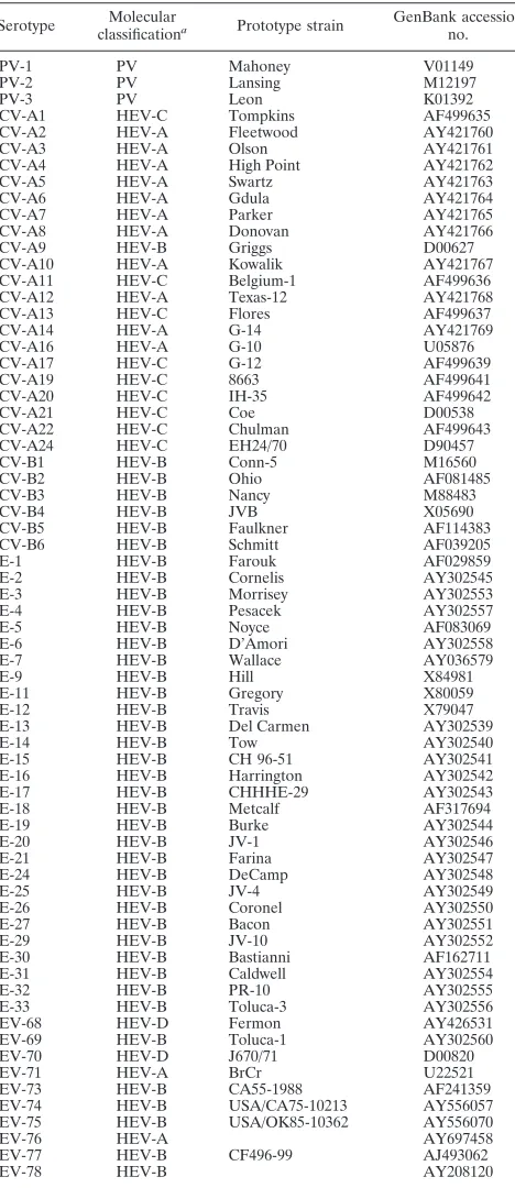
Prototype strains.The prototype strains of the 68 serotypes of HEV are listed in Table 1. Their VP2 coding sequences, retrieved from the GenBank database, were used for designing primers able to match all the serotypes of HEV. In addition, 12 prototype strains, namely, A6, A13, A14, B2, CV-B4, CV-B5, CV-B6, E-2, and E-6 and the 3 Sabin vaccinal strains of PV, were typed by the VP2 method.
The specificity of the VP2 method was tested by using a clinical isolate of rhinovirus (strain 1789).
Virus isolation of field isolates and typing by neutralization. The typing method was applied to 104 strains of HEV isolated from clinical samples sent to the Virology Unit of the University Hospital of Saint-Etienne (France) from 1977 to 2006 and to 12 strains of HEV recovered from environmental samples at the Faculty of Pharmacy of Monastir (Tunisia) from 1995 to 1997. The isolation technique was performed in both laboratories according to standard protocols as previously described (4, 40). Once the enteroviral cytopathic effect involved more than 50% of the cell monolayer, the cells were scraped and an indirect immu-nofluorescence assay using the panenterovirus monoclonal antibody 5-D8/1 (DakoCytomation, Trappes, France) was performed to confirm the identification at the genus level (40). The titer of each cell culture supernatant was first determined, and then the neutralization assay was performed in 96-well tissue culture plates by using A to H and, in case of failure, J to P Lim-Benyesh Melnick intersecting pools of hyperimmune horse sera (Statens Serum Institut, Copen-hagen, Denmark) against 100 50% tissue culture infective doses of each isolate as previously described (18). A further identification was performed using a monovalent horse antiserum corresponding to the serotype identified by the intersecting pools (Statens Serum Institut, Copenhagen, Denmark). HEV iso-lates that failed to be identified by this standard seroneutralization procedure were qualified as “untypeable” strains.
RNA extraction and reverse transcription.Viral RNA was extracted from 140
l of cell culture supernatant using a QIAamp viral RNA mini kit (QIAGEN, Courtaboeuf, France) according to the manufacturer’s recommendations. Ten microliters of extracted RNA was reverse transcribed into cDNA at 42°C for 45 min using 200 units of SuperScriptIII reverse transcriptase and 2.5 ng/l of random primers (Invitrogen, Cergy Pontoise, France) in the presence of 10 units of RnaseOUT recombinant RNase inhibitor (Invitrogen, Cergy Pontoise, France).
Amplification experiments. (i) General precautions and conditions.To pre-vent the occurrence of amplicon contamination, the different steps of the PCR assays (extraction, mixture preparation, amplification, purification, and revealing of PCR products) were performed in separate rooms, gloves and masks were worn during the critical steps, and negative controls consisting of water samples were included in all experiments, according to the standard guidelines for mo-lecular biology.
All the amplification experiments were performed in a Mastercycler gradient thermal cycler (Eppendorf, Hamburg, Germany). The PCR products were re-vealed by agarose gel electrophoresis.
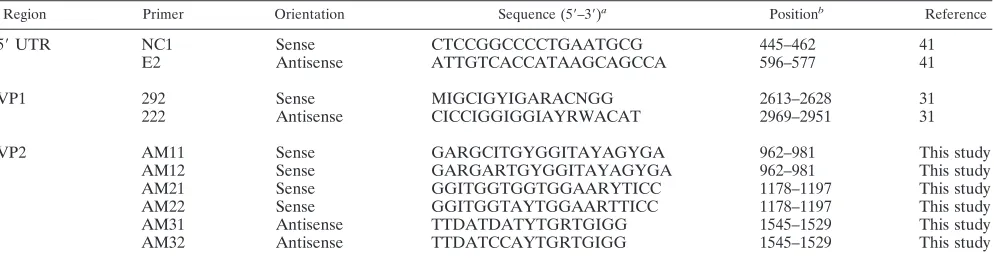
(ii) 5ⴕUTR.The presence of enteroviral RNA in the field isolates was sys-tematically confirmed by a PCR test in the 5⬘untranslated region (5⬘UTR). The cDNA was amplified by using 10 pmol of the primers (Table 2) described by Zoll et al. (41) and 1.25 U of recombinantTaqDNA polymerase (Invitrogen, Cergy Pontoise, France) in 50l of reaction mixture. A band of the expected size of 152 bp (in reference to the PV-1 sequence, GenBank accession no. V01149) was observed.
(iii) VP1 region.Five microliters of cDNA was amplified using 50 pmol of the 292 and 222 primers (Table 2) and 1.25 units of PlatinumTaqDNA polymerase (Invitrogen, Cergy Pontoise, France) in 50l of reaction mixture, according to the protocol described by Oberste et al. (31). A band of the expected size of 357 bp (with reference to PV-1) was observed after agarose gel electrophoresis.
(iv) VP2 region.A mixture of two pairs of sense (AM11 and AM12) and antisense (AM31 and AM32) primers (Table 2) was used for the first run of PCR. Five microliters of cDNA was amplified using 80 pmol of each primer and 1.25 units of PlatinumTaqDNA polymerase in 50l of reaction mixture (Invitrogen, Cergy Pontoise, France). Amplification included an initial cycle of 95°C for 5 min; 40 further cycles of denaturation at 94°C for 30 s, annealing at 48°C for 45 s, and extension at 72°C for 45 s; and a final extension cycle at 72°C for 5 min. A
band of the expected size of 584 bp (with reference to PV-1) was observed after electrophoresis in an agarose gel. For PV, the annealing temperature was low-ered to 42°C.
[image:2.585.303.537.80.620.2]In case of failure of this first PCR, a seminested amplification was performed using 2l of the first PCR mixture, a mixture of two pairs of sense (AM21 and AM22) and antisense (AM31 and AM32) primers (40 pmol each) (Table 2), and 1.25 units of PlatinumTaqDNA polymerase in 50l of reaction mixture (In-vitrogen, Cergy Pontoise, France). Amplification included an initial cycle of 95°C
TABLE 1. Prototype strains of HEV used in this study
Serotype Molecular
classificationa Prototype strain
GenBank accession no.
PV-1 PV Mahoney V01149
PV-2 PV Lansing M12197
PV-3 PV Leon K01392
CV-A1 HEV-C Tompkins AF499635
CV-A2 HEV-A Fleetwood AY421760
CV-A3 HEV-A Olson AY421761
CV-A4 HEV-A High Point AY421762
CV-A5 HEV-A Swartz AY421763
CV-A6 HEV-A Gdula AY421764
CV-A7 HEV-A Parker AY421765
CV-A8 HEV-A Donovan AY421766
CV-A9 HEV-B Griggs D00627
CV-A10 HEV-A Kowalik AY421767
CV-A11 HEV-C Belgium-1 AF499636
CV-A12 HEV-A Texas-12 AY421768
CV-A13 HEV-C Flores AF499637
CV-A14 HEV-A G-14 AY421769
CV-A16 HEV-A G-10 U05876
CV-A17 HEV-C G-12 AF499639
CV-A19 HEV-C 8663 AF499641
CV-A20 HEV-C IH-35 AF499642
CV-A21 HEV-C Coe D00538
CV-A22 HEV-C Chulman AF499643
CV-A24 HEV-C EH24/70 D90457
CV-B1 HEV-B Conn-5 M16560
CV-B2 HEV-B Ohio AF081485
CV-B3 HEV-B Nancy M88483
CV-B4 HEV-B JVB X05690
CV-B5 HEV-B Faulkner AF114383
CV-B6 HEV-B Schmitt AF039205
E-1 HEV-B Farouk AF029859
E-2 HEV-B Cornelis AY302545
E-3 HEV-B Morrisey AY302553
E-4 HEV-B Pesacek AY302557
E-5 HEV-B Noyce AF083069
E-6 HEV-B D’Amori AY302558
E-7 HEV-B Wallace AY036579
E-9 HEV-B Hill X84981
E-11 HEV-B Gregory X80059
E-12 HEV-B Travis X79047
E-13 HEV-B Del Carmen AY302539
E-14 HEV-B Tow AY302540
E-15 HEV-B CH 96-51 AY302541
E-16 HEV-B Harrington AY302542
E-17 HEV-B CHHHE-29 AY302543
E-18 HEV-B Metcalf AF317694
E-19 HEV-B Burke AY302544
E-20 HEV-B JV-1 AY302546
E-21 HEV-B Farina AY302547
E-24 HEV-B DeCamp AY302548
E-25 HEV-B JV-4 AY302549
E-26 HEV-B Coronel AY302550
E-27 HEV-B Bacon AY302551
E-29 HEV-B JV-10 AY302552
E-30 HEV-B Bastianni AF162711
E-31 HEV-B Caldwell AY302554
E-32 HEV-B PR-10 AY302555
E-33 HEV-B Toluca-3 AY302556
EV-68 HEV-D Fermon AY426531
EV-69 HEV-B Toluca-1 AY302560
EV-70 HEV-D J670/71 D00820
EV-71 HEV-A BrCr U22521
EV-73 HEV-B CA55-1988 AF241359
EV-74 HEV-B USA/CA75-10213 AY556057
EV-75 HEV-B USA/OK85-10362 AY556070
EV-76 HEV-A AY697458
EV-77 HEV-B CF496-99 AJ493062
EV-78 HEV-B AY208120
aAccording to Stanway et al. (36).
on May 16, 2020 by guest
http://jcm.asm.org/
for 5 min; 30 further cycles of denaturation at 94°C for 30 s, annealing at 56°C for 45 s, and extension at 72°C for 45 s; and a final extension cycle at 72°C for 5 min. A band of the expected size of 368 bp (with reference to PV-1) was observed after electrophoresis in an agarose gel.
Template purification and sequencing.The amplicons were purified using Montage PCR centrifugal filter devices (Millipore) or a Qiaquick gel extraction kit (QIAGEN, Courtaboeuf, France), depending on the presence of single or multiple bands, respectively. The purified products were sequenced using 0.5 pmol/l of primer and the GenomeLab Dye Terminator Cycle Sequencing Quick Start kit (Beckman Coulter, Villepinte, France) according to the manufacturer’s instructions. The sequencing primers were those used in the seminested PCR assay described for the VP2 region and 292/222 for the VP1 region (Table 2). The electrophoresis and analysis of DNA sequence reactions were performed with the automated DNA sequencer CEQ8000 (Beckman Coulter, Villepinte, France).
Sequence analysis and phylogeny. To determine the enterovirus type, the obtained consensus sequences of 357 bp for VP1 and 368 bp for VP2 were compared to all the corresponding HEV sequences available in GenBank for each region by using BLAST software (1). As proposed by Oberste et al. (22), nucleotide sequence homology of at least 75% was required for assignment to the same serotype. The prototype with the highest identity score calculated with FASTA software (33) was concluded to be the strongest candidate for identifi-cation.
Sequence alignments were performed by using the Clustal W program (version 1.81) (39). Phylogenetic and molecular evolutionary analyses were conducted using MEGA 3.1 (15). Genetic distances were calculated using the Tamura-Nei method (37), and phylogenetic trees were constructed using the neighbor-joining method (35) with 1,000 bootstrap pseudoreplicates (12).
Nucleotide sequence accession numbers. The sequences described in the present report are available in the GenBank database under accession numbers DQ869680 to DQ869857.
RESULTS
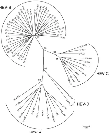
Phylogenic analysis of the VP2 coding region, design of
primers, and PCR strategy.
The VP2 coding regions of the 68
prototypes strains (Table 1) were aligned using Clustal W
software. The region between nucleotides 1178 and 1545 (with
reference to PV-1) was chosen for its high variability between
strains. As shown in Fig. 1, all 68 prototype strains of HEV
were clustered into the four species HEV-A, -B, -C, and -D;
the three PV serotypes were found to be close to HEV-C, as
previously described after VP1 analysis.
Two pairs of sense (AM11/AM12) and antisense (AM31/
AM32) primers were designed to allow the amplification of the
N-terminal and central parts of the VP2 coding regions (584 bp
with reference to PV-1) of all the serotypes of HEV (Table 2).
For most strains, the optimal annealing temperature of the
PCR was 48°C; however, for PV strains, it was lowered to 42°C.
A few strains needed a second round of amplification by a
seminested PCR assay (368 bp with reference to PV-1) with
antisense primers (AM31/AM32) and an internal pair of
prim-ers (AM21/AM22) (Table 2). The sensitivities of the test, as
evaluated on three clinical isolates belonging to different
se-rotypes (CV-B4, E-6, and E-11), were shown to be
approxi-mately 10
⫺1and 10
⫺450% tissue culture infective dose per
reaction tube for the first and seminested PCR assays,
respec-tively.
For all the sequence and phylogenetic analyses, only the
central part of VP2, corresponding to a 368-bp fragment of
PV-1, was taken into consideration because of its high
vari-ability among HEV strains (Fig. 1).
Typing of well-characterized strains of HEV by VP2
se-quencing.
A total of 61 field isolates, most of them belonging
to the HEV-B species, were selected because of their previous
identification at the serotype level by a seroneutralization test.
The VP2 regions of all the strains were successfully amplified
by using the two pairs of primers AM11/AM12 and AM31/
AM32. As shown in Table 3, total concordance was observed
between seroneutralization and VP2 sequencing for the
deter-mination of HEV serotypes. Thirteen of these isolates were
also typed by the VP1 sequencing assay with identical results.
With the exception of four strains (two typed E-11, one E-14,
and another CV-A21), all the strains in Table 3 exhibited a
highest identity score of more than 75% and a second-highest
score of less than 75% with regard to the respective prototype
strains. For the amino acid sequence, the homology was at
least 84% with the closest prototype for all the tested strains
(Table 3).
In addition, a few prototype strains, including the Sabin vaccine
strains of the three PV serotypes, three CV-A strains propagated
in suckling mice (CV-A6, CV-13, and CV-A14), and six HEV-B
strains (CV-B2, CV-B4, CV-B5, CV-B6, E-2, and E-6) were
cor-rectly typed by using the VP2 assay. However, as stated above, the
temperature of hybridization of primers in the PCR was lowered
to 42°C for the three Sabin PV strains in order to obtain a
successful amplification. No amplification was obtained for a field
isolate of rhinovirus (data not shown).
[image:3.585.45.542.81.210.2]Typing by VP1 and VP2 sequencing of “untypeable” strains
of HEV.
A total of 55 field isolates were selected because they
could not be identified by the intersecting pools of
hyperim-mune horse antisera: 12 strains exhibited titers too low to be
TABLE 2. Primers used in this study
Region Primer Orientation Sequence (5⬘–3⬘)a Positionb Reference
5
⬘
UTR
NC1
Sense
CTCCGGCCCCTGAATGCG
445–462
41
E2
Antisense
ATTGTCACCATAAGCAGCCA
596–577
41
VP1
292
Sense
MIGCIGYIGARACNGG
2613–2628
31
222
Antisense
CICCIGGIGGIAYRWACAT
2969–2951
31
VP2
AM11
Sense
GARGCITGYGGITAYAGYGA
962–981
This study
AM12
Sense
GARGARTGYGGITAYAGYGA
962–981
This study
AM21
Sense
GGITGGTGGTGGAARYTICC
1178–1197
This study
AM22
Sense
GGITGGTAYTGGAARTTICC
1178–1197
This study
AM31
Antisense
TTDATDATYTGRTGIGG
1545–1529
This study
AM32
Antisense
TTDATCCAYTGRTGIGG
1545–1529
This study
aThe following standard ambiguity codes were used: D⫽G, A, or T; R⫽A or G; Y⫽T or C; N⫽A, T, C, or G; M⫽A or C; and I⫽deoxyinosine. bWith reference to the sequence of PV-1.
on May 16, 2020 by guest
http://jcm.asm.org/
tested by the seroneutralization test, 26 strains could be
neu-tralized retrospectively by using the monovalent antiserum
cor-responding to the type identified by sequencing assays, and 17
strains failed to be neutralized even by the antiserum-matching
molecular typing.
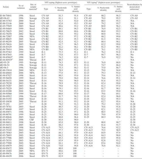
These 55 strains were typed by sequencing both the VP1 and
VP2 regions. As shown in Table 4, concordant results were
ob-served for 48 of them (87.3%). For six strains (four identified as
EV-71, one as E-9, and one as E-30 by the VP2 method), no
amplification was obtained by the VP1 method. The last
discor-dant strain was typed as CV-B4 by the VP1 method and CV-B3
by the VP2 method and monovalent antisera; preliminary results
suggest that this strain is a CV-B3/CV-B4 recombinant strain in
the capsid region (data not shown).
As shown in Table 4, both typing methods exhibited highest
nucleotide and amino acid identity scores of more than 75%
and 85%, respectively, for all but four of the tested strains (one
typed E-7, one E-11, and another CV-A16 by the VP2 method
and one typed E-11 by the VP1 method); of note, three of
these strains were not neutralized by the corresponding
mono-valent antiserum.
Only two strains (SE-98-85865/E-7 and SE-04-86919/E-9 in
Table 4) needed to be amplified by seminested PCR before
being sequenced.
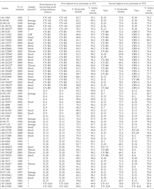
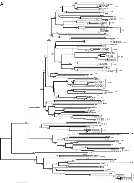
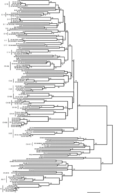
[image:4.585.114.477.68.505.2]Using Clustal W, the sequences of prototype and tested field
strains were combined to construct a phylogenetic tree for each
of the VP1 and VP2 regions. As shown in Fig. 2, despite higher
bootstrap values for VP1, the two phylogenies gave very close
results. All the field strains were clustered with their
corre-sponding prototype, with the exception of one strain of E-14
FIG. 1. Phylogenetic tree depicting the relationships among the 68 prototype strains of HEV serotypes listed in Table 1 and based on the
alignment by Clustal W of the central part (nucleotides 1178 to 1545, with reference to PV-1) of the VP2 coding region. The tree was constructed
using MEGA 3.1 software according to the neighbor-joining method. Only bootstrap values over 70 are shown.
on May 16, 2020 by guest
http://jcm.asm.org/
TABLE 3. Comparative results between neutralization and VP2 sequencing for typing field isolates of HEV
Isolate Yr of isolation
Sample materiala
Neutralization by intersecting pools of hyperimmune
antisera
First highest-score prototype in VP2 Second highest-score prototype in VP2
Type % Nucleotide identity
% Amino acid identity
Type % Nucleotide
identity Type
% Amino acid identity
SE-81-3363
1981
?
CV-A9
CV-A9
82.7
93.1
E-24
73.9
E-30
76.2
MO-98-86
1998
Sewage
CV-A9
CV-A9
82.1
90.3
E-24
72.9
E-30
73.0
MO-98-141
1998
Sewage
CV-A9
CV-A9
83.0
93.2
E-24
73.9
E-30
75.9
SE-01-58710
2001
Saliva
CV-A9
CV-A9
82.8
93.2
E-24
73.9
E-30
75.9
SE-05-20257
b2005
Stool
CV-A9
CV-A9
78.1
91.4
E-13
71.2
E-30
76.1
SE-89-5145
1989
?
CV-B1
CV-B1
79.8
93.2
CV-B4
72.4
CBV-3
79.8
SE-02-71582
2002
CSF
CV-B1
CV-B1
80.8
94.3
CV-B4
74.5
CBV-3
80.3
SE-02-70595
2002
Stool
CV-B1
CV-B1
80.8
93.4
CV-B3
73.0
CBV-3
79.4
SE-02-71028
2002
CSF
CV-B1
CV-B1
81.1
94.3
CV-B4
74.2
CBV-3
80.3
SE-04-12537
2004
Stool
CV-B2
CV-B2
81.4
92.1
CV-B1
70.2
CBV-4
74.5
SE-01-59691
2001
Stool
CV-B2
CV-B2
84.9
94.2
CV-B1
71.2
CBV-4
75.0
SE-98-82668
1998
Stool
CV-B2
CV-B2
84.5
96.2
CV-B1
72.4
CBV-4
77.5
SE-02-72206
b2002
Throat
CV-B3
CV-B3
81.6
96.2
CV-B1
68.6
CBV-1
76.6
SE-03-78384
b2003
Stool
CV-B3
CV-B3
81.5
96.2
E-29
69.8
CBV-1
76.4
SE-03-78350
b2003
Stool
CV-B3
CV-B3
82.1
96.0
E-29
70.1
CBV-1
75.2
SE-05-23233
b2005
Stool
CV-B3
CV-B3
80.2
96.2
CV-B4
70.0
CBV-1
76.4
SE-05-23218
b2005
Nose
CV-B3
CV-B3
80.3
96.2
CV-B1
69.2
CBV-1
76.8
SE-05-23212
b2005
Throat
CV-B3
CV-B3
80.6
96.0
CV-B4
70.2
CBV-1
76.4
SE-94-51301
1994
Stool
CV-B3
CV-B3
81.5
95.0
CV-B1
70.0
CBV-1
78.4
SE-03-78346
b2003
Stool
CV-B4
CV-B4
83.1
99.0
CV-B1
72.4
CBV-2
77.7
SE-04-84828
2004
Stool
CV-B4
CV-B4
80.7
98.0
CV-B1
72.4
CBV-2
77.7
SE-04-85460
2004
Stool
CV-B4
CV-B4
80.1
96.2
E-12
71.5
CBV-2
76.1
MO-98-74
1998
Sewage
CV-B5
CV-B5
77.2
93.2
E-11
71.3
CV-B4
75.7
SE-03-78505
b2003
Stool
CV-B5
CV-B5
81.2
93.8
E-25
70.5
CBV-4
76.5
SE-03-78619
b2003
Stool
CV-B5
CV-B5
80.8
94.2
E-25
70.7
CBV-4
78.8
SE-03-79895
2003
Stool
CV-B5
CV-B5
80.7
93.2
CV-B4
72.5
CBV-4
78.6
SE-88-11044
1988
?
E-3
E-3
83.2
99.0
E-7
73.2
E-12
80.5
MO-95-130
1995
Sewage
E-3
E-3
82.4
99.0
E-7
72.7
E-12
80.7
SE-86-8934
1986
?
E-4
E-4
82.4
93.2
E-32
73.3
E-1
76.9
SE-84-4559
1984
?
E-6
E-6
99.6
99.0
E-11
72.0
E-29
80.0
SE-02-70337
2002
Stool
E-6
E-6
80.0
95.9
E-31
73.7
E-29
79.5
SE-85-4680
1985
?
E-7
E-7
78.2
94.2
E-32
72.9
E-32
85.7
SE-83-3808
1983
?
E-9
E-9
80.3
94.2
E-7
71.1
E-11
76.1
SE-03-81103
2003
Stool
E-9
E-9
80.5
93.3
E-13
71.3
E-11
76.1
SE-04-22413
2004
Stool
E-9
E-9
80.3
93.2
E-13
71.3
E-11
75.7
SE-87-3289
1987
?
E-11
E-11
75.1
89.2
E-19
74.0
E-19
84.3
MO-95-38
1995
Sewage
E-11
E-11
76.2
87.3
E-19
73.3
E-19
80.5
MO-96-75
1996
Sewage
E-11
E-11
74.6
86.4
E-19
73.0
E-19
79.6
MO-97-135
1997
Sewage
E-11
E-11
75.5
87.7
E-19
71.7
E-19
79.2
SE-02-70530
2002
Stool
E-11
E-11
74.5
84.3
E-19
74.3
E-12
78.4
SE-00-52798
2000
Stool
E-13
E-13
78.8
96.0
E-27
71.7
EV-69
77.4
SE-00-53170
2000
Stool
E-13
E-13
77.0
94.2
E-32
71.8
EV-69
78.8
SE-02-71406
2002
Stool
E-14
E-14
79.3
96.1
E-2
75.2
E-2
80.9
SE-89-9796
1989
?
E-17
E-17
79.4
94.0
E-13
73.2
E-7
72.5
SE-85-6268
1985
?
E-18
E-18
81.3
94.1
E-2
72.2
E-32
75.2
SE-88-8012
1988
?
E-20
E-20
83.7
97.1
E-24
68.1
E-24
72.1
SE-94-50463
b1994
Stool
E-20
E-20
79.3
97.1
EV-73
71.3
E-24
73.3
SE-03-78301
b2003
Throat
E-20
E-20
80.1
92.3
CV-B6
70.3
E-6
72.1
SE-03-78928
b2003
Throat
E-20
E-20
78.7
90.2
CV-B6
69.2
E-6
70.5
SE-03-79470
2003
Stool
E-20
E-20
80.1
92.3
CV-B6
70.3
E-6
72.1
SE-84-4617
1984
?
E-21
E-21
80.1
98.0
E-30
72.8
E-30
78.4
SE-86-7617
1986
?
E-24
E-24
80.8
93.1
E-19
71.4
E-20
73.5
SE-03-78187
2003
Throat
E-25
E-25
78.0
95.2
E-19
71.2
E-29
79.0
SE-83-3259
1983
?
E-29
E-29
81.1
94.2
E-25
70.5
E-6
78.0
SE-87-7460
1987
?
E-30
E-30
85.5
96.1
E-21
72.1
E-21
77.6
MO-97-128
1997
Sewage
E-30
E-30
86.6
98.0
E-21
72.0
E-21
78.0
SE-99-93442
1999
Throat
E-30
E-30
84.3
96.1
E-25
71.3
E-21
78.8
SE-05-15103
2005
Stool
E-30
E-30
87.2
98.1
E-29
72.0
E-21
78.5
SE-82-3851
1982
?
E-33
E-33
79.0
88.7
E-13
69.5
E-12
78.5
SE-87-13658
1987
?
CV-A16
CV-A16
88.7
94.0
CV-A7
70.0
EV-71
85.0
SE-88-11568
1988
?
CV-A21
CV-A21
89.6
98.3
CV-A24
75.6
CV-A24
88.5
a
?, sample origin not available.
b
Strain also typed by VP1 sequencing (with a concordant result in all cases).
on May 16, 2020 by guest
http://jcm.asm.org/
TABLE 4. Comparative results between VP1 and VP2 sequencing for typing field isolates of HEV not typeable by intersecting pools of
hyperimmune horse sera
Isolate Yr of isolation
Site or sample materiala
VP2 typing (highest-score prototype) VP1 typingb(highest-score prototype)
Neutralization by monovalent
antiserumc
Type % Nucleotide identity
% Amino acid identity
Type % Nucleotide identity
% Amino acid identity
SE-96-70841
1996
Stool
CV-A9
81.3
91.4
CV-A9
80.2
96.3
CV-A9
MO-96-63
1996
Sewage
CV-A9
81.1
91.1
CV-A9
78.9
95.3
CV-A9
SE-00-53763
2000
Stool
CV-A9
81.3
92.0
CV-A9
80.5
95.3
NA
SE-00-53146
2000
Stool
CV-A9
82.0
93.3
CV-A9
79.2
95.7
NA
SE-03-77049
2003
Stool
CV-A9
80.6
93.2
CV-A9
80.0
96.2
NA
SE-99-94869
1999
Stool
CV-B1
80.1
94.3
CV-B1
76.9
92.3
CV-B1
SE-02-70625
2002
Stool
CV-B1
80.0
88.6
CV-B1
80.0
93.3
CV-B1
SE-02-70250
2002
Stool
CV-B1
79.8
93.2
CV-B1
80.0
94.1
CV-B1
SE-96-72087
1996
NPA
CV-B2
85.5
97.2
CV-B2
80.2
99.0
CV-B2
SE-05-90025
2005
Stool
CV-B2
83.2
95.3
CV-B2
81.5
99.1
CV-B2
SE-05-90249
2005
Throat
CV-B2
82.4
92.3
CV-B2
80.4
98.1
CV-B2
SE-03-78616
2003
Stool
CV-B3
81.7
96.3
CV-B4
82.2
99.0
CV-B3
SE-04-85429
2004
Stool
CV-B4
82.2
96.1
CV-B4
81.3
98.1
CV-B4
SE-06-70414
2006
NPA
CV-B5
79.6
92.4
CV-B5
76.1
97.2
CV-B5
SE-99-97363
1999
Stool
E-1
75.3
91.1
E-1
76.2
91.8
E-1
SE-98-87643
1998
Stool
E-7
75.8
87.3
E-7
80.7
96.3
No
SE-98-85865
d1998
Stool
E-7
74.2
82.8
E-7
76.9
92.2
No
SE-04-86919
d2004
Throat
E-9
80.3
93.2
-
-
-
NA
MO-95-29
1995
Sewage
E-11
74.3
85.5
E-11
76.9
88.9
No
MO-96-54
1996
Sewage
E-11
75.0
87.1
E-11
74.7
86.4
No
MO-96-5
1996
Sewage
E-11
75.3
88.3
E-11
75.0
86.7
No
SE-00-53010
2000
NPA
E-13
78.9
96.1
E-13
79.0
94.0
NA
SE-00-60604
2006
NPA
E-13
77.8
95.1
E-13
79.3
93.4
E-13
SE-98-87927
1998
Stool
E-14
80.5
95.0
E-14
79.6
91.5
NA
SE-99-96858
1999
Stool
E-14
79.8
96.0
E-14
76.6
95.1
E-14
SE-00-56151
2000
Stool
E-14
78.6
96.0
E-14
81.6
95.3
No
SE-03-79230
2003
Throat
E-16
78.9
93.3
E-16
82.4
99.0
NA
SE-03-79287
2003
Stool
E-16
79.0
92.3
E-16
81.3
97.4
NA
SE-03-79229
2003
Stool
E-16
79.1
93.3
E-16
81.7
98.1
NA
SE-04-83200
2004
Stool
E-16
79.8
93.5
E-16
83.5
99.0
E-16
SE-99-94960
1999
Throat
E-17
80.8
97.0
E-17
79.6
89.1
No
SE-96-69912
1996
Throat
E-18
81.7
94.2
E-18
82.4
96.0
NA
SE-00-55193
2000
Stool
E-18
81.6
95.1
E-18
82.2
95.3
No
SE-05-10030
2005
Throat
E-18
81.1
93.2
E-18
82.7
95.4
NA
SE-77-193
1977
?
E-20
78.2
91.3
E-20
79.2
92.5
E-20
SE-03-78761
2003
Stool
E-20
80.3
92.2
E-20
78.6
91.8
E-20
SE-97-80688
1997
Stool
E-25
82.5
97.1
E-25
82.1
95.2
E-25
SE-98-89071
1998
Stool
E-25
81.5
97.1
E-25
80.5
95.2
E-25
SE-99-90791
1999
Stool
E-25
79.5
98.0
E-25
79.0
87.9
E-25
SE-03-80646
2003
Stool
E-25
80.0
96.4
E-25
80.3
92.6
E-25
SE-98-87543
1998
CSF
E-30
85.8
98.0
-
-
-
E-30
SE-98-96811
1998
Stool
E-30
85.9
98.0
E-30
80.8
90.7
E-30
SE-05-50152
2005
Stool
E-30
88.2
97.8
E-30
81.6
91.7
E-30
SE-98-86832
1998
Stool
CV-A13
79.5
95.8
CV-A13
76.6
92.2
CV-A13
SE-03-75245
2003
Stool
CV-A13
77.7
93.3
CV-A13
75.5
90.9
CV-A13
SE-03-78463
2003
Stool
CV-A14
84.1
97.9
CV-A14
84.9
97.9
No
SE-03-78693
2003
Stool
CV-A14
84.2
95.8
CV-A14
84.3
98.0
No
SE-03-77906
2003
Stool
CV-A14
85.3
98.9
CV-A14
83.9
95.0
No
SE-03-78331
2003
Stool
CV-A14
83.9
95.9
CV-A14
82.8
93.2
No
SE-03-77098
2003
Stool
CV-A14
84.1
97.1
CV-A14
83.6
94.0
No
SE-02-69079
2002
Stool
CV-A16
73.0
94.8
CV-A16
76.8
92.1
NA
SE-04-85672
2004
Stool
EV-71
80.8
100
-
-
-
No
SE-05-18404
2005
NPA
EV-71
80.6
100
-
-
-
No
SE-00-99676
2000
Stool
EV-71
81.9
100
-
-
-
No
SE-06-40439
2006
Stool
EV-71
82.9
100
-
-
-
No
aNPA, nasopharyngeal aspirate; CSF, cerebrospinal fluid. b-, no amplification despite at least three different attempts.
cSerotype, neutralization with the corresponding antiserum; No, no neutralization with the corresponding monovalent antiserum; NA, not applicable because of the
viral titer was too low.
dStrain needing a further seminested round to be typed.
on May 16, 2020 by guest
http://jcm.asm.org/
FIG. 2. Phylogenetic trees depicting the relationships among HEV serotypes based on alignment by Clustal W of (A) VP1 and (B) VP2 coding
regions of the 68 prototype strains of HEV listed in Table 1 (boldface) and of the field strains of this study typed in both regions. The trees were
constructed according to the neighbor-joining method using MEGA 3.1 software. Only bootstrap values over 70 are shown.
on May 16, 2020 by guest
http://jcm.asm.org/
FIG. 2—
Continued
.
on May 16, 2020 by guest
http://jcm.asm.org/
grouped with the E-5 prototype in the VP1 tree and two strains
of E-7 grouped with the E-19 prototype in the VP2 tree.
DISCUSSION
While serotyping of HEV has no significant impact on the
clinical management of patients, identification at the serotype
level is important for several reasons: (i) in the context of
worldwide polio eradication, it is essential to develop tools able
to delineate polio and nonpolio HEV; (ii) from the
epidemi-ological point of view, it is important to identify which serotype
is responsible for clusters of cases, especially when large
out-breaks (i.e., EV-70 or CV-A24 and epidemic hemorrhagic
con-junctivitis) or severe clinical conditions (i.e., EV-71 and fatal
neurological disease) occur; (iii) as with all RNA viruses, HEV
serotypes display a high potential for genetic diversity that
requires powerful molecular tools to detect variants,
recombi-nants, and newly emerging serotypes (26).
Molecular methods are now recognized as alternative
tech-niques to seroneutralization for modern serotyping of HEV
(22). Because VP1 contains most of the neutralizing epitopes,
the sequencing of a part of this coding region is not far from
being considered the gold standard (7, 20, 24, 25). The study of
Casas et al. (8) showed that sequencing of the VP1 coding
region was a more discriminant technique for HEV typing than
analysis of a part of the VP2 coding region or of the
polymer-ase gene. However, recent studies have shown that VP1-bpolymer-ased
sequencing methods could fail to amplify some E and CV
strains (5, 13, 14). In addition, primers targeting the VP1
re-gion and designed to amplify all the HEV serotypes did not
recognize some serotypes not belonging to the HEV-B species
(i.e., EV-68, EV-70, and EV-71) because of some mismatch; to
circumvent this problem, Oberste et al. (28) recently proposed
a first round of amplification using primers located within the
3
⬘
UTR in order to identify the strain at the species level,
followed by a second PCR assay using species-specific primers
that allow a proper identification at the serotype level.
In this report, we propose a new strategy for typing HEV
using a sequencing assay based on primers targeting the VP2
region. This region had previously been recommended for
typing purposes (2, 9, 34). In contrast to other studies that
amplified different fragments of the N-terminal part of the
VP2 coding region (8, 16, 27), which is not highly divergent in
HEV serotypes, the technique described here focused on a
368-bp fragment (with reference to PV-1) of the central part of
VP2 that was found to be able to segregate correctly all the
serotypes of HEV (Fig. 1) in relation to the presence in this
region of neutralizing epitopes, particularly the EF loop (17).
This structure, constituted of approximately 50 amino acids in
the center of VP2, is characterized by high variability among
HEV and is probably responsible for a great part of the
dis-criminatory power of this typing method.
All the prototype and field strains that were evaluated could
be typed successfully by the VP2 assay with good confidence
(the percent nucleotide homology was
ⱖ
75% [Tables 3 and
4]), with the exception of one E-7, three E-11, and one
CV-A16 strains, which were shown to be slightly more distant from
the prototype strain although they were correctly typed,
prob-ably because of the high evolution rate of these serotypes
compared to others. Actually, the cutoff of 75% for delineating
a serotype is not an absolute limit, as previously exemplified by
using the VP1 typing assay, which showed nucleotide
diver-gences of up to 27% for E-11 (30) and E-30 (23) strains.
Consequently, for strains that do not reach the 75% nucleotide
homology with the prototype strain exhibiting the first highest
score in the VP2 typing assay, we propose to retain the
iden-tification as the more probable and to confirm the result by
measuring sequence homology with other strains belonging to
the same serotype.
Only 36 of the 68 serotypes of HEV were tested, most of
them belonging to the HEV-B species, which corresponds to
the isolates most frequently recovered in clinical virology.
Se-rotypes of the HEV-D species (EV-68 and EV-70), as well as
newly described serotypes of HEV (EV-74 to -78) (21, 26, 29),
were not available, but the primers described here matched
perfectly with their published sequences (Fig. 1). Concerning
the Sabin PV strains, it must be stressed that the hybridization
temperature of primers should be lowered to 42°C to obtain an
efficient rate of amplification, despite the risk of enhancing
nonspecific reactions. Further controls on wild isolates of PV
are needed to control this effect.
Globally, the following strategy could be evaluated in case of
unsuccessful amplification with the standard VP2 assay
de-scribed here: either use seminested amplification in order to
increase the sensitivity of the typing method or lower the
tem-perature of the hybridization step to 42°C if the
epidemiolog-ical context is compatible with a PV isolate.
The VP2 typing assay described here was shown to exhibit at
least three interesting features compared to previously
de-scribed methods spanning other regions of the HEV genome:
(i) the use of a pair of primers with a limited number of
degenerate positions allowed the correct identification of four
untypeable strains of EV-71 that were not recognized by the
VP1 primers described by Oberste et al. (31), probably because
of a mismatch (T/C) with primer 292 at position 2593
(accord-ing to prototype strain EV-71, accession no. U22521); (ii) the
ability to perform a seminested PCR assay for a few strains
exhibiting low infectious titers enhances the sensitivity of the
typing method, as exemplified by strain SE-04-86919, which
was nontypeable by neutralization and VP1 assays but was
typed as E-9 by VP2 (Table 4 and Fig. 2); (iii) the discrepant
typing results obtained for strain SE-03-78616 (CV-B4 by VP1
and CV-B3 by VP2 and monovalent antiserum) (Table 4)
illustrate the interest of combining different typing methods to
identify interserotypic recombinants in the capsid region.
Strain SE-98-87543, which exhibited a correct infectious titer
and was typed E-30 by VP2 and neutralized by the E-30
mono-valent antiserum but was not typed by VP1, could represent
another recombinant strain in the capsid region.
In conclusion, the VP2 assay described in this study is
pro-posed as an additional tool for typing HEV strains on a routine
basis. Since most strains were shown to be typed by using a
single round of PCR with two pairs of primers, this method
could be used as a screening test for the typing of field isolates.
Further experiments on a wider spectrum of strains are needed
to verify its ability to actually identify most HEV strains. In
addition, the seminested version of the test should be
evalu-ated for its capacity to directly type HEV in clinical samples
without using the cell culture step, as previously suggested for
other regions (8, 11).
on May 16, 2020 by guest
http://jcm.asm.org/
ACKNOWLEDGMENTS
Dorsaf Nasri is supported by funds from the “Comite
´ Mixte de
Coope
´ration Universitaire (CMCU) Franco-Tunisien” (program no.
03S0804) and the Doctoral School of Monastir.
Jean-Luc Bailly, Christophe Ginevra, Florence Grattard, and Philip
Lawrence are acknowledged for helpful discussions of phylogenetic
studies. The technical assistance of Nelly Bonnet was appreciated.
REFERENCES
1.Altschul, S. F., W. Gish, W. Miller, E. W. Myers, and D. J. Lipman.1990. Basic local alignment search tool. J. Mol. Biol.215:403–410.
2.Arola, A., J. Santti, O. Ruuskanen, P. Halonen, and T. Hyypia.1996. Iden-tification of enteroviruses in clinical specimens by competitive PCR followed by genetic typing using sequence analysis. J. Clin. Microbiol.34:313–318. 3.Bailly, J. L., A. Beguet, M. Chambon, C. Henquell, and H. Peigue-Lafeuille.2000.
Nosocomial transmission of echovirus 30: molecular evidence by phylogenetic anal-ysis of the VP1 encoding sequence. J. Clin. Microbiol.38:2889–2892.
4.Belguith, K., A. Hassen, and M. Aouni.2006. Comparative study of four extraction methods for enterovirus recovery from wastewater and sewage sludge. Bioresour. Technol.97:414–419.
5.Bolanaki, E., C. Kottaridi, P. Markoulatos, L. Margaritis, and T. Katsorchis. 2005. A comparative amplification of five different genomic regions on Cox-sackie A and B viruses. Implications in clinical diagnostics. Mol. Cell Probes 19:127–135.
6.Brown, B., M. S. Oberste, K. Maher, and M. A. Pallansch.2003. Complete genomic sequencing shows that polioviruses and members of human entero-virus species C are closely related in the noncapsid coding region. J. Virol. 77:8973–8984.
7.Caro, V., S. Guillot, F. Delpeyroux, and R. Crainic.2001. Molecular strategy for “serotyping” of human enteroviruses. J. Gen. Virol.82:79–91. 8.Casas, I., G. F. Palacios, G. Trallero, D. Cisterna, M. C. Freire, and A.
Tenorio.2001. Molecular characterization of human enteroviruses in clinical samples: comparison between VP2, VP1, and RNA polymerase regions using RT nested PCR assays and direct sequencing of products. J. Med. Virol. 65:138–148.
9.Hyypia, T., T. Hovi, N. J. Knowles, and G. Stanway.1997. Classification of entero-viruses based on molecular and biological properties. J. Gen. Virol.78:1–11. 10.Ishiko, H., Y. Shimada, M. Yonaha, O. Hashimoto, A. Hayashi, K. Sakae, and N.
Takeda.2002. Molecular diagnosis of human enteroviruses by phylogeny-based classification by use of the VP4 sequence. J. Infect. Dis.185:744–754.
11.Iturriza-Gomara, M., B. Megson, and J. Gray.2006. Molecular detection and characterization of human enteroviruses directly from clinical samples using RT-PCR and DNA sequencing. J. Med. Virol.78:243–253. 12.Kimura, M.1980. A simple method for estimating evolutionary rate of base
substitutions through comparative studies of nucleotide sequences. J. Mol. Evol.16:111–120.
13.Kottaridi, C., E. Bolanaki, and P. Markoulatos.2004. Amplification of echoviruses genomic regions by different RT-PCR protocols—a comparative study. Mol. Cell Probes18:263–269.
14.Kottaridi, C., E. Bolanaki, N. Siafakas, and P. Markoulatos.2005. Evalua-tion of seroneutralizaEvalua-tion and molecular diagnostic methods for echovirus identification. Diagn. Microbiol. Infect. Dis.53:113–119.
15.Kumar, S., K. Tamura, and M. Nei.2004. MEGA3: integrated software for Molecular Evolutionary Genetics Analysis and sequence alignment. Brief. Bioinform.5:150–163.
16.Manzara, S., M. Muscillo, G. La Rosa, C. Marianelli, P. Cattani, and G. Fadda.2002. Molecular identification and typing of enteroviruses isolated from clinical samples. J. Clin. Microbiol.40:4554–4560.
17.Mateu, M. G.1995. Antibody recognition of picornaviruses and escape from neutralization: a structural view. Virus Res.38:1–24.
18.Melnick, J. L., and I. L. Wimberly.1985. Lyophilized combination pools of enterovirus equine antisera: new LBM pools prepared from reserves of antisera stored frozen for two decades. Bull. W. H. O.63:543–550. 19.Muir, P., U. Ka¨mmerer, K. Korn, M. N. Mulders, T. Po¨yry, B. Weissbrich,
R. Kandolf, G. M. Cleator, and A. M. van Loon for The European Union Concerted Action on Virus Meningitis and Encephalitis.1998. Molecular typing of enteroviruses: current status and future requirements. Clin. Micro-biol. Rev.11:202–227.
20.Norder, H., L. Bjerregaard, and L. O. Magnius.2001. Homotypic echovi-ruses share aminoterminal VP1 sequence homology applicable for typing. J. Med. Virol.63:35–44.
21.Norder, H., L. Bjerregaard, L. O. Magnius, B. Lina, M. Aymard, and J. J.
Chomel.2003. Sequencing of “untypable” enteroviruses reveals two new types, EV-77 and EV-78, within human enterovirus type B and substitutions in the BC loop of the VP1 protein for known types. J. Gen. Virol.84:827–836. 22.Oberste, M. S., K. Maher, M. R. Flemister, G. Marchetti, D. R. Kilpatrick, and
M. A. Pallansch.2000. Comparison of classic and molecular approaches for the identification of untypeable enterovirus. J. Clin. Microbiol.38:1170–1174. 23.Oberste, M. S., K. Maher, M. L. Kennett, J. J. Campbell, M. S. Carpenter,
D. Schnurr, and M. A. Pallansch.1999. Molecular epidemiology and genetic diversity of echovirus type 30 (E30): genotypes correlate with temporal dynamics of E30 isolation. J. Clin. Microbiol.37:3928–3933.
24.Oberste, M. S., K. Maher, D. R. Kilpatrick, M. R. Flemister, B. A. Brown, and M. A. Pallansch.1999. Typing of human enterovirus by partial sequenc-ing of VP1. J. Clin. Microbiol.37:1288–1293.
25.Oberste, M. S., K. Maher, D. R. Kilpatrick, and M. A. Pallansch.1999. Molecular evolution of the human enteroviruses: correlation of serotype with VP1 sequence and application to picornavirus classification. J. Virol.73:1941–1948.
26.Oberste, M. S., K. Maher, S. M. Michele, G. Belliot, M. Uddin, and M. A. Pallansch.2005. Enteroviruses 76, 89, 90 and 91 represent a novel group within the speciesHuman enterovirus A.J. Gen. Virol.86:445–451. 27.Oberste, M. S., K. Maher, and M. A. Pallansch.1998. Molecular phylogeny
of all human enterovirus serotypes based on comparison of sequences at the 5⬘end of the region encoding VP2. Virus Res.58:35–43.
28.Oberste, M. S., K. Maher, A. J. Williams, N. Dybdahl-Sissoko, B. A. Brown, M. S. Gookin, S. Penaranda, N. Mishrik, M. Uddin, and M. A. Pallansch. 2006. Species-specific RT-PCR amplification of human enteroviruses: a tool for rapid species identification of uncharacterized enteroviruses. J. Gen. Virol.87:119–128.
29.Oberste, M. S., S. M. Michele, K. Maher, D. Schnurr, D. Cisterna, N. Junttila, M. Uddin, J. J. Chomel, C. S. Lau, W. Ridha, S. al-Busaidy, H. Norder, L. O. Magnius, and M. A. Pallansch.2004. Molecular identification and characterization of two proposed new enterovirus serotypes, EV74 and EV75. J. Gen. Virol.85:3205–3212.
30.Oberste, M. S., W. A. Nix, D. R. Kilpatrick, M. R. Flemister, and M. A. Pallansch. 2003. Molecular epidemiology and type-specific detection of echovirus 11 isolates from the Americas, Europe, Africa, Australia, Southern Asia and the Middle East. Virus Res.91:241–248.
31.Oberste, M. S., W. A. Nix, K. Maher, and M. A. Pallansch.2003. Improved molecular identification of enteroviruses by RT-PCR and amplicon sequenc-ing. J. Clin. Virol.26:375–377.
32.Pallansch, M. A., and R. Roos.2007. Enteroviruses: polioviruses, coxsackie viruses, echoviruses, and newer enteroviruses, p. 839–893.InD. M. Knipe and P. M. Howley (ed.), Fields virology, 5th ed., vol. 1. Lippincott Williams and Wilkins, Philadelphia, PA.
33.Pearson, W. R., and D. J. Lipman.1988. Improved tools for biological sequence comparison. Proc. Natl. Acad. Sci. USA85:2444–2448. 34.Poyry, T., L. Kinnunen, T. Hyypia, B. A. Brown, C. Horsnell, T. Hovi, and G.
Stanway.1996. Genetic and phylogenetic clustering of enteroviruses. J. Gen. Virol.77:1699–1717.
35.Saitou, N., and M. Nei.1987. The neighbor-joining method: a new method for reconstructing phylogenetic trees. Mol. Biol. Evol.4:406–425. 36.Stanway, G., F. Brown, and P. Christian.2005. Picornaviridae, p. 757–778.In
C. M. Fauquet, M. A. Mayo, J. Maniloff, U. Desselberger, and L. A. Ball (ed.), Virus taxonomy: classification and nomenclature of viruses. Eighth report of the International Committee on the Taxonomy of Viruses. Elsevier Academic Press, Amsterdam, The Netherlands.
37.Tamura, K., and M. Nei.1993. Estimation of the number of nucleotide substitutions in the control region of mitochondrial DNA in humans and chimpanzees. Mol. Biol. Evol.10:512–526.
38.Thoelen, I., P. Lemey, I. Van der Donck, K. Beuselinck, A. M. Lindberg, and M. Van Ranst.2003. Molecular typing and epidemiology of enteroviruses identified from an outbreak of aseptic meningitis in Belgium during the summer of 2000. J. Med. Virol.70:420–429.
39.Thompson, J. D., D. G. Higgins, and T. J. Gibson.1994. CLUSTAL W: improving the sensitivity of progressive multiple sequence alignment through sequence weighting, position-specific gap penalties and weight matrix choice. Nucleic Acids Res.22:4673–4680.
40.Trabelsi, A., F. Grattard, M. Nejmeddine, M. Aouni, T. Bourlet, and B. Pozzetto. 1995. Evaluation of an enterovirus group-specific anti-VP1 monoclonal antibody, 5-D8/1, in comparison with neutralization and PCR for rapid identification of en-teroviruses in cell culture. J. Clin. Microbiol.33:2454–2457.
41.Zoll, G. J., W. J. Melchers, H. Kopecka, G. Jambroes, H. J. van der Poel, and J. M. Galama.1992. General primer-mediated polymerase chain reaction for detection of enteroviruses: application for diagnostic routine and persistent infections. J. Clin. Microbiol.30:160–165.