0095-1137/08/$08.00
⫹
0
doi:10.1128/JCM.02248-07
Copyright © 2008, American Society for Microbiology. All Rights Reserved.
RNase-Resistant Virus-Like Particles Containing Long Chimeric RNA
Sequences Produced by Two-Plasmid Coexpression System
䌤
Yuxiang Wei,
1,2† Changmei Yang,
1,2† Baojun Wei,
1,2Jie Huang,
1,2Lunan Wang,
2Shuang Meng,
2Rui Zhang,
2and Jinming Li
1,2*
Graduate School, Peking Union Medical College, Chinese Academy of Medical Sciences, Beijing, People’s Republic of China,
1and
Department of Immunoassay and Molecular Diagnosis, National Center for Clinical Laboratory, Beijing Hospital, Beijing,
People’s Republic of China
2Received 20 November 2007/Returned for modification 28 December 2007/Accepted 16 February 2008
RNase-resistant, noninfectious virus-like particles containing exogenous RNA sequences (armored RNA) are
good candidates as RNA controls and standards in RNA virus detection. However, the length of RNA packaged
in the virus-like particles with high efficiency is usually less than 500 bases. In this study, we describe a method
for producing armored L-RNA. Armored L-RNA is a complex of MS2 bacteriophage coat protein and RNA
produced in
Escherichia coli
by the induction of a two-plasmid coexpression system in which the coat protein
and maturase are expressed from one plasmid and the target RNA sequence with modified MS2 stem-loop (pac
site) is transcribed from another plasmid. A 3V armored L-RNA of 2,248 bases containing six gene fragments—
hepatitis C virus, severe acute respiratory syndrome coronavirus (SARS-CoV1, SARS-CoV2, and SARS-CoV3),
avian influenza virus matrix gene (M300), and H5N1 avian influenza virus (HA300)—was successfully
ex-pressed by the two-plasmid coexpression system and was demonstrated to have all of the characteristics of
armored RNA. We evaluated the 3V armored L-RNA as a calibrator for multiple virus assays. We used the
WHO International Standard for HCV RNA (NIBSC 96/790) to calibrate the chimeric armored L-RNA, which
was diluted by 10-fold serial dilutions to obtain samples containing 10
6to 10
2copies. In conclusion, the
approach we used for armored L-RNA preparation is practical and could reduce the labor and cost of quality
control in multiplex RNA virus assays. Furthermore, we can assign the chimeric armored RNA with an
international unit for quantitative detection.
Armored RNA is a complex of MS2 bacteriophage coat
protein and RNA produced in
Escherichia coli
by the induction
of an expression plasmid that encodes the bacteriophage
se-quence consisting of the maturase, the coat protein, the pac
site, and an exogenous RNA sequence. This method produces
recombinant virus-like particles that are noninfectious and
contain predefined RNA (2–6, 8, 11, 12, 15, 16, 28). These
armored RNAs are RNase resistant by virtue of their
encap-sulation within an MS2 coat protein, and they have been widely
used as controls, standards, or calibrators for the detection of
hepatitis C virus (HCV) (11, 12, 28), human
immune-defi-ciency virus (11, 15), severe acute respiratory syndrome
coro-navirus (SARS-CoV) (3, 11), enterovirus (2, 5), avian influenza
virus 5 (4), and West Nile virus (6) using reverse
transcription-PCR (RT-transcription-PCR), real-time RT-transcription-PCR, and branched DNA
as-says (2–6, 11, 12, 15, 28).
The MS2 bacteriophage consists of 180 U of the bacteriophage
coat protein that encapsulates the bacteriophage genome (25).
The MS2 phage RNA genome comprises a single plus-sense
strand encoding 3,569 nucleotides. The genes are organized from
the 5
⬘
end as follows: the maturase or A protein, the
bacterio-phage coat protein, a 75-amino-acid lysis protein, and a replicase
subunit. Packaging of the RNA genome by coat protein is
initi-ated by high-specificity binding to a unique site on the RNA, a
single stem-loop structure, containing the initiation codon of the
gene for the viral replicase. The armored RNA contains
approx-imately 1.7 kb of bacteriophage RNA sequence encoding the
maturase, the coat protein, and the pac site. The wild-type MS2
bacteriophage contains an RNA genome of approximately 3.6 kb.
In addition, the extensively folded nature of MS2 RNA (22) may
make it particularly suitable for uptake into the confines of a small
capsid. Thus, theoretically, at most, 1.9 kb of nonbacteriophage
RNA sequence might be encapsulated by this method. Practically,
the packaging of 500 bases of RNA has been demonstrated to be
very efficient; however, packaging of 1- and 1.5-kb amounts of
RNA is inefficient (15). Recently, Huang et al. (11) used armored
RNA technology to package a 1,200-nucleotide foreign RNA
sequence by deleting some disposable sequences between the
multiple cloning site and the transcription terminator; however, to
date, there have been no reports of armored RNA with sizes
greater than 1,200 bp.
Although most RT-PCR assays do not target RNA
se-quences longer than 500 bases, there are some advantages if
longer target RNA sequences are packaged. For example, the
human immunodeficiency virus Quantiplex assay (Chiron
Corp.) uses a standard that is approximately 3 kb in length;
consequently, it is not possible to produce a single armored
RNA standard for this assay using routine armored RNA
tech-nology. A further advantage of using armored RNA of several
kilobases is that PCR primers for different regions of these
genes may be used with a single armored RNA standard.
Ac-cordingly, it is not necessary to construct a different armored
* Corresponding author. Mailing address: Department of
Immuno-assay and Molecular Diagnosis, National Center for Clinical
Labora-tory, 1 Dahua Road, Dongdan, Beijing 100730, People’s Republic of
China. Phone: 86-10-58115053. Fax: 86-10-65212064. E-mail: ljm63hn
@yahoo.com.cn.
† Y.W. and C.Y. contributed equally to this study.
䌤Published ahead of print on 27 February 2008.
1734
on May 16, 2020 by guest
http://jcm.asm.org/
(20, 21).
In this article, we describe a method for packaging a long
(
⬎
2,000 bp) RNA sequence, which is referred to as armored
L-RNA technology. We sought to determine whether the
bac-teriophage sequences encoding the maturase and the coat
pro-tein could be replaced with nonbacteriophage RNA sequences,
thereby enabling long-fragment RNA sequences to be
pack-aged. In order to achieve this, we took advantage of a
two-plasmid coexpression system in which the maturase and coat
protein were expressed from one plasmid [pET-28(b)] and the
target RNA containing a modified stem-loop (pac site) of MS2
was produced by a second plasmid (pACYCDuet-1). The pac
site was located in the middle of the target sequence. In the
present study, we used a C-5 variant of the wild-type stem-loop
in which uridine had been substituted by cytosine. The
replace-ment of the wild-type uridine with a cytosine at position
⫺
5
significantly increases the affinity of the RNA for the coat
protein dimer (9, 19, 27, 29). The affinity increase has been
estimated to be 6-fold (26) or even as high as 50-fold (13). The
stronger binding of the C-5 variant compared to the wild-type
sequence has been suggested to be due to an interaction
in-volving the donation of a hydrogen by the amino group of the
cytosine or the corresponding hydroxyl group of the uracil enol
tautamer (24).
MATERIALS AND METHODS
Construction of pET-MC.MS2 maturase and coat protein genes were
ampli-fied by using primers S-MC and A-MC (Table 1) from the pMS27plasmid (kindly
provided by D. S. Peabody) containing nucleotides (nt) 81 to 1749 of the MS2 bacteriophage gene (GenBank accession no. V00642). Sense and reverse primers contained BamHI and HindIII restriction sites (underlined), respectively. These primers correspond to nt 81 to 101 and nt 1721 to 1741 of the MS2 phage genome sequence. The 1.7-kb PCR-amplified DNA fragments were gel purified, digested with BamHI and HindIII, and then ligated to a linearized pET28b (Novagen) vector to generate the recombinant plasmid pET-MC. This plasmid was
trans-formed into competentE. coliDH5␣ cells according to the manufacturer’s
instructions. The pET-MC plasmids in positive clones that could replicate in LB
agar in the presence of 100g of kanamycin ml⫺1
were isolated by using a Takara MiniBEST plasmid purification kit (TaKaRa). The DNA insert was sequenced with vector-specific primers using an automated fluorescent DNA sequencer (model 3730XL; Applied Biosystems). The resulting sequences were identified by a search of the NCBI databases for homologous sequences using BLAST.
Construction of pACYC-3V.An exogenous chimeric sequence 2,248 bp in length comprising the following sequences was inserted into a pACYCDuet-1
plasmid (p15A-type replication origin; Novagen): M-300 (nt 17⬃373, 357 bp
from avian influenza virus matrix gene; GenBank accession no. DQ864720), SARS-CoV1 (nt 15224 to 15618, 395 bp from SARS-CoV; GenBank accession no. AY864806), SARS-CoV2 (nt 18038 to 18340, 303 bp from SARS-CoV; GenBank accession no. AY864806), SARS-CoV3 (nt 328110 to 28692, 583 bp
from SARS-CoV; GenBank accession no. AY864806), a pac site (19 bp), HCV
(nt 18 to 310, 293 bp from HCV 5⬘UTR; GenBank accession no. AF139594), and
HA300 (nt 295 to 611, 317 bp from H5N1 avian influenza virus; GenBank accession no. DQ864720). The target sequence included the forward and reverse primer sites, flanking regions, and probe-binding sites previously published or described. A 19-mer pac site was placed between SARS-CoV3 and HCV (Fig. 1). We spliced the six target DNA sequences using overlapping extensions (10). During the first-round PCR, these six small fragments were amplified as fol-lows. SARS-CoV1 was amplified from a pBSSR-V6 plasmid (kindly provided by the Chinese Academy of Medical Sciences and Peking Union Medical College, Institute of Basic Medical Sciences), containing nt 13785 to 16051 of the SARS-CoV gene, using the primers S-SARS1 and LAP-SARS1. SARS-SARS-CoV2 was amplified from a pNCCL-SARS plasmid (constructed by our laboratory), con-taining nt 18038 to 18340 of the SARS-CoV gene, using the primers LAP-SARS2 and A-SARS2. SARS-CoV3 was amplified from a pBSSR7-8 plasmid (kindly provided by the Chinese Academy of Medical Sciences and Peking Union Med-ical College, Institute of Basic MedMed-ical Sciences), containing nt 27730 to 29212 of the SARS-CoV gene, using the primers LAP-SARS3 and A-SARS3. The HCV fragment was amplified from a pNCCL-HCV plasmid (constructed by our laboratory), containing nt 18 to 310 of the HCV gene, using the primers S-HCV and HCV-LAP1(underlined for 19mer pac site). HA300 was amplified from a
TAAAATGTCTGATAATGGA
CCCC
LAP-SARS2
⫹...ATCAGACATTTTAATTTGTTAACC
AGTCGGTACAGCTACTAAG
S-HCV ...
ACATGAGGATCACCCATGT
GGCGAC
ACTCCACCATAGATCACTC
HCV-LAP1...ATGTAAGACCATTCCGGCTCGCAA
GCACCCTATCAGGCAGTAC
A-HA300 ...GAATCCGTCTTCCATCTTTCCCCCA
CAGTACCAAAAGATCTTC
HA300LAP...CTGATAGGGTGCTTGCGAGCCGG
AATGGTCTTACATAGTGGAG
M
⫹
SLAP1...ATGGCTCTGTCACATTTTGGATAG
AGTAGCTGAGTGCGACCTCC
TTAG
M
⫹
SLAP2...AAGGAGGTCGCACTCAGCTACTCT
ATCCAAAATGTGACAGAGC
CATG
FIVELAP1 ...CATGGGTGATCCTCATGTACTACG
TGATGAGGAGCGAGAAGAG
FIVELAP2 ...CGCTCCTCATCACGTAGTACATGA
GGATCACCCATGTGGC
OverlapA
⬘
...CCTTAATTAA CCCACAGTACCAAA
AGATCTTCTTG
M300-S
⬘
...TTGGCCGGCC GAGTCTTCTAACC
GAGGTCGAAACG
overlap-A ...CCCACAGTACCAAAAGATCTT
CTTG
M300RT-S ...GGATTTGTATTCACGCTCACC
HA300RT-A...TGGGGATGATCTGAATTTTCTC
aBamHI, HindIII, FseI, and PacI restriction sites are indicated by
underscor-ing; a C vairiant is indicated in boldface type.
on May 16, 2020 by guest
[image:2.585.300.542.79.491.2]pNCCL-H5N1 plasmid (constructed by our laboratory) using the primers HA300LAP and A-HA300. M300 was amplified from a pNCCL-H5N1 plasmid
(constructed by our laboratory) using the primers M300-S⬘and M⫹SLAP1. The
PCR products from the first-round amplifications were gel purified and used, together with outside primers, in the overlap extension PCR. In the second-round PCR, amplified SARS-CoV1 plus SARS-CoV2 and HCV plus HA300 were
amplified using the primers pairs S-SARS1–LAP-SARS2⫹and
FIVELAP2-Over-lapA⬘, respectively. The PCR products from the second round were gel purified. The
third-round PCR amplified SARS-CoV1 plus SARS-CoV2 plus SARS-CoV3 using
the primers M⫹SLAP2 and FIVELAP1. The PCR products from the third round
were gel purified. The fourth-round PCR amplified SARS-CoV1 plus SARS-CoV2
plus SARS-CoV3 plus HCV plus HA300 using primers M⫹SLAP2 and OverlapA⬘.
The fifth-round PCR amplified M300 plus SARS-CoV1 plus SARS-CoV2 plus
SARS-CoV3 plus HCV plus HA300 using the primers M300-S⬘and OverlapA⬘.
Sense and reverse primers contained FseI and PacI restriction sites (underlined in Table 1), respectively. The fifth-round PCR products were gel purified. The purified fragments were cloned into a pGEM-T Easy vector (Promega) and then excised from the resulting recombinant plasmid with the FseI and PacI restriction enzymes. Simultaneously, the pACYCDuet-1 plasmid was digested with FseI and PacI, and the resulting fragments were ligated into the linearized pACYCDuet-1 plasmid to produce a new donor plasmid (pACYC-3V). pACYC-3V plasmids in positive
clones, which could replicate on LB agar in the presence of 100g of
chloramphen-icol ml⫺1, were confirmed by PCR and sequencing.
Construction of pET-MS2-3V.In order to compare armored RNA particles and armored L-RNA particles, we constructed pET-MS2-3V according to rou-tine armored RNA technology (15). The DNAs encoding the maturase; the coat protein; the pac site; and the exogenous chimeric sequence (1,900 bp) containing SARS-CoV1, SARS-CoV2, SARS-CoV3, HCV, and HA300 were cloned down-stream of the inducible T7 promoter of pET28b.
Expression and purification of virus-like particles. Both pET-MC and
pACYC-3V plasmids were cotransformed intoE. colistrain BL21(DE3). The 3V
armored L-RNA was expressed as described previously (16). The cells were harvested by centrifugation and then washed three times with phosphate-buff-ered saline. The cells were pelleted and then resuspended in 20 ml of sonication
buffer (5 mM MgSO4, 0.1 M NaCl, 50 mM Tris [pH 8.0]). The cells were
sonicated (Branson Sonifier 350) by using a small sonication probe operating at 50% duty cycle (unit 5 power) for five pulses. The sonicate was placed on ice for 1 min, and then the sonication step was repeated another five times. The sonicate was centrifuged in order to pellet the cell debris. A total of 20 ml of supernatant
was then incubated with 1,000 U ofE. coliRNase 1 and 200 U of bovine
pancreatic DNase 1 at 37°C for 40 min in order to eliminateE. coliRNA and
DNA. After nuclease treatment, 5l of supernatant was electrophoresed on an
agarose gel in TAE buffer and stained with ethidium bromide to assay for armored L-RNA. A CsCl gradient was then performed as the standard method (16). In order to compare the densities of 3V armored L-RNA and 3V armored RNA particles, the 3V armored RNA from pET-MS2-3V was expressed as described previously (16). Each RNA was loaded on separate gradients. After ultracentrifugation, the ultracentrifugation tube was stabilized in an upright position. An 18-gauge needle was slowly inserted into the bottom of the tube, and 0.5-ml fractions were collected and weighed in order to determine the density of the CsCl. The optical density of each fraction at 260 nm was measured in order
to quantify the 3V armored L-RNA and 3V armored RNA particles. A 5-l
portion of each fraction was electrophoresed on an agarose gel in TAE buffer and stained with ethidium bromide in order to determine the fractions contain-ing armored L-RNA and those containcontain-ing armored RNA. The fractions were then pooled and dialyzed against sonication buffer in order to remove CsCl. The dialysate was collected and stored at 4°C.
RNA extraction.RNA was extracted from 140l of the purified armored L-RNA by using a QIAamp viral RNA minikit (Qiagen) according to the
man-ufacturer’s instructions. The extracted RNA was eluted in 60l of diethyl
pyrocarbonate-treated H2O and then used as a template for RT-PCR and RNA
electrophoresis.
Identification of armored L-RNA by RT-PCR.RT of the 3V armored L-RNA was carried out by using the Overlap-A downstream primer; this primer corre-sponds to nt 581 to 605 of the H5N1 gene. PCR was carried out with the primers M300RT-S and HA300RT-A in order to amplify the full length of the 3V L-RNA. RT-PCR was conducted in separate (two-step) reactions by using an Eppendorf PCR system Autorisierter thermal cycler (Eppendorf). For the RT
step, each reaction mixture (20l) contained 4l of first-strand buffer
(Invitro-gen), 1l of 10 mM deoxynucleoside triphosphates, 1l of RNaseOUT, 1l of
0.1 mM dithiothreitol, 1l of reverse primer (Overlap-A), 1l of SuperScript III
reverse transcriptase (Invitrogen), 5l of RNA, and sterile distilled water to 20
l. The reaction mixture was incubated initially at 55°C for 50 min and then at
70°C for 15 min. An aliquot (5l) of the resulting cDNA was amplified by PCR
using a 25-l mixture that contained 1⫻PCR buffer (Promega), 1.5 mM MgCl2,
300M deoxynucleoside triphosphate, 1.5M concentrations of each primer,
and 1.25 U ofTaqDNA polymerase (Promega). After an initial incubation at
95°C for 3 min, 40 cycles of the following temperature conditions were used: 95°C for 30 s, 56°C for 30 s, and 72°C for 140 s. A final extension at 72°C for 10 min was performed. Several controls, including a negative control with no template, a positive control with DNA from pACYC-3V, and a negative control with supernatant of the virus-like particles without RT, were tested simultaneously.
PCR products (5l) were analyzed by electrophoresis on agarose gels containing
[image:3.585.139.448.70.287.2]ethidium bromide.
FIG. 1. Armored L-RNA packaging system. Two expressing vectors were constructed, in which the maturase and the coat protein were
expressed from one plasmid [pET-28(b)] and the pac site and the six-target chimeric RNA sequence were produced from the second plasmid
(pACYCDuet-1). The pac site was located between SARS3 and HCV. 3V armored L-RNA was produced by inducing and expressing the
two-plasmid system.
on May 16, 2020 by guest
http://jcm.asm.org/
In order to increase the sensitivity of the RT-PCR, a second amplification was
performed in a 25-l reaction mixture containing 5 l of the amplification
product under the same PCR conditions.
The identity of the amplification products was confirmed by agarose gel elec-trophoresis. The strands of full-length 3V PCR product were cloned into pGEM-T Easy vectors (Promega), and then the recombinant AT clones were sent to Beijing Sunbiotech Co. to be sequenced using T7 and SP6 primers. The sequences obtained were compared to the target sequences.
In order to determine the nature of the RNA packaged into 3V armored RNA, RT of the RNA was carried out with three downstream primers: Overlap-A, HCV-LAP1, and A-SARS3. PCR was carried out with the primers S-SARS1 and HA300RT-A in order to amplify the full length of the 3V RNA and with the primer pairs S-SARS1 and HCV-LAP1, S-SARS1 and A-SARS3, and S-SARS1
and A-SARS2 to amplify the SARS-CoV1⫹SARS-CoV2⫹SARS-CoV3⫹HCV
sequence, the SARS-CoV1⫹SARS-CoV2⫹SARS-CoV3 sequence, and the
SARS-CoV1⫹SARS-CoV2 sequence, respectively.
Stability of 3V armored L-RNA.The armored L-RNA was examined for stability in newborn calf serum. Initially, the purified 3V armored L-RNA prep-aration was quantified, in duplicate, with an HCV RNA PCR fluorescence quantitative diagnostic kit (Shanghai Kehua Bio-Engineering Co., Ltd.). The quantified 3V armored L-RNA was serially diluted 10-fold with the newborn calf serum to obtain 10,000 and 1,000,000 copies/ml. For each stability study, a single
batch was separated into aliquots in individual time point samples of 100l. The
samples were then incubated at 4°C, 37°C, or room temperature. 3V armored L-RNA plasma samples were removed at each time point and were stored at
⫺80°C until completion of the experiment. All samples were quantified by using
an HCV RNA RT-PCR fluorescence quantitative diagnostic kit (Shanghai Kehua) and LightCycler thermal cycler (Roche). The data were then analyzed by using LightCycler software (Roche).
Calibration of the chimeric armored RNA against an international standard for HCV RNA.Initially, the purified 3V armored L-RNA preparation was quan-tified, in duplicate, using a HCV RNA PCR fluorescence quantitative diagnostic kit (Shanghai Kehua). The quantified 3V armored L-RNA was diluted with
cence quantitative diagnostic kit. We then used the calibrated chimeric armored RNA to prepare calibrators of the SARS-CoV2 and HA300 real-time RT-PCR
assays. Samples were assayed in three replicates in a 25-l final volume
contain-ing 12.5l of extracted RNA and 12.5l of the master mix supplied with the
respective kits. The thermal cycling conditions used for the three different kits are given in Table 2.
RESULTS
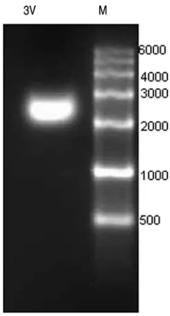
Homogeneity of armored L-RNA.
RNA was isolated from
purified armored L-RNA particles. The majority of the 3V
L-RNA packaged was approximately 2,200 bases in length, as
detected by ethidium bromide staining (Fig. 2).
Density gradient analysis of armored RNA and armored
L-RNA particles.
Armored RNA and armored L-RNA
parti-cles form narrow bands at 1.37 and 1.36 g ml
⫺1, respectively,
[image:4.585.43.282.89.396.2]when sedimented to their buoyant density in CsCl density
gradients. This compares favorably with the previously
re-ported value of 1.35 g ml
⫺1(15).
FIG. 2. Characterization of the recombinant RNA packaged in
armored L-RNA. Recombinant RNA was isolated from 3V armored
L-RNA, fractionated in a denaturing 3% agarose gel, stained with
ethidium bromide, and detected by using UV fluorescence.
Abbrevi-ations: M, RNA marker; 3V, 3V armored L-RNA recombinant RNA.
H5N1
1
1
42
30:00
20
None
2
1
92
3:00
20
None
3
5
92
10:00
20
None
45
30:00
20
None
72
1:00
20
None
4
40
92
10:00
20
None
60
30:00
20
Single
5
1
40
0:00
20
None
SARS-Cov2
1
1
42
30:00
20
None
2
1
92
3:00
20
None
3
5
92
10:00
20
None
52
20:00
2
None
72
30:00
20
None
40
92
5:00
20
None
4
60
30:00
20
Single
5
1
40
10:00
20
None
on May 16, 2020 by guest
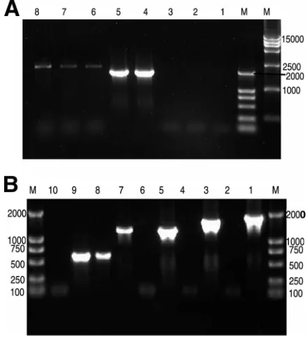
[image:4.585.359.482.451.679.2]Cloning and sequencing of the RT-PCR products.
The size
of the RT-PCR amplification products of the RNA extracted
from 3V armored L-RNA was full length (2,248 bp), whereas
the size of RNA extracted from 3V armored RNA was
be-tween 1,000 and 2,000 bp (Fig. 3). The sequencing result
dem-onstrated that the size was 1,200 bp.
Durability of armored L-RNA.
The armored L-RNA was
completely resistant to DNase and RNase treatment under
conditions in which naked DNA and RNA are both degraded
rapidly (data not shown).
Armored L-RNA plasma stability.
The 3V armored L-RNA
in newborn calf serum incubated at 4, 37, and 25°C was stable
over 2 months (Fig. 4).
Calibration of the chimeric armored RNA.
In order to
eval-uate the 3V armored L-RNA as a calibrator for multiple virus
assays, we used the National Reference HCV RNA assigned
the HCV International Standard (NIBSC 96/790) to calibrate
the serially diluted chimeric armored L-RNA. The
concentra-tions of the chimeric armored L-RNA for the five samples (10
6,
10
5, 10
4, 10
3, and 10
2) were 1.354
⫻
10
7IU ml
⫺1, 5.740
⫻
10
5IU ml
⫺1, 6.580
⫻
10
4IU ml
⫺1, 5.428
⫻
10
3IU ml
⫺1, and
9.613
⫻
10
2IU ml
⫺1, respectively (Fig. 5).
DISCUSSION
The armored L-RNA (2,248 bp) expressed by our
two-plas-mid coexpression system differs in several respects from the
virus-like particles previously described by Pickett and
Pea-body (17). These authors also used a two-plasmid expression
system; their goal was to determine whether the 21-nt
Opera-tor (pac site) would confer MS2-specific packageability on
non-bacteriophage RNA in vivo. The
E. coli
was induced such that
the Operator-
lacZ
hybrid RNA was coexpressed with the MS2
coat protein. The specificity of the Pickett and Peabody
bac-teriophage packaging system, however, was poor since the host
E. coli
RNA was packaged in preference to the Operator-
lacZ
RNA. In other studies, Pickett and Peabody modified the
packaging of the Operator-
lacZ
RNA by changing the ratios of
coat protein to Operator-
lacZ
RNA produced in
E. coli
. By
increasing the concentration of the Operator-
lacZ
RNA and
decreasing the concentration of the coat protein, these
re-searchers were able to encapsulate mainly the Operator-
lacZ
[image:5.585.317.522.68.231.2]RNA. These results suggest that the original Pickett and
Pea-body packaging strategy lacked specificity because they were
unable to determine an appropriate ratio of coat protein to
Operator-
lacZ
RNA. Furthermore, the size of the
lacZ
RNA
FIG. 3. Ethidium bromide-stained 1% agarose gel of RT-PCR
am-plification products of RNA extracted from 3V armored L-RNA and
3V armored RNA. (A) RT-PCR amplification products of RNA
ex-tracted from 3V armored L-RNA. Lane 1, negative control with no
template; lanes 2 and 3, negative control without RT; lanes 4 and 5,
positive control of pACYC-3V plasmid; lanes 6 to 8, RT-PCR of 3V
full-length L-RNA. (B) RT-PCR amplification products of RNA
ex-tracted from 3V armored RNA: lane 1, positive control of
pET-MS2-3V plasmid using the primers S-SARS1 and HA300RT-A; lane 2,
RT-PCR of SARS-CoV1 plus SARS-CoV2 plus SARS-CoV3 plus
HCV
⫹
HA300 using the primers S-SARS1 and HA300RT-A; lane 3,
positive control of pET-MS2-3V plasmid using the primers S-SARS1
and HCV-LAP1; lane 4, RT-PCR of SARS-CoV1 plus SARS-CoV2
plus SARS-CoV3 plus HCV using the primers S-SARS1 and
HCV-LAP1; lane 5, positive control of pET-MS2-3V plasmid using the
primers S-SARS1 and A-SARS3; lane 6, negative control without RT
using primers S-SARS1 and A-SARS3; lane 7, RT-PCR of
SARS-CoV1 plus SARS-CoV2 plus SARS-CoV3 using the primers S-SARS1
and A-SARS3; lane 8, RT-PCR of SARS-CoV1 plus SARS-CoV2
using the primers S-SARS1 and A-SARS2; lane 9, positive control of
pET-MS2-3V plasmid using the primers S-SARS1 and A-SARS2; lane
10, negative control without reverse transcription using the primers
S-SARS1 and A-SARS2.
FIG. 4. Stability study of 3V armored L-RNA. 3V armored L-RNA
was added to newborn calf serum to a final concentration of 10,000 and
10,000,000 copies/ml. Samples were incubated at 4°C, 37°C, or room
temperature for 0, 1, 2, 4, and 8 weeks. Samples were removed at each
time point and were stored at
⬃
80°C until the completion of the
experiment. From these materials, we isolated template RNA for
real-time RT-PCR assays. Water was used as a negative control. All RNA
templates were assayed in a single run by using an HCV RNA PCR
fluorescence quantitative diagnostic kit (Shanghai Kehua
Bio-Engi-neering Co., Ltd.). Real-time RT-PCR was conducted by using
Light-Cycler technology (Roche). The mean for low-copy samples was 67,226
IU/ml (4.83 log
10; range, 50,100 to 79,400 IU/ml [range, 4.70 to 4.92
log
10]), and the coefficient of variation was 12.9%. The mean for
high-copy samples was 29,060,000 IU/ml (7.45 log
10; range, 22,900,000
to 41,700,000 IU/ml [range, 7.36 to 7.62 log
10]), and the coefficient of
variation was 22%.
on May 16, 2020 by guest
http://jcm.asm.org/
[image:5.585.52.272.70.312.2]purified from these virus-like particles was approximately 500
bases as opposed to the expected full-length 3,000 bases. These
authors were, however, unable to determine whether the
deg-radation occurred before or after encapsulation. In their
sec-ond set of packaging studies, the RNA was not assessed for size
by gel electrophoresis. The MS2 bacteriophage sequence of the
RNA of Pickett and Peabody’s particles consisted only of the
site. Since the MS2 bacteriophage RNA genome is
approxi-mately 3.6 kb, it is likely that the maximal size of target RNA
packaged will be approximately 2.0 kb in armored RNA;
how-ever, to date, there have been no reports of armored RNA of
more than 1,200 bp using the method proposed by Pasloske
et al.
In order to arrive at an appropriate ratio of coat protein to
the six-target chimeric sequence, we selected the pET28b and
pACYCDuet-1 plasmids as expression vectors. These two
plas-mids are members of different compatibility groups. Therefore,
they can be stably maintained together in the same bacterial
host. These plasmids contain the same T7 bacteriophage
pro-moter, and they are both low-copy plasmids, having almost
equivalent copy numbers. Given this equivalence, the ratio of
coat protein from pET28b to the six-target chimeric RNA
sequence from pACYCDuet-1 should be appropriate.
Compared to armored RNA of approximately 1,200 bp
ob-tained using the original armored technology, our work
indi-cates that long-fragment (2,248 bp) RNA sequences can be
encapsulated by using the two-plasmid coexpression method in
conjunction with the C-variant of the wild-type stem-loop. The
armored L-RNA particles have all of the characteristics of
armored RNA. The technology allows the user to precisely
define the control RNA’s sequence. The armored L-RNA
comprising a 2,248-nt foreign RNA sequence, which includes
three SARS-CoV fragments, one HCV fragment, and two
H5N1 fragments, can be used as a control or calibrator for
SARS-CoV, HCV, and H5N1 qualitative or quantitative
de-tection by RT-PCR. The inclusion of the HCV 5
⬘
UTR made it
easy to assign an international (IU) value to the SARS-CoV
and H5N1 RNAs within the armored L-RNA and avoided
complex procedures involved in value assignment of
calibra-tors or standards in situations where their international
stan-dard (IS) are not available (20, 21). Moreover, the
metrolog-ical traceability of nucleic acid measurement of all RNA
viruses without IS could be solved by the same model as
chi-meric armored L-RNA.
From Fig. 5, it can be seen that the highest copy number of
chimeric armored L-RNA used was calibrated higher than
ex-pected and was not near a 1:1 correlation, a finding that might be
explained as follows. First, an error in the first step of dilution
could have occurred. Second, it was acceptable that the detection
deviation for the samples was in the range of the target value
⫾
0.27 log
10(14).
[image:6.585.66.258.69.487.2]In theory, the length of armored L-RNA expressed by the
two-plasmid system could be as much as approximately 3.6 kb
FIG. 5. Calibration of the real-time RT-PCR assay for HCV,
SARS-CoV2, and HA300 (H5N1). First, the quantified 3V armored
L-RNA was diluted with newborn calf serum 10-fold serially to obtain
100, 1,000, 10,000, 100,000, and 1,000,000 copies/ml. We used the
National Reference material for HCV RNA (GBW09151, 2.26
⫻
10
3IU ml
⫺1to 4.22
⫻
10
7IU ml
⫺1) to calibrate the serial dilutions of
chimeric armored L-RNA and then used the calibrated L-RNA to
prepare calibrators for the two real-time RT-PCR assays. From these
materials, we isolated RNA template for RT-PCR assays. Newborn
calf serum was used as a negative control. Real-time RT-PCR was
conducted on a LightCycler thermal cycler (Roche). (A) Log
concen-tration of the international standard for HCV RNA versus the cycle
number for the HCV RT-PCR; (B) amplification curve for the HCV
RT-PCR assay; (C) amplification curve for the SARS-CoV2 RT-PCR
assay; (D) amplification curve for the HA300 (H5N1) RT-PCR assay.
on May 16, 2020 by guest
since the MS2 bacteriophage RNA genome is 3,569 bp in
length. The results presented here indicate that at least a
2,248-bp armored L-RNA can be expressed with high
effi-ciency. We have also successfully expressed an approximate
2,700-bp chimeric armored RNA by the two-plasmid system
(data not shown), and the construction of an expression vector
for a chimeric armored L-RNA of more than 3,000 bp in length
is under way.
Because the pac site is a key point in the interaction between
the MS2 coat protein and exogenous RNA, it is believed that
the expression efficiency and package capacity could be further
enhanced if the number of pac sites were to be increased within
the chimeric RNA.
In conclusion, the results presented here demonstrate that
the two-plasmid expression system for armored L-RNA
(
⬎
2,000 bp) is effective. The chimeric armored L-RNA, which
exhibits RNase resistance and stability properties similar to
armored RNA, can be used as a calibrator in SARS-CoV,
H5N1, and HCV RT-PCR assays.
ACKNOWLEDGMENTS
This study was supported in part by the SEPSDA project of the
European Commission (under no. Sp22-CT-2004-003831), the
Na-tional Natural Science Foundation of China (30371365 and 30571776),
and the Capital Medicine Development Foundation of Beijing
(2002-3041).
REFERENCES
1.Argetsinger, J., and G. Gussin.1966. Intact ribonucleic acid from defective
particles of bacteriophage R17. J. Mol. Biol.21:421–424.
2.Beld, M., R. Minnaar, J. Weel, C. Sol, M. Damen, H. van der Avoort, P. Wertheim-van Dillen, A. van Breda, and R. Boom.2004. Highly sensitive assay for detection of enterovirus in clinical specimens by reverse
transcrip-tion-PCR with an armored RNA internal control. J. Clin. Microbiol.42:
3059–3064.
3.Bressler, A. M., and F. S. Nolte.2004. Preclinical evaluation of two real-time, reverse transcription-PCR assays for detection of the severe acute
respira-tory syndrome coronavirus. J. Clin. Microbiol.42:987–991.
4.Das, A., E. Spackman, D. Senne, J. Pedersen, and D. L. Suarez.2006. Development of an internal positive control for rapid diagnosis of avian influenza virus infections by real-time reverse transcription-PCR with
lyoph-ilized reagents. J. Clin. Microbiol.44:3065–3073.
5.Donia, D., M. Divizia, and A. Pana.2005. Use of armored RNA as a standard to construct a calibration curve for real-time RT-PCR. J. Virol. Methods
126:157–163.
6.Eisler, D. L., A. McNabb, D. R. Jorgensen, and J. L. Isaac-Renton.2004. Use of an internal positive control in a multiplex reverse transcription-PCR to
detect West Nile virus RNA in mosquito pools. J. Clin. Microbiol.42:841–
843.
7.Heisenberg, M.1966. Formation of defective bacteriophage particles by fr
amber mutants. J. Mol. Biol.17:136–144.
8.Hietala, S. K., and B. M. Crossley.2006. Armored RNA as virus surrogate in a real-time reverse transcriptase PCR assay proficiency panel. J. Clin.
Microbiol.44:67–70.
9.Horn, W. T., M. A. Convery, N. J. Stonehouse, C. J. Adams, L. Liljas, S. E. Phillips, and P. G. Stockley.2004. The crystal structure of a high affinity RNA stem-loop complexed with the bacteriophage MS2 capsid: further
challenges in the modeling of ligand-RNA interactions. RNA10:1776–1782.
10.Horton, R. M., H. D. Hunt, S. N. Ho, J. K. Pullen, and L. R. Pease.1989. Engineering hybrid genes without the use of restriction enzymes: gene
splic-ing by overlap extension. Gene77:61–68.
11.Huang, Q., Y. Cheng, Q. Guo, and Q. Li.2006. Preparation of a chimeric Armored RNA as a versatile calibrator for multiple virus assays. Clin. Chem.
52:1446–1448.
12.Konnick, E. Q., S. M. Williams, E. R. Ashwood, and D. R. Hillyard.2005. Evaluation of the COBAS hepatitis C virus (HCV) TaqMan analyte-specific reagent assay and comparison to the COBAS Amplicor HCV Monitor V2.0
and Versant HCV bDNA 3.0 assays. J. Clin. Microbiol.43:2133–2140.
13.Lowary, P. T., and O. C. Uhlenbeck.1987. An RNA mutation that increases
the affinity of an RNA-protein interaction. Nucleic Acids Res.15:10483–
10493.
14.Oliver, A. R., S. F. Pereira, and D. A. Clark.2007. Comparative evaluation of the automated Roche TaqMan real-time quantitative human immunode-ficiency virus type 1 RNA PCR assay and the Roche AMPLICOR version 1.5
conventional PCR assay. J. Clin. Microbiol.45:3616–3619.
15.Pasloske, B. L., C. R. Walkerpeach, R. D. Obermoeller, M. Winkler, and D. B. DuBois.1998. Armored RNA technology for production of
ribonucle-ase-resistant viral RNA controls and standards. J. Clin. Microbiol.36:3590–
3594.
16.Pasloske, B. L., D. DuBois, D. Brown, and M. Winkler.April 2001. Ribo-nuclease-resistant RNA preparation and utilization. U.S. patent 6,214,982. 17.Pickett, G. G., and D. S. Peabody.1993. Encapsidation of heterologous
RNAs by bacteriophage MS2 coat protein. Nucleic Acids Res.21:4621–4626.
18.Romaniuk, P. J., P. Lowary, H. N. Wu, G. Stormo, and O. C. Uhlenbeck.
1987. RNA binding site of R17 coat protein. Biochemistry26:1563–1568.
19.Rowsell, S., N. J. Stonehouse, M. A. Convery, C. J. Adams, A. D. Ellington, I. Hirao, D. S. Peabody, P. G. Stockley, and S. E. Phillips.1998. Crystal structures of a series of RNA aptamers complexed to the same protein
target. Nat. Struct. Biol.5:970–975.
20.Saldanha, J., and A. Heath.2003. Collaborative study to calibrate hepatitis C virus genotypes 2-6 against the HCV International Standard, 96/790
(ge-notype 1). Vox Sang84:20–27.
21.Saldanha, J., A. Heath, C. Aberham, J. Albrecht, G. Gentili, M. Gessner, and G. Pisani.2005. World Health Organization collaborative study to establish a replacement WHO international standard for hepatitis C virus RNA
nu-cleic acid amplification technology assays. Vox Sang88:202–204.
22.Scheuermann, R. H., and H. Echols.1984. A separate editing exonuclease
for DNA replication: the epsilon subunit ofEscherichia coliDNA
polymer-ase III holoenzyme. Proc. Natl. Acad. Sci. USA81:7747–7751.
23.Shiba, T., and Y. Suzuki.1981. Localization of A protein in the RNA-A
protein complex of RNA phage MS2. Biochim. Biophys. Acta654:249–255.
24.Stockley, P. G., N. J. Stonehouse, C. Walton, D. A. Walters, G. Medina, J. M. Macedo, H. R. Hill, S. T. Goodman, S. J. Talbot, and H. K. Tewary.1993. Molecular mechanism of RNA-phage morphogenesis. Biochem. Soc. Trans.
21:627–633.
25.Stockley, P. G., N. J. Stonehouse, and K. Valegard.1994. Molecular
mech-anism of RNA phage morphogenesis. Int. J. Biochem.26:1249–1260.
26.Talbot, S. J., S. Goodman, S. R. Bates, C. W. Fishwick, and P. G. Stockley.
1990. Use of synthetic oligoribonucleotides to probe RNA-protein
interac-tions in the MS2 translational operator complex. Nucleic Acids Res.18:
3521–3528.
27.Valegard, K., J. B. Murray, N. J. Stonehouse, S. van den Worm, P. G. Stockley, and L. Liljas.1997. The three-dimensional structures of two com-plexes between recombinant MS2 capsids and RNA operator fragments
reveal sequence-specific protein-RNA interactions. J. Mol. Biol.270:724–
738.
28.WalkerPeach, C. R., M. Winkler, D. B. DuBois, and B. L. Pasloske.1999. Ribonuclease-resistant RNA controls (armored RNA) for reverse transcrip-tion-PCR, branched DNA, and genotyping assays for hepatitis C virus. Clin.
Chem.45:2079–2085.
29.Witherell, G. W., J. M. Gott, and O. C. Uhlenbeck.1991. Specific interaction between RNA phage coat proteins and RNA. Prog. Nucleic Acid Res. Mol.
Biol.40:185–220.
![FIG. 1. Armored L-RNA packaging system. Two expressing vectors were constructed, in which the maturase and the coat protein wereexpressed from one plasmid [pET-28(b)] and the pac site and the six-target chimeric RNA sequence were produced from the second plasmid](https://thumb-us.123doks.com/thumbv2/123dok_us/8266448.841096/3.585.139.448.70.287/packaging-expressing-constructed-maturase-wereexpressed-chimeric-sequence-produced.webp)