www.impactjournals.com/oncotarget/ Oncotarget, Vol. 6, No.2
The microRNA-218~Survivin axis regulates migration, invasion,
and lymph node metastasis in cervical cancer
Ryunosuke Kogo1, Christine How1, Naz Chaudary1, Jeff Bruce1, Wei Shi1, Richard P. Hill1,2,3, Payam Zahedi1, Kenneth W. Yip1 and Fei-Fei Liu1,2,3,4
1 Ontario Cancer Institute, University Health Network (UHN), Toronto, Ontario, Canada 2 Department of Medical Biophysics, University of Toronto, Toronto, Canada
3 Department of Radiation Oncology, University of Toronto, Toronto, Canada
4 Department of Radiation Oncology, Princess Margaret Cancer Centre, UHN, Toronto, Canada
Correspondence to: Fei-Fei Liu, email: Fei-Fei.Liu@rmp.uhn.on.ca
Keywords: miR-218, survivin, cervical cancer, migration, invasion
Received: September 30, 2014 Accepted: November 24, 2014 Published: November 25, 2014
This is an open-access article distributed under the terms of the Creative Commons Attribution License, which permits unrestricted use, distribution, and reproduction in any medium, provided the original author and source are credited.
ABSTRACT
Cervical cancer is the third most common cancer in women worldwide. In the
present study, global microRNA profiling for 79 cervical cancer patient samples led to the identification of miR-218 down-regulation in cervical cancer tissues compared to normal cervical tissues. Lower miR-218 expression was associated significantly with
worse overall survival (OS), disease-free survival (DFS), and pelvic/aortic lymph node recurrence. In vitro, miR-218 over-expression decreased clonogenicity, migration, and
invasion. Survivin (BIRC5) was subsequently identified as an important cervical cancer
target of miR-218 using in silico prediction, mRNA profiling, and quantitative
real-time PCR (qRT-PCR). Concordant with miR-218 over-expression, survivin knockdown by siRNA decreased clonogenicity, migration, and invasion. YM155, a small molecule
survivin inhibitor, significantly suppressed tumor growthand lymph node metastasis
in vivo. Our findings demonstrate that the miR-218~survivin axis inhibits cervical
cancer progression by regulating clonogenicity, migration, and invasion, and suggest that the inhibition of survivin could be a potential therapeutic strategy to improve outcome in this disease.
INTRODUCTION
Cervical cancer is the third most common cancer in women globally [1]. In patients with locally advanced cervical cancer, cisplatin-based concurrent chemoradiotherapy can improve overall survival, progression free survival, and recurrence rates [2-4]; however, the 5-year survival rates for stage III and IV patients remains at less than 40% [5]. Moreover, approximately 30% of patients experience lymph node recurrence and distant metastasis after primary treatment [6]. The ability to prevent lymph node and distant metastasis remains an important yet unresolved therapeutic goal for these patients. Recent molecularly targeted therapeutics have shown potential in decreasing metastasis and improving survival for several human malignancies [7]; however, no proven drugs yet exist for cervical cancer.
MicroRNAs are small, non-coding RNAs that post-transcriptionally down-regulate the expression of multiple target genes [8]. MicroRNA dysregulation occurs in numerous human malignancies and is associated with altered malignant potential; affecting survival, proliferation, apoptosis, and invasion [9]. Recently, global profiling studies have enabled microRNA-based stratifications of cancer types and patient outcomes [10, 11]. However, the function and target genes of many microRNAs remain to be elucidated; this characterization is fundamentally necessary in order to acquire a deeper understanding of cancer progression.
described to be associated with tumor invasion [16]. Thus, miR-218 is a clinically important and interesting microRNA for investigation.
In the current study, new miR-218-related associations were identified in clinically annotated cervical cancer samples. Furthermore, the cellular and molecular functions of miR-218 and one of its key targets, survivin (BIRC5), were elucidated. Lastly, we validated these observations in vitro and in vivo using a small molecule survivin suppressant (YM155), and provide data in support of targeting the miR-218~survivin axis in cancer therapy and preventing metastasis.
RESULTS
miR-218 down-regulation was associated with reduced survival in cervical cancer patients
Analysis of Taqman Low Density Array (TLDA) data determined that expression of miR-218 was significantly reduced in 79 cervical cancer tissues compared to 11 normal cervix tissues (P<0.001; Figure 1A). Further details of this study have been described in How et al. [17]; in brief, these patients have all been treated for cure (radiation and chemotherapy)
with a median follow-up time of 6 years. We therefore investigated the association between miR-218 expression with patient survival. Initially, the median miR-218 expression value was utilized to divide the 79 cervical cancer patients into high vs. low expression groups (miR-218 highmedian, n=39; miR-218 lowmedian, n=40). The miR-218 low expression group experienced a worse overall survival (OS), and disease-free survival (DFS) (OS P=0.074; DFS P=0.079, Figure S1), but the data were of borderline statistical significance. The groups were then re-divided, based on the lowest level of miR-218 expression measured in the normal cervix population. This resulted in 35 patients with high miR-218 expression
vs. 44 with low miR-218 expression. Using this new cut-off level, the low miR-218 expression group experienced a significantly poorer outcome with regards to both OS and DFS (OS P=0.009; DFS P=0.014; Figure 1B). These data suggest that cervical cancer patients with lower miR-218 expression levels than detected in normal cervical epithelium tissues will experience a poor outcome.
[image:2.612.114.461.390.630.2]Clinical factors were also analyzed for the miR-218 high vs. low expression groups (Table 1). The two groups did not differ in age, tumor size, International Federation of Gynecology and Obstetrics (FIGO) staging, or distant metastasis. Of note however, miR-218 down-regulation was strongly associated with pelvic and para-aortic lymph node recurrence (P=0.032 and P=0.013, respectively), as
Figure 1: miR-218 down-regulation is associated with poor survival in cervical cancer patients. A) miR-218 expression in 79 cervical cancer patient samples and 11 normal cervix epithelial samples. miR-218 expression (log2) was measured using Taqman
Low Density Array (TLDA) Human MicroRNA A Arrays V2.0 for 79 cervical cancer tissues and 11 normal cervix tissues. B) Kaplan-Meier analysis of overall (left) and disease-free survival (right) in 79 cervical cancer patients. Solid line: miR-218 high expression group (n=35); dotted line: miR-218 low expression group (n=44). C) Genomic alteration of miR-218 loci (left: hsa-miR-218-1, chromosome 4p15.31; right: hsa-miR-218-2, chromosome 5q34) using copy number data from 105 cervical squamous cell carcinoma samples generated by TCGA using SNP 6.0 arrays. Genomic alteration was visualized using the IGV (Integrative Genomic Viewer, Broad Institute). Blue
well as an association with lymph node metastasis at the time of diagnosis (P=0.053).
Deletion of miR-218 genomic loci in cervical cancer
In other cancers, miR-218 down-regulation is known to occur through promoter hypermethylation or genomic loss [12, 13, 18]. In order to elucidate the mechanism of miR-218 down-regulation in cervical cancer, The Cancer Genome Atlas (TCGA) genomic data (SNP arrays) and epigenetic data (methylation profiles) from 105 cervical squamous cell carcinoma samples were analyzed. Copy number data indicated that most patients’ miR-218 genomic loci were deleted (hsa-miR-218-1: 50%, hsa-miR-218-2: 31%, Figure 1C). Methylation and microRNA sequencing data were also analyzed, but no correlations were observed between miR-218 methylation and miR-218 expression level (data not shown). Overall, these data suggest that in cervical cancer, reduced miR-218 expression level is likely related to deletion of the miR-218 loci.
miR-218 reduced cell survival, migration, and invasion in vitro
In order to elucidate the biological significance of miR-218 down-regulation, SiHa and ME-180 cells, which are both human papillomavirus (HPV) positive cervical squamous cell carcinoma lines, were transfected with pre-miR negative control (pre-miR-NC) or pre-pre-miR-218 (pre-miR- (miR-218). Forty-eight hours post-transfection, miR-218 was over-expressed by more than 200-fold in SiHa and ME-180 cells (P<0.01, Figure S2). In both cell lines, this over-expression significantly reduced clonogenicity (P<0.05, Figure 2A).
Because miR-218 down-regulation was observed to be associated with lymph node metastasis and recurrence in our patients, we performed migration and invasion assays. Consistent with the clinical data, miR-218 over-expression reduced migration and invasion capacities of both SiHa and ME-180 cells (P<0.05; Figures 2B and 2C).
Survivin is an important target of miR-218 in cervical cancer cells
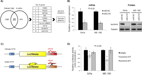
In order to identify biologically relevant miR-218 targets, we first examined in silico predicted targets using miRDB (http://mirdb.org/miRDB/) [19, 20]. These data were combined with mRNA array (GeneChip Human Genome U133 Plus 2.0) data generated from the same 79 cervical cancer tissues and 11 normal cervix tissues used for TLDA [17]. At the intersection between the in silico predicted targets and mRNAs that were up-regulated
by greater than 2 fold were 35 candidate targets (Figure 3A; Table S1). For these candidate targets, their in silico
prediction scores and expression levels were used to rank the genes independently, then these ranks were summed for a cumulative final rank (Table S1).
The top 10 candidate target genes from Table S1 were then assayed in the SiHa and ME-180 cells using qRT-PCR after 48 hrs of 218 transfection, with miR-NC transfection as a control (Figure S3). Survivin was the most consistently and significantly reduced target after miR-218 transfection in both cell lines (Figure S3; re-presented in Figure 3B, left panel). Correspondingly, miR-218 over-expression also reduced survivin protein expression (Figure 3B, right panel).
In order to confirm direct targeting and binding between miR-218 and the survivin 3’-untranslated region (3’-UTR), we cloned the survivin 3’-UTR (which included a miR-218 predicted binding site) into the pMIR-REPORT luciferase vector (Figure 3C). Cells transfected with wild type survivin 3’-UTR pMIR-REPORT vector (survivin-WT) showed a significant reduction in luciferase activity in both SiHa and ME-180 cells (P<0.05 relative to empty pMIR-REPORT vector, Figure 3D). These inhibitory effects were not observed with a mutant survivin 3’-UTR pMIR-REPORT vector (survivin-MT; containing a mutation in the miR-218 binding site), thereby confirming specific and direct survivin 3’-UTR targeting by miR-218.
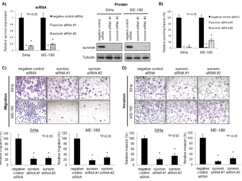
Survivin knockdown reduced survival, migration, and invasion in cervical cancer cells
Survivin is the smallest member of the inhibitor of apoptosis (IAP) family, and is mainly associated with the regulation of mitosis and inhibition of apoptosis [21]. Some functions of survivin still remain unknown because survivin interacts with numerous proteins. In order to characterize survivin function in cervical cancer cells, SiHa and ME-180 cells were transfected with survivin siRNAs. Survivin knockdown was confirmed both at the mRNA and protein level (Figure 4A). This down-regulation was accompanied by a significant reduction in clonogenicity compared to control cells transfected with negative control siRNA (P<0.05; Figure 4B). Additionally, survivin knockdown also reduced migration and invasion capacities, phenocopying miR-218 over-expression (P<0.05; Figures 4C and 4D). Overall, these data support the postulate that the miR~218-survivin axis regulates clonogenicity, migration, and invasion in cervical cancer.
Efficacy of a survivin targeting compound in cervical cancer
miR-Table 1: miR-218 expression and clinical factors
miR-218 low (n=44) miR-218 high (n=35)
Factors number % number % P value
Age (mean ± SD) 49.1 ± 2.1 years 51.7 ± 2.4 years 0.41
Tumor size
< 5cm 27 61.4 21 60.0 0.9
> 5cm 17 38.6 14 40.0
FIGO staging
Stage I 15 34.1 9 25.7 0.42
Stage II, III 29 65.9 26 74.3
Lymph node metastasis
Absent 18 40.9 22 62.9 0.053*
Present 26 59.1 13 37.1
Recurrence
Pelvic LN recurrence
Absent 30 68.2 31 88.6 0.032**
Present 14 31.8 4 11.4
Para-aortic LN recurrence
Absent 37 84.1 35 100.0 0.013**
Present 7 15.9 0 0.0
Distant metastasis recurrence
Absent 36 81.8 32 91.4 0.22
Present 8 18.2 3 8.6
[image:4.612.87.507.71.558.2]SD: Standard deviation, LN: Lymph node, *P<0.1, **P<0.05.
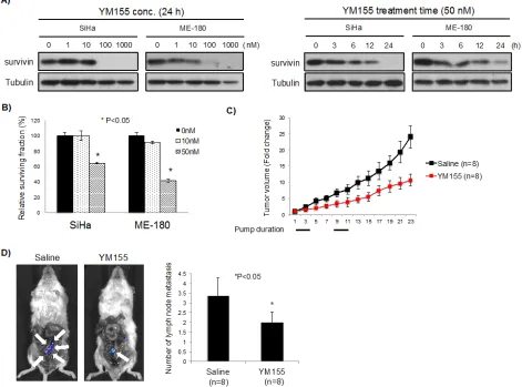
[image:4.612.85.524.383.620.2]218~survivin axis regulates cervical cancer clonogenicity, migration, and invasion, then in turn, a survivin suppressant/inhibitor could be therapeutically important in this disease. Although several survivin inhibitors exist [22], we utilized the readily available YM155. This small molecule suppresses survivin transcription and is currently being assessed in several clinical trials for lymphoma, prostate cancer, malignant melanoma, and NSCLC [23].
YM155 decreased survivin expression in both a concentration and time dependent manner (Figure 5A). As well, YM155 significantly reduced clonogenicity of SiHa and ME-180 cells, which was comparable to survivin siRNA (Figures 5B and 4B). In order to examine the effects of YM155 against tumor growth and lymph node metastasis in cervical cancer cells, we generated luciferase-expressing ME-180 cells (Luc-ME-180 cells), and evaluated in vivo anti-tumor activity. Mice treated with YM155 had significantly reduced tumor growth compared to control mice treated with saline (53% reduction at day 22, P<0.05, Figure 5C). We evaluated intratumoral survivin protein expression at days 0, 3, and 7, and confirmed inhibition of survivin expression during YM155 treatment (compared to saline controls; Figure
S4A). Next, the inhibitory effect of YM155 on lymph node metastases was examined using an orthotopic xenograft model of cervical cancer [24]. After 28 days implantation, the number of lymph node metastases in YM155 vs.
saline treated mice was evaluated using bioluminescence. All metastatic lymph nodes detected by bioluminescence were confirmed using histological analyses (Figure S4B). Importantly, YM155 significantly reduced the number of lymph node metastasis (P<0.05, Figure 5D), without affecting the large primary tumor transplanted into the cervix (data not shown).
DISCUSSION
[image:5.612.60.548.342.599.2]The current study demonstrated that miR-218 is significantly down-regulated in human cervix cancer, which in turn, was associated with poor OS and DFS, as well as increased risk of lymph node recurrence. The identified miR-218~survivin axis regulated clonogenicity, migration, and invasion of cervical cancer cells in vitro. Furthermore, a small molecule survivin suppressant, YM155 was able to reduce tumor growth and lymph node metastasis in vivo.
Figure 3: Survivin is a direct target of miR-218. A) Identification of miR-218 targets in cervical cancer. Cancer (Up): mRNA
expression greater than 2-fold compared to normal cervix, from GeneChip Human Genome U133 Plus 2.0 Array data for 79 cervical cancer tissues and 11 normal cervix tissues; in silico: predicted targets of miR-218 by miRDB (http://mirdb.org/miRDB/). B) mRNA (left) and protein (right) survivin expression after 48 hrs miR-218 or miR-NC (10 nM each) transfection in SiHa and ME-180 cells. Survivin mRNA expression levels were normalized to GAPDH. *P<0.05, bars represent mean ± SEM from triplicates. C) Schema of pMIR-REPORT vectors for the luciferase assay. Wild type (upper) and/or Mutant (lower) survivin 3’UTR were cloned downstream of firefly luciferase
miR-218 down-regulation has been reported in several cancers [12-15]; it has been shown to inhibit migration and invasion in gastric cancer [15], glioma [25], head & neck [26], renal cell [27], and cervical cancers [28]. Consistent with previous reports, in this current study, we observed that miR-218 inhibited migration, and invasion in cervical cancer cells. Our study further validated and extended upon these findings both clinically, by identifying the association with lymph node involvement at presentation, nodal recurrence, OS, DFS; and experimentally, by elucidating the role of survivin as a downstream mediator of miR-218 under-expression.
The mechanism of miR-218 down-regulation may depend on the type of cancer. To date, promoter
hypermethylation in nasopharyngeal carcinoma [12] and genomic deletion in non-small cell lung [18] and bladder cancers [13] have been reported. Our analysis of the data obtained from TCGA SNP arrays suggested that genomic deletion of 218 loci is likely a mechanism for miR-218 under-expression in cervical cancer. miR-miR-218 is located in two genomic loci; 4p15.31 (hsa-miR-218-1) and 5q34 (hsa-miR-218-2), which are located within the intronic regions of SLIT2 and SLIT3, respectively. Loss of chromosome 4p has been previously reported in cervical cancer [29], which interestingly, was also noted to be associated with lymph node metastasis [29].
[image:6.612.63.553.230.597.2]Several studies have identified miR-218 targets, such as ROBO1 [15], IKK-β [25], Laminin-332 [26],
Figure 4: Survivin knockdown reduced survival, migration, and invasion in cervical cancer cells. A) mRNA (left) and protein (right) survivin expression after 48 hrs negative control siRNA or survivin siRNAs (10 nM each) transfection in SiHa and ME-180 cells. Survivin mRNA expression levels were normalized to GAPDH, relative to cells transfected with negative control siRNA. B) Clonogenic assays were performed by seeding SiHa and ME-180 cells transfected with negative control siRNA or survivin siRNAs (10 nM each). At 48 hrs post-transfection, cells were re-seeded at low density in 6-well plates. After 10-14 days incubation, colonies were stained and counted. C) Representative image (left) and quantification bar graph (right) of migrated SiHa and ME-180 cells. Migration assays were performed by seeding SiHa and ME-180 cells transfected with negative control siRNA or survivin siRNAs (10 nM each) in trans-well chambers. After 48 hrs incubation, migrated cells were stained and counted. D) Representative image (left) and quantification bar graph
Rictor [30], and survivin [12]. We chose to focus only on a single, key target of miR-218 in cervical cancer, identified by combining in silico prediction and global mRNA array data in order to evaluate this axis in some depth. Future studies should delve into the relevance of the other targets in similar model systems. For example, bioinformatics-related analyses may be expanded, and survivin-lacking cells may be utilized to compare the contributing effects of survivin vs. other targets in miR-218-induced phenotypes. Survivin is an IAP and functions as an oncogene in cancer cells due to its anti-apoptotic properties. The main functions of survivin include inhibition of caspase-dependent apoptosis and caspase-incaspase-dependent cell death, as well as in the regulation of mitosis [21]. However, a complete understanding of all of survivin’s functions
remains unclear since this protein interacts with a number of other proteins with multi-functional effects [21].
Our data showed that survivin knockdown by siRNA significantly reduced clonogenicity, migration, and invasion in SiHa and ME-180 cells, phenocopying the results of miR-218 over-expression. Survivin is known to promote tumor cell invasion (in vitro) and metastasis (in vivo), in cooperation with XIAP, another IAP family member [22, 31]. Future work will be required to further elucidate the detailed mechanisms by which survivin affects migration and invasion.
[image:7.612.67.538.248.597.2]Survivin is typically absent in normal adult cells (except for germ cells), and is highly over-expressed in cancer cells, thereby serving as a drug target. YM155 leads to the repression of survivin promoter activity by binding
Figure 5: Effect of YM155, a survivin suppressant, in vitro and in vivo. A) YM155 inhibited survivin expression. (Left) SiHa and ME-180 cells were treated with YM155 (0, 1, 10, 100, and 1000 nM) for 24 hrs. Survivin expression was analyzed by Western blotting. (Right) SiHa and ME-180 cells were treated with YM155 (50 nM) for the indicated period of time. Survivin expression was analyzed by Western blotting. B) Clonogenic assays were performed by seeding SiHa and ME-180 cells treated with YM155 (0, 10, and 50 nM) for 24 h. After 14 days incubation, colonies were stained and counted. *P<0.05, bars represent mean ± SEM from triplicates. C) Luc-ME-180 cell subcutaneous xenografts were treated with YM155 (10mg/kg/day). Saline or YM155 was administered as a 3-day continuous infusion per week for 2 weeks. Tumor volume (mm3) was normalized to tumor volume at beginning of treatment (day 1) *P<0.05, bars represent mean
to the transcription factor ILF3 and disrupting the ILF/p54 complex [32]. Phase II clinical trials have been conducted with YM155 alone or as part of combination therapy in prostate cancer, lymphoma, melanoma, and non-small cell lung cancer patients [23]. Our data demonstrated that YM155 significantly reduced survivin expression in a concentration and time-dependent manner, and reduced cell proliferation in vitro. We present the first orthotopic metastatic model for YM155 evaluation, and demonstrated that YM155 indeed reduced lymph node metastasis. Primary tumor growth was reduced subcutaneously, but not orthotopically. We speculate that the tumor size (large donor tumors needed to be transplanted in the orthotopic model), and/or the microenvironment might well account for this discrepant observation. Other survivin-inhibiting compounds are currently in development and might be even more effective inhibitors [22]. Nonetheless, we have demonstrated the proof-of-concept that such inhibitors should be further explored in the prevention of metastasis in this disease.
The role of the mir-218∼survivin axis in potentially promoting nodal metastasis may suggest a therapeutic opportunity for survivin inhibitors in treating node-positive, locally advanced cancers, and/or cancers with occult nodal involvement. Survivin inhibition may be particularly helpful for adjuvant therapy used to inhibit lymph node metastasis after primary tumor resection. Further work will be required to investigate the use of survivin inhibitors with existing standard treatments (e.g., chemotherapy and/or radiation) for such disease. Moreover, whether this therapeutic opportunity exists in other cancers (e.g., head and neck, lung) remains to be investigated.
In conclusion, the miR-218~survivin axis is pivotal, both on a clinical and basic cellular level, in regulating clonogenicity, migration, and invasion in cervical cancer. Anti-survivin therapy might provide a potentially useful strategy in partially restoring this axis, and thereby improve outcome for patients with cervical cancer.
METHODS
Ethics Statement
Written informed consent was obtained from patients according to a protocol approved by the University Health Network (UHN) Research Ethics Board. Animal experiments were performed in strict accordance with the protocols approved by the Animal Care Committee (ACC) of the Ontario Cancer Institute (OCI), UHN.
Patient Samples and microRNA/mRNA Profiling
Seventy-nine cervical cancer tissues and 11 normal cervix tissues were collected from fresh frozen punch biopsies. Total RNA was isolated using the Total RNA Purification Kit (Norgen Biotek), according to the manufacturer’s protocol. Global microRNA and mRNA profiles were analyzed with the TLDA Human MicroRNA A Array V2.0 (Life Technologies), and GeneChip Human Genome U133 Plus 2.0 Array (Affymetrix), respectively.
Copy Number Analysis of TCGA Data
Level 3 segmented copy number data for 105 cervical squamous cell carcinoma samples generated by TCGA using SNP 6.0 arrays were downloaded from the Broad Firehose website (2012_11_02 stdata Run). Copy number data for miR-218 encoding loci were then visualized using the integrated genome viewer (igv; https://www.broadinstitute.org/software/igv) [33].
Cell Lines and Transfections
Cervical cancer cell lines, SiHa and ME-180, were obtained from American Type Culture Collection (ATCC) and cultured in α-MEM with 10% Fetal Bovine Serum at 37˚C, 5% CO2. These cells were authenticated every six months at the Centre for Applied Genomics (Hospital for Sick Children, Toronto, Canada) using the AmpF/STR Identifier PCR Amplification Kit (Applied Biosystems); as well, they were determined to be mycoplasma free every 3 months using the MycoAlert Mycoplasma Detection Kit (Lonza).
SiHa and ME-180 cells were transfected with Lipofectamine RNAiMAX (Life Technologies), according to the manufacturer’s protocol. Pre-miR miRNA negative control, pre-miR-218 (Life Technologies), AllStars Negative Control siRNA, and two survivin siRNAs (Qiagen) were transfected at 10 nM.
Quantitative Real-Time PCR (qRT-PCR) Analysis
Total RNA was isolated using the Total RNA Purification Kit (Norgen Biotek), and miR-218 expression levels were measured using the TaqMan MicroRNA Assays (Life Technologies). The 2-ΔΔ Ct method was used
to calculate relative miR-218 expression, using RNU44 as a reference gene.
expression.
Western Blot Analysis
Total protein was extracted using RIPA buffer (50 mM TrisHCl, 150 mM NaCl, 2 mM EDTA, 1% NP-40, 0.1% SDS), then separated using a Novex 4-20% Tris-Glycine Gel (Life Technologies). Proteins were detected using survivin (1:1000 dilution, Novus Biologicals) and α-Tubulin (loading control, 1:40000 dilution, Sigma) antibodies. Specific proteins were detected using SuperSignal West Pico Chemiluminescent Substrate (Thermo Scientific).
Viability and Clonogenic Assays
Cell viability was examined using the CellTiter 96 Non-Radioactive Cell Proliferation Assay (MTS assay, Promega), according to the manufacturer’s protocol. For clonogenic assays, transfected cells were re-seeded 48 hrs post-transfection at low density in 6-well plates. Cells were incubated for 10-14 days, then fixed and stained with 0.2% methylene blue in 50% methanol. The surviving fraction was calculated by comparison with control cells.
Invasion and Migration Assays
Invasion and migration assays were performed using the BD BioCoat Matrigel Invasion Chambers and Control Inserts (BD Biosciences), respectively. Briefly, cells (1 × 10⁵ cells/well) were seeded with medium containing low serum (1%) in the upper chamber. The lower chamber was filled with medium containing high serum (20%) as a chemoattractant. Cells were incubated for 48 hrs and then membranes were stained using Diff-Quick (Siemens). A light microscope was used to count the number of migrating and invading cells.
Luciferase microRNA Binding Assay
Wild-type (WT) or mutant (MT) fragments of the survivin 3’-untranslated region (3’UTR) containing the predicted miR-218 binding site were amplified by Platinum Taq DNA polymerase (Life Technologies). Primer sequences are listed in Table S2. Amplified PCR products were cloned in the pMIR-REPORT miRNA expression reporter vector (Life Technologies). Cells were co-transfected with 20 nM pre-miR, 100 ng pMIR-REPORT vector (Firefly luciferase), and 50 ng pRL-SV40 vector (Renilla luciferase, Promega) as a reference control using Lipofectamine 2000 (Life Technologies). Luciferase activities were measured using Dual-Glo Luciferase Assay System (Promega) at 24 hrs post-transfection.
Generation of Luciferase Expressing ME-180 Cells (Luc-ME-180)
Luciferase containing lentivirus (Lenti-Luc) was generated by transient transfection of 293T cells with psPAX2, pCMV-VSVG (Addgene), and CSII-CMV-Bsd-Luciferase plasmids using Lipofectamine 2000 (Life Technologies). At 48 hrs after co-transfection, lentivirus-containing supernatant was collected, and then passed through a 0.45 µm filter. Cultured ME-180 cells were incubated for 48 hrs with Lenti-Luc, then cultured for 2 weeks in the presence of 4.0 µg/mL Blasticidin S (Life Technologies).
In Vivo Experiments
Six to eight week-old severe combined immunodeficient (SCID) female mice were utilized for all experiments. ME-180 or Luc-ME-180 (1 × 10⁷) cells were injected into the left flank or left gastrocnemius muscle subcutaneously or intramuscularly as indicated. Tumor growth was monitored by measuring tumor volume (length × width2/2, mm3) or tumor plus leg diameter (mm). YM155 treatment commenced once the tumor volume reached 50 mm3 or the leg diameter reached 8 mm. YM155 or saline control (mice randomized) was administered subcutaneously as a 3-day per week continuous infusion for 2 weeks using the Alzet Osmotic Pump® (Model 1003D).
For the orthotopic xenograft model, Luc-ME-180 (1 × 10⁷) cells were injected into the left gastrocnemius muscle intramuscularly (donor mice). Once the leg diameter reached 9-12mm, tumors were excised and dissected. The tumors were then cut into 2-3 mm3 fragments in α-MEM media and placed on ice. Recipient mice were anesthetized and their uteruses exposed. A small incision was made in the cervix and a tumor fragment was sutured in place using a single 8-0 silk suture. The peritoneal membrane was closed in two layers using 8-0 silk sutures followed by skin closure using wound clips [24].
Bioluminescence Imaging
Statistical Analysis
All experiments have been performed at least three independent times, and the data are presented as the mean ± standard error of the mean (SEM). Statistical significance between treatment groups was determined using the Student’s t-test or Χ2 test. Overall survivalcurves were plotted according to the Kaplan-Meier method, withthe log-rank test applied for comparison. Statistical analyses were performed using JMP5 (SAS Institute).
ACKNOWLEDGEMENTS
This work has been supported by a grant from the Ontario Institute for Cancer Research, and also in part from the Campbell Family Institute for Cancer Research, and the Ministry of Health and Long-Term Planning.
REFERENCES
1. Jemal A, Bray F, Center MM, Ferlay J, Ward E and Forman D. Global cancer statistics. CA Cancer J Clin. 2011; 61: 69-90.
2. Green JA, Kirwan JM, Tierney JF, Symonds P, Fresco L, Collingwood M and Williams CJ. Survival and recurrence after concomitant chemotherapy and radiotherapy for cancer of the uterine cervix: a systematic review and meta-analysis. Lancet. 2001; 358: 781-786.
3. Keys HM, Bundy BN, Stehman FB, Muderspach LI, Chafe WE, Suggs CL, 3rd, Walker JL and Gersell D. Cisplatin, radiation, and adjuvant hysterectomy compared with radiation and adjuvant hysterectomy for bulky stage IB cervical carcinoma. N Engl J Med. 1999; 340: 1154-1161. 4. Morris M, Eifel PJ, Lu J, Grigsby PW, Levenback C,
Stevens RE, Rotman M, Gershenson DM and Mutch DG. Pelvic radiation with concurrent chemotherapy compared with pelvic and para-aortic radiation for high-risk cervical cancer. N Engl J Med. 1999; 340: 1137-1143.
5. American Cancer Society. Cervical Cancer. Atlanta, Ga: American Cancer Society. 2013.
6. Waggoner SE. Cervical cancer. Lancet. 2003; 361: 2217-2225.
7. Sethi N and Kang Y. Unravelling the complexity of metastasis - molecular understanding and targeted therapies. Nat Rev Cancer. 2011; 11: 735-748.
8. Bartel DP. MicroRNAs: genomics, biogenesis, mechanism, and function. Cell. 2004; 116: 281-297.
9. Calin GA and Croce CM. MicroRNA signatures in human cancers. Nat Rev Cancer. 2006; 6: 857-866.
10. Hu X, Schwarz JK, Lewis JS, Jr., Huettner PC, Rader JS, Deasy JO, Grigsby PW and Wang X. A microRNA expression signature for cervical cancer prognosis. Cancer Res. 2010; 70: 1441-1448.
11. Lui WO, Pourmand N, Patterson BK and Fire A. Patterns
of known and novel small RNAs in human cervical cancer. Cancer Res. 2007; 67: 6031-6043.
12. Alajez NM, Lenarduzzi M, Ito E, Hui AB, Shi W, Bruce J, Yue S, Huang SH, Xu W, Waldron J, O’Sullivan B and Liu FF. MiR-218 suppresses nasopharyngeal cancer progression through downregulation of survivin and the SLIT2-ROBO1 pathway. Cancer Res. 2011; 71: 2381-2391.
13. Davidson MR, Larsen JE, Yang IA, Hayward NK, Clarke BE, Duhig EE, Passmore LH, Bowman RV and Fong KM. MicroRNA-218 is deleted and downregulated in lung squamous cell carcinoma. PLoS One. 2010; 5: e12560. 14. Li CH, To KF, Tong JH, Xiao Z, Xia T, Lai PB, Chow SC,
Zhu YX, Chan SL, Marquez VE and Chen Y. Enhancer of zeste homolog 2 silences microRNA-218 in human pancreatic ductal adenocarcinoma cells by inducing formation of heterochromatin. Gastroenterology. 2013; 144: 1086-1097 e1089.
15. Tie J, Pan Y, Zhao L, Wu K, Liu J, Sun S, Guo X, Wang B, Gang Y, Zhang Y, Li Q, Qiao T, Zhao Q, Nie Y and Fan D. MiR-218 inhibits invasion and metastasis of gastric cancer by targeting the Robo1 receptor. PLoS Genet. 2010; 6: e1000879.
16. Yu J, Wang Y, Dong R, Huang X, Ding S and Qiu H. Circulating microRNA-218 was reduced in cervical cancer and correlated with tumor invasion. J Cancer Res Clin Oncol. 2012; 138: 671-674.
17. How C, Hui AB, Alajez NM, Shi W, Boutros PC, Clarke BA, Yan R, Pintilie M, Fyles A, Hedley DW, Hill RP, Milosevic M and Liu FF. MicroRNA-196b regulates the homeobox B7-vascular endothelial growth factor axis in cervical cancer. PLoS One. 2013; 8: e67846.
18. Tatarano S, Chiyomaru T, Kawakami K, Enokida H, Yoshino H, Hidaka H, Yamasaki T, Kawahara K, Nishiyama K, Seki N and Nakagawa M. miR-218 on the genomic loss region of chromosome 4p15.31 functions as a tumor suppressor in bladder cancer. Int J Oncol. 2011; 39: 13-21.
19. Wang X. miRDB: a microRNA target prediction and functional annotation database with a wiki interface. RNA. 2008; 14: 1012-1017.
20. Wang X and El Naqa IM. Prediction of both conserved and nonconserved microRNA targets in animals. Bioinformatics. 2008; 24: 325-332.
21. Altieri DC. Survivin, cancer networks and pathway-directed drug discovery. Nat Rev Cancer. 2008; 8: 61-70.
22. Altieri DC. Targeting survivin in cancer. Cancer Lett. 2013; 332: 225-228.
23. Rauch A, Hennig D, Schafer C, Wirth M, Marx C, Heinzel T, Schneider G and Kramer OH. Survivin and YM155: how faithful is the liaison? Biochim Biophys Acta. 2014; 1845: 202-220.
treatment. Curr Protoc Pharmacol. 2011; Chapter 14: Unit 14 19.
25. Song L, Huang Q, Chen K, Liu L, Lin C, Dai T, Yu C, Wu Z and Li J. miR-218 inhibits the invasive ability of glioma cells by direct downregulation of IKK-beta. Biochem Biophys Res Commun. 2010; 402: 135-140.
26. Kinoshita T, Hanazawa T, Nohata N, Kikkawa N, Enokida H, Yoshino H, Yamasaki T, Hidaka H, Nakagawa M, Okamoto Y and Seki N. Tumor suppressive microRNA-218 inhibits cancer cell migration and invasion through targeting laminin-332 in head and neck squamous cell carcinoma. Oncotarget. 2012; 3: 1386-1400.
27. Yamasaki T, Seki N, Yoshino H, Itesako T, Hidaka H, Yamada Y, Tatarano S, Yonezawa T, Kinoshita T, Nakagawa M and Enokida H. MicroRNA-218 inhibits cell migration and invasion in renal cell carcinoma through targeting caveolin-2 involved in focal adhesion pathway. J Urol. 2013; 190: 1059-1068.
28. Yamamoto N, Kinoshita T, Nohata N, Itesako T, Yoshino H, Enokida H, Nakagawa M, Shozu M and Seki N. Tumor suppressive microRNA-218 inhibits cancer cell migration and invasion by targeting focal adhesion pathways in cervical squamous cell carcinoma. Int J Oncol. 2013; 42: 1523-1532.
29. van den Tillaart SA, Corver WE, Ruano Neto D, ter Haar NT, Goeman JJ, Trimbos JB, Fleuren GJ and Oosting J.
Loss of heterozygosity and copy number alterations in
flow-sorted bulky cervical cancer. PLoS One. 2013; 8: e67414. 30. Uesugi A, Kozaki K, Tsuruta T, Furuta M, Morita K,
Imoto I, Omura K and Inazawa J. The tumor suppressive microRNA miR-218 targets the mTOR component Rictor and inhibits AKT phosphorylation in oral cancer. Cancer Res. 2011; 71: 5765-5778.
31. Mehrotra S, Languino LR, Raskett CM, Mercurio AM, Dohi T and Altieri DC. IAP regulation of metastasis. Cancer Cell. 2010; 17: 53-64.
32. Yamauchi T, Nakamura N, Hiramoto M, Yuri M, Yokota H, Naitou M, Takeuchi M, Yamanaka K, Kita A, Nakahara T, Kinoyama I, Matsuhisa A, Kaneko N, Koutoku H, Sasamata M, Kobori M, et al. Sepantronium bromide (YM155) induces disruption of the ILF3/p54(nrb) complex, which is required for survivin expression. Biochem Biophys Res Commun. 2012; 425: 711-716.