Biology Dissertations Department of Biology
8-11-2015
Combating the Epigenome: Elucidation of
Mechanisms Underlying Chemoresistance and
Enhancing Tumor Immunogenicity
Ercan Cacan
Georgia State University
Follow this and additional works at:https://scholarworks.gsu.edu/biology_diss
This Dissertation is brought to you for free and open access by the Department of Biology at ScholarWorks @ Georgia State University. It has been accepted for inclusion in Biology Dissertations by an authorized administrator of ScholarWorks @ Georgia State University. For more information, please [email protected].
Recommended Citation
Cacan, Ercan, "Combating the Epigenome: Elucidation of Mechanisms Underlying Chemoresistance and Enhancing Tumor Immunogenicity." Dissertation, Georgia State University, 2015.
by
ERCAN CACAN
Under the Direction of Susanna F. Greer, PhD
ABSTRACT
Chemotherapy and radiation therapy remain the backbone of cancer treatments, and now
cancer immunotherapy offers promising new approaches for the treatment of malignancies. One
of the major obstacles for chemo-based therapies is acquired chemoresistance. We find
Regulator of G-protein signaling protein (RGS10) is an important regulator of cell survival and
chemoresistance in ovarian cancer. Our findings further indicate RGS10 transcript expression is
suppressed by DNA hypermethylation and histone deacetylation during acquired
chemoresistance in ovarian cancer. We identify two important epigenetic regulators, HDAC1
and cisplatin-mediated cell death.
We further focus on modulation of death receptors in advanced colorectal cancer (CRC)
cells. We use a combination treatment of irradiation and proteasome inhibition to further induce
activation of tumor-specific immune responses. We investigate the effect of the 26S proteasome
inhibitor bortezomib alone or in combination with radiotherapy, on the expression of death
receptors in normal colon and in colorectal cancer cell lines. Our results indicate a combination
of 26S proteasome inhibition and sub-lethal radiation significantly increases the sensitivity of
carcinoma cells to apoptosis. Combination treatment up-regulates cell surface expression of
DR4, DR5 and Fas by increasing their transcriptional activation. Thus, the combination
treatment enhanced sensitivity to killing through FAS and TRAIL receptors by CD8+ T cells. We
further characterized the mechanisms by which radiation controls CRC expression of death
receptors. We have shown that sub-lethal irradiation increases expression of our target molecules
by enhancing histone acetylation at promoter regions through decreasing binding of HDAC2 and
HDAC3, and by DNA hypomethylation, via decreasing binding of DNMT1. In sum, our studies
provide insight into the alteration of molecular pathways involved in cancer cell death and
survival.
by
ERCAN CACAN
A Dissertation Submitted in Partial Fulfillment of the Requirements for the Degree of
Doctor of Philosophy
in the College of Arts and Sciences
Georgia State University
Copyright by Ercan Cacan
by
ERCAN CACAN
Committee Chair: Susanna F. Greer
Committee: Timothy L. Denning
Charlie Garnett-Benson
Electronic Version Approved:
Office of Graduate Studies
College of Arts and Sciences
Georgia State University
DEDICATION
This dissertation is dedicated to my both daughters Hazal and Hilal, and my wife Hatun.
This work would not have been possible without their perpetual love, faith, support, patience and
I would like to express my deepest appreciation to my advisor, Dr. Susanna F. Greer for
guiding, supporting and encouraging me throughout my PhD study. I would like to thank her for
her unconditional availability and scientific inputs for every step in the research and providing an
excellent laboratory atmosphere that is positive and very supportive, where an independent
thought is encouraged but guidance is always available. Greer lab has been definitely a place that
I have matured as a scientist and had a chance to interact with a wonderful group of people.
My thesis committee has been very helpful and always accessible. I would like to thank
Dr. Charlie Benson for her enormous contributions, experimental helps and availability and Dr.
Timothy Denning for his encouragement, feedback and suggestions on my research.
I would like to extent my thanks to all my labmates for their encouragement, collegiality
and for being such a huge family. I would like to thank the departmental and university staff for
rendering services cordially and efficiently
I owe a huge debt of gratitude to my family for their constant love and support that allow
TABLE OF CONTENTS
ACKNOWLEDGEMENTS ... v
LIST OF FIGURES ... xi
1 INTRODUCTION ... 1
1.1 Ovarian Cancer and Chemoresistance ... 2
1.2 Regulators of G-protein Signaling (RGS) Proteins and their Regulation in Cancer ... 4
1.3 RGS10 and its Regulation in Cancer ... 6
1.4 Chromatin Organization and Accessibility ... 6
1.5 Histone Acetylation ... 7
1.6 DNA Methylation ... 8
1.7 The Immune System ... 9
1.8 Tumor Immunosurveillance and Immunoediting ... 11
1.9 Roles of CTLs and NK Cells in Antitumor Immunity ... 11
1.10 Apoptosis and Death Receptors ... 12
1.11 Radiation-Induced Gene Expression ... 14
1.12 The 26S Proteasome ... 15
1.13 Summary ... 16
2.1.1 RGS10 Expression is Suppressed in Ovarian Cancer Cells ... 28
2.1.2 Histone Modifications at RGS10 Promoters in Ovarian Cancer Cells ... 28
2.1.3 HDAC1 and DNMT1 Suppress RGS10 Expression in Chemoresistant Ovarian Cancer Cells ... 31
2.1.4 Inhibition of HDAC and DNMT Activity Enhances RGS10 Expression and Decreases Ovarian Cancer Cell Viability ... 32
2.1.5 Knocking Down HDAC1 Enhances Cisplatin-Stimulated Apoptosis in Chemoresistant Cells ... 34
2.1.6 DNMT1 Knock Down Decreases HDAC1 Binding to the RGS10 Promoter in Chemoresistant Ovarian Cancer Cells ... 35
2.2 DISCUSSION ... 36
2.3 MATERIALS AND METHODS ... 40
2.3.1 Cell Lines and Reagents ... 40
2.3.2 siRNA Constructs and Transient Transfection ... 41
2.3.3 RNA Expression and Quantitative Real-Time PCR ... 41
2.3.4 Chromatin Immunoprecipitation (ChIP) Assay ... 42
2.3.5 Chromatin Immunoprecipitation Assay in siRNA Treated Cells ... 43
2.3.6 RNA Expression in siRNA Treated Cells ... 44
3 COMBINATION TREATMENT WITH SUB-LETHAL IONIZING
RADIATION AND THE PROTEASOME INHIBITOR, BORTEZOMIB, ENHANCES
TUMOR IMMUNOGENICITY AND ANTI-TUMOR IMMUNE ATTACK ... 60
3.1 RESULTS ... 62
3.1.1 Effects of Combination Treatment on Colorectal Cancer Cell Viability .... 62
3.1.2 Combined Treatment does not Inhibit the Initial DNA Repair Response .. 63
3.1.3 Inhibition of the 26S Proteasome Further Enhances Transcript Expression of DR4, DR5 and Fas Over Radiation Alone Treated Carcinoma Cells ... 65
3.1.4 Combination Treatment with Bortezomib and Radiation Up-regulates Cell Surface Protein Expression of Death Receptors Over Either Modality Alone ... 66
3.1.5 Proteasome Inhibition can Further Increase Radiation-Induced Sensitivity to Killing Through FasL and TRAIL Receptors ... 67
3.1.6 Proteasome Inhibition can Further Increase Radiation-Induced Sensitivity to Killing of CRC CELLS by CD8+ T Cells ... 68
3.2 DISCUSSION ... 69
3.3 MATERIAL AND METHODS ... 73
3.3.1 Reagents and Cell Lines ... 73
3.3.2 Irradiation ... 73
3.3.3 Apoptosis Assay ... 73
3.3.4 Comet Assay ... 74
3.3.7 Functional Death Receptor Assay ... 76
3.3.8 CTL Killing Assay ... 77
3.3.9 Statistical Analyses ... 78
4 RADIATION ENHANCES EXPRESSION OF DEATH RECEPTORS THROUGH EPIGENETIC MECHANISMS ... 88
4.1 RESULTS ... 91
4.1.1 Surface expression of death receptors increase when DNMTs and HDACs are inhibited ... 91
4.1.2 Radiation increased histone acetylation at Fas and DR5 promoters ... 92
4.1.3 Radiation treatment decreased binding of HDAC2 and HDAC3 to Fas promoter ... 93
4.1.4 Radiation treatment decreased DNMT1 binding to Fas promoter ... 94
4.2 DISCUSSION ... 94
4.3 MATERIAL AND METHODS ... 98
4.3.1 Reagents and Cell Lines ... 98
4.3.2 Irradiation ... 98
4.3.3 Cell Surface Staining and Flow Cytometry Analysis ... 99
4.3.6 Statistics ... 100
5 CONCLUSIONS ... 105
Figure 1.1 GPCR-mediated cell survival and acquired chemoresistance. ... 19
Figure 1.2 Chromatin organization and accessibility. ... 20
Figure 1.3 Histone Acetylation ... 21
Figure 1.4 DNA Methylation ... 22
Figure 1.5 Cancer immunotherapy ... 23
Figure 1.6 Activation of death receptor mediated apoptosis. ... 24
Figure 1.7 Structure of the 26S proteasome ... 25
Figure 2.1 Loss of RGS10 transcript expression in ovarian cancer cells. ... 45
Figure 2.2 Histone acetylation at RGS10 promoters in parental A2780 cells and chemoresistant A2780-AD cells. ... 46
Figure 2.3 HDAC binding at RGS10 promoters in chemoresistant A2780-AD cells and parental A2780 cells. ... 47
Figure 2.4 Histone H3 and acetylation levels at RGS10 promoters in chemoresistant C13 cells and parental OV2008 cells ... 48
Figure 2.5 Histone acetylation and HDAC binding at RGS10 promoters in IOSE-80 and CAOV-3 ovarian cells. ... 49
Figure 2.6 HDAC1 knockdown significantly increases RGS10 expression while HDAC1 overexpression decreases RGS10 expression in chemoresistant ovarian cancer cells. ... 50
Figure 2.8 Effects of HDAC inhibitor trichostatin A (TSA) and DNMT inhibitor
5-Aza-2′-deoxycytidine (5-Aza-dC) on RGS10 transcript expression. ... 54
Figure 2.9 Effects of combination treatment of TSA and 5-Aza-dC on RGS10
transcript expression and cell viabilities. ... 55
Figure 2.10 HDAC1 knockdown increases cisplatin stimulated apoptosis in
chemoresistant cells. ... 56
Figure 2.11 DNMT1 knock down decreases HDAC1 binding to RGS10 promoters in
chemoresistant ovarian cancer cells. ... 58
Figure 3.1 Tumor cells remain viable after a combination treatment of sub-lethal
irradiation and proteasome inhibitor ... 79
Figure 3.2 Initial DNA damage response is not inhibited by 26S proteasome
inhibition. ... 80
Figure 3.3 Inhibition of the 26S proteasome enhances transcript expression of death
receptors with combination of radiation in tumor cells. ... 81
Figure 3.4 A combination treatment of sub-lethal dose of radiation and proteasome
inhibition up-regulates cell surface expression of death receptors. ... 82
Figure 3.5 Inhibition of the 26S proteasome and sub-lethal irradiation can enhance
sensitivity to killing through FAS in HCT116 cells, but not in CCD-18Co cells. ... 83
Figure 3.6 Sub-lethal irradiation and inhibition of the 26S proteasome can enhance
sensitivity to killing through TRAIL receptors in both HCT116 and SW620 colorectal
cancer cell lines, but not in normal CCD-18Co colon cells. ... 85
Figure 3.7 Combination of sub-lethal irradiation and bortezomib or irradiation
remodeling enzymes in HCT116 cells. ... 101
Figure 4.2 IR, TSA or 5-Aza-dC up-regulates the cell surface expression of death
receptors in SW620 cells. ... 102
Figure 4.3 Histone acetylation at DR5 and FAS promoters in non-radiated and
irradiated cells. ... 103
Figure 4.4 Histone deacetylases binding to Fas promoters in non-radiated and
irradiated cells. ... 104
Figure 4.5 DNA methyltransferases binding to Fas promoters in non-radiated and
1 INTRODUCTION
Cancer is characterized by uncontrolled cell growth, increased cell proliferation and
migration, and decreased apoptosis (Hanahan and Weinberg 2000). How cancer is managed
clinically depends on the type and stage of the disease with surgery, chemotherapy, radiotherapy
and immunotherapy being the most common cancer treatment modalities (Joensuu 2000).
Surgery can help to prevent cancer recurrence, but only in the unlikely scenario that the tumor
has not spread to other locations within the body. Chemotherapy is a major therapeutic strategy
which uses a variety of toxic drugs to interfere with the basic replication machinery of tumor
cells; however, cancer cells can develop resistance to chemotherapeutic drugs (McGuire 2003).
Accumulating evidences suggest that one of the mechanisms behind chemoresistance is genetic
and epigenetic alternations of crucial genes in cancer cells during the recurring treatment of
chemotherapy (Brown, Curry et al. 2014). These alterations can cause the dysregulation of tumor
suppressor or oncogenes in cancer cells and gradually induce tumor cells to acquire
chemoresistance against chemotherapeutic agents (Wang and Zhong 2015). Radiotherapy is
widely used as the standard of care for multiple cancers due to its ability to kill cancer cells
directly through via cytotoxic effects. Recent findings suggest that irradiation also modifies the
tumor microenvironment, leading to the recruitment of immune cells which can further target
tumors for immune regulated killing (Kwilas, Donahue et al. 2012, Kumari, Cacan et al. 2013,
Garnett-Benson, Hodge et al. 2015). Cancer immunotherapy approaches are now increasingly
being investigated for the treatment of malignancy. Although the immune system has an intrinsic
ability to recognize and eliminate tumor cells, tumors frequently interfere with the development
and function of anti-tumor immune responses (Murphy 2010, Vitale, Cantoni et al. 2014).
et al. 2014).
The focus of this dissertation will be elucidation of potential mechanisms for ovarian
cancer chemoresistance, particularly epigenetic regulation of Regulators of G-protein Signaling
10 (RGS10) protein in chemoresistant ovarian cancer cells, and enhancing tumor
immunogenicity against immune responses by using a combination treatment of sub-lethal
ionizing radiation and proteasome inhibitor. Our particular interest will be on death receptors.
Finally, we will focus on potential mechanisms that sub-lethal radiation use to enhance
expression of death receptors.
1.1 Ovarian Cancer and Chemoresistance
Ovarian cancer is one the most deadly gynecological cancer worldwide (Leung,
Diamandis et al. 2014). Majority of ovarian cancer patients are diagnosed at advanced stages
where the tumor start to spread to other locations, which makes adjuvant chemotherapy
necessary for the clinical management of patients (Colombo, Labaki et al. 2014). Platinum based
combination chemotherapy is the standard treatment strategy for ovarian cancer patients
(Bookman 2012, Luvero, Milani et al. 2014). First line therapeutic drugs such as cisplatin are
often initially quite effective and the majority of patients with ovarian cancer respond to the
drug, however more than 60% of patients whose tumors initially respond to cisplatin relapse
within two-three years with drug-resistant, terminal disease (Galluzzi, Senovilla et al. 2012).
Thus, chemoresistance is the major clinical obstacle for the treatment of ovarian cancer patients.
The platinum compound cisplatin is a cytotoxic anti-tumor drug used as first-line
preferentially with the N7 position of guanine base and form variety of monofunctional and
bifunctional adducts which cause crosslinks and prevents normal DNA function (Siddik 2003).
Damage in DNA blocks replication machinery. This initiates a DNA damage response, leading
to activation of p53 tumor suppressor and subsequent G2/M cell cycle arrest and activation of
caspase-dependent apoptosis (Dasari and Tchounwou 2014). Cisplatin resistance can be due to
limitations in the formation of cytotoxic platinum-DNA adducts and DNA damage tolerance by
cellular proteins (Helm and States 2009). DNA adducts activate several signal transduction
pathways, including p53 apoptotic pathway. However, mechanisms that inhibit DNA damage
signal to apoptosis such as activation of phosphatidylinositide 3-kinase/protein kinase B (also
known PI3-K/Akt) pathway interfere with caspase activation (Siddik 2003). Thus, the
development of resistance to cisplatin, in part, reflects changes in the signaling pathways that
link DNA damage to cell cycle arrest and initiation of apoptosis.
Accumulating evidences suggest that some cancer cells are resistant to chemotherapeutic
drugs from the very beginning of anti-cancer drug treatment (Gottesman 2002). These types of
cancer cells have the capacity of drug-resistance such as limiting drug uptake and enhancing
efflux (Vinogradov and Wei 2012). On the other hand, genetic and epigenetic alternations of
crucial genes in cancer cells can also acquire chemoresistance during the recurring treatment of
chemotherapy (Brown, Curry et al. 2014, Wang and Zhong 2015). The genetic and epigenetic
alterations result in the dysregulation of tumor suppressor or oncogenes in ovarian cells and
gradually induce tumor cells to acquire chemoresistance against chemotherapeutic agents. These
changes are one mechanism by which ovarian cancer cells undergo the ability to survive in the
presence of chemotherapeutic drugs (Berry and Bapat 2008, Karaca, Atmaca et al. 2013).
resistance in ovarian cancer (Yang, Fraser et al. 2008, Zhang, Zhang et al. 2009, Hooks, Callihan
et al. 2010, Peng, Wang et al. 2010). Akt (also known as protein kinase B) is a master regulator
of multiple cell signaling pathways that involved in cell survival, proliferation and death
(Cecconi, Mauro et al. 2012). Akt activation is a potential mechanism for enhanced survival and
acquired chemoresistance (Ali, Farrand et al. 2012, Singh, Chaudhry et al. 2013). The Akt
signaling pathway is activated by G-protein coupled receptor (GPCR) mediated cell survival
signals (Radeff-Huang, Seasholtz et al. 2004) in response to growth factor such as
lysophosphatidic acid (LPA).
1.2 Regulators of G-protein Signaling (RGS) Proteins and their Regulation in Cancer
GPCRs are a diverse family of cell surface signaling receptors that regulate a wide
variety of cellular functions, including cell proliferation, migration, survival and the development
of cancer (Lappano and Maggiolini 2011). Ligand binding to GPCR results in conformational
changes in G-protein, which promotes the exchange guanosine triphosphate (GTP) binding to the
G-proteins lead to activation of these proteins by splitting into the active Gα and Gβγ subunits
(Tuteja 2009). Active G-proteins can activate downstream effector proteins PI3K and Akt
(Cheaib, Auguste et al. 2015).
Signaling of G-proteins in the cell is terminated by hydrolysis of GTP to guanosine
diphosphate (GDP); however, the hydrolysis of GTP to GDP is very slow in the basal state. RGS
proteins have profound physiological effects on GPCR signaling (Neitzel and Hepler 2006) and
the hydrolysis of GTP to GDP is dramatically accelerated by RGS proteins through their GTPase
expression and activity of RGS proteins can result in profound effects on the G-proteins
mediated cellular signaling pathways.
There are over 30 members of the mammalian RGS protein family (Traynor 2010). RGS
proteins are composed of variety of sizes and domain, but they share a conserved 120 amino
acid, which is known as the RGS domain (RGS box) (Kimple, Bosch et al. 2011). This RGS
domain contains the canonical GAP activity of RGS proteins and accelerates the hydrolysis of
GTP into GDP up to 1000-fold (Popov, Yu et al. 1997); thus rapid hydrolysis of GTP results in
G-protein down-regulating GPCR mediated signaling.
The RGS protein family is classified into eight subfamilies based on their domain
structure and sequence homology (Wieland and Mittmann 2003). Thus, this diverse protein
family has been associated with different types of cancer. The ability of RGS proteins to
attenuate GPCR mediated signaling leads to inhibition of tumor cell migration, proliferation and
survival (Hurst, Henkel et al. 2008). Among members of the RGS protein family, RGS2 shows
tumor suppressor like activity, particularly in prostate cancer cells (Cao, Qin et al. 2006). RGS5
plays roles in vessel remodeling during carcinogenesis (Holobotovskyy, Manzur et al. 2013).
RGS5-deficient mice display vessels morphological changes and improved blood flow (Manzur,
Hamzah et al. 2009). RGS16 suppresses the activity of PI3K by sequestering its p85alpha
subunit, thus attenuating PI3K-mediated growth signaling pathways in breast cancer cells (Liang,
Bansal et al. 2009). RGS10 has been identified as an inhibitor of GPCR mediated cell survival
and proliferation signaling in ovarian cancer cells (Hooks, Callihan et al. 2010, Ali, Cacan et al.
2013). It has further been shown that RGS2 is a Gq selective GAP, however; RGS10 and RGS17
specifically display GAP activity against Gi family G-proteins (Hurst and Hooks 2009). In
highly diverse structures and functions in different cancer models, the main role of RGS proteins
remains to regulate the duration and amplitude of G-protein signaling through their GAP activity.
1.3 RGS10 and its Regulation in Cancer
It has recently shown however that RGS10 function as tumor suppressor gene that
down-regulates LPA stimulated cell survival signaling pathways (Hooks, Callihan et al. 2010). In our
study, we will focus on RGS10, which is a relatively small protein and lacks the multiple
regulatory domains found in other RGS proteins. Our data demonstrates that RGS10 expression
significantly is suppressed in chemoresistant ovarian cancer cells. We further demonstrate that
suppressed-RGS10 expression in chemoresistant cells indirectly amplifies receptor-stimulated
cell survival signaling to allow cancer cells to escape chemotherapeutic drug-stimulated cell
death (Figure 1.1) (Ali, Cacan et al. 2013, Cacan, Ali et al. 2014).
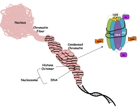
1.4 Chromatin Organization and Accessibility
Eukaryotic cellular DNA is tightly packed in a highly organized form; differential
packaging of DNA with histone and non-histone proteins into chromatin determines DNA
accessibility during transcription (Andersson, Bjorkroth et al. 1984, Yaniv 2014). The
fundamental particle of chromatin is the nucleosome. Histones are integral components of
nucleosomes structures as they provide a scaffold for double stranded DNA to wrap around.
H2A, H2B, H3 and H4 are four histone proteins, two copies of which form the octameric
2014). Histone tails are subject to several modifications that influence the ability of nucleosomes
to form stable higher chromatin structures (Kouzarides 2007).
Compacted chromatin structures are generally repressive to DNA interactions which
prevent the movement of RNA polymerases as well as the binding of transcription factors
(Venters and Pugh 2009). DNA is negatively charged while histones are positively charged, and
thus double stranded DNA is wrapped tightly around histones due to charge differences
(Morales, Giamarchi et al. 2001). Histone tails are subject to several modifications that alter
N-terminal tails of histones and create the epigenetic code, which influences the accessibility of
chromatin (Morales and Richard-Foy 2000). Epigenetic mechanisms cause heritable changes in
gene expression without altering the primary DNA sequence (Ptashne 2007). Along with histone
modifications, DNA methylation also regulates gene expression. DNA methylation occurs by
covalent modification of cytosines to CpG dinucleotides (Strathdee and Brown 2002). DNA
methylation and histone acetylation have been heavily studied due to their significant impact on
gene expression.
1.5 Histone Acetylation
Histone acetylation generally facilitates gene expression whereas histone deacetylases
(HDACs) return DNA to a less accessible conformation by removing acetyl groups from
histones (Figure 1.3.) (Kuo and Allis 1998). Histone acetylation is carried out by a group of
enzymes called histone acetyltransferases (HATs), which catalyze the transfer of an acetyl group
from acetyl-CoA to the amino groups of lysine residues on the N-terminal tails of histones
the stability of chromatin structures (Zhang and Reinberg 2001).
HDACs are a class of enzymes that remove acetyl groups from lysine residues on
histones, allowing histones to wrap DNA more tightly (Secrist, Zhou et al. 2003). HDACs are
classified into four groups. Class I HDACs (HDAC1, HDAC2, HDAC3 and HDAC8) are found
primarily in the nucleus, while HDAC3 is able to shuttle between the nucleus and the cytoplasm
(Nakagawa, Oda et al. 2007). Therefore, members of class I HDACs are mainly responsible for
regulating gene expression. Pharmacological HDAC inhibitors are widely used to enhance
histone acetylation and increase gene expression. For example, Trichostatin A (TSA) selectively
inhibits class I and class II HDACs. HDAC inhibitors bind the active site of HDACs to inhibit
their function, and have been used for cancer therapy in the last decade (Delcuve, Khan et al.
2013). For instance, the FDA has approved two HDAC inhibitors, Vorinostat and Romidepsin,
for the treatment of cutaneous T cell lymphoma and peripheral T-cell lymphoma (Rangwala,
Zhang et al. 2012).
Besides the roles of histone acetylation and deacetylation in terms of promoter
accessibility, it is also suggested that acetylated or deacetylated histone tails could serve as
signals for the binding of other proteins (Braunstein, Sobel et al. 1996). In this case, histone
acetylation or deacetylation is targeted by proteins that could lead to alteration in chromatin
structures.
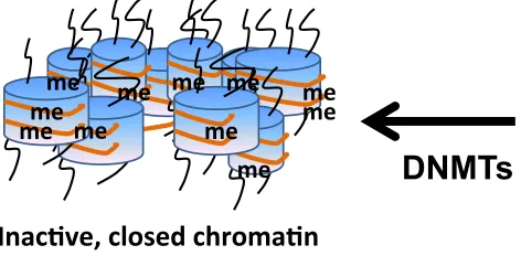
1.6 DNA Methylation
DNA methylation is the most studied epigenetic mechanism associated with gene
(Strathdee and Brown 2002) and is carried out by DNA methyltransferases (DNMTs) by
catalyzing transfer of the methyl groups to cytosine residues in DNA form methyl cytosine by
using S-adenosylmethionine (SAMe) as a methyl donor (Rhee, Jair et al. 2000). In mammalian
cells, three active DNMTs have been identified, DNMT1, DNMT3a, and DNMT3b. DNMT3a
and DNMT3b facilitate formation of de novo DNA methylation patterns while DNMT1 is
required for maintenance of the established patterns of DNA methylation (Jin, Li et al. 2011).
Methylated promoters preferentially bind to methyl CpG binding proteins, which inhibits
their recognition by transcription factors and RNA polymerase. Accumulating evidence has
shown DNA methylation to play significant roles in cancer by activating oncogenes and
silencing tumor suppressors (Wajed, Laird et al. 2001). DNA hypermethylation of CpG
dinucleotides frequently contributes to loss of tumor suppressor genes by accumulating in their
promoter regions (Figure 1.4.) (Esteller 2002), and aberrant DNA methylation is now associated
with drug resistance in cancer (Maeda, Ando et al. 2014). DNA methylation is reversible, and
thus, DNA demethylating agents are in clinical trials for several cancer types, with the
supposition that DNA demethylation may reactivate tumor suppressor genes (Seidel, Florean et
al. 2012, Glasspool, Brown et al. 2014, Navada, Steinmann et al. 2014). Two cytosine analogs,
5-azacytidine and 5-aza-2'-deoxycytidine, have been approved by the FDA for the treatment of
myelodysplastic syndrome, a heterogeneous bone marrow disorder, and acute myeloid leukemia
(Muller, Ruter et al. 2006).
1.7 The Immune System
The immune system is a complex network of molecules, cells, tissues and organs that
organized into multiple levels of defense. The skin and mucosal surfaces represent the first
defense level. If this first level of defense is broken, our body use two equally important types of
defense against infections; innate and adaptive immunity.
Innate immunity is fast and broadly effective. Any pathogen that able to penetrate an
epithelial surface is immediately encountered by the effector cells and molecules of the innate
immune response. Following the infections, resident macrophages phagocytize the pathogen and
release inflammatory cytokines that recruit other component of innate immunity such as
neutrophils and natural killer (NK) cells to the infected area to terminate the infection
(Diefenbach and Raulet 2002, Kobayashi, Voyich et al. 2005, Kumar, Kawai et al. 2009). While
a majority of infections are cleared by innate immunity, some pathogens escape from innate
immune responses. In this case, the pathogen faces as combination of innate and adaptive
immunity.
Adaptive immunity is the third line immune defense of the body and adds specificity to
the immune response. Adaptive immunity has two essential components, B and T lymphocytes.
B and T cells use highly diverse surface receptors to recognize a vast variety of antigens. B cells
can intact pathogens while T cells recognizes only antigens that are presented by major
histocompatibility complex (MHC) class I or II molecules (Klug, Miller et al. 2009). CD4+ T
cells recognize peptides that are presented by MHC class II and help in the activation of antigen
presenting cells (macrophages, dendritic and B cells) (Steinitz, van Helden et al. 2012). CD8+
cytotoxic T cells recognize peptides that are presented by MHC class I and kill infected cells and
tumor cells (Foss 2002). Two major mechanisms of CD8+ cytotoxic T cells killing will be
1.8 Tumor Immunosurveillance and Immunoediting
Tumor immunosurveillance is the process of identification and elimination of tumor cells
by the immune system. Immune responses to cancer cells are similar have to those of virally
infected cells (Figure 1.5). Cancer immunosurveillance is an important host protection process
that inhibits carcinogenesis and maintains cellular homeostasis (Smyth, Dunn et al. 2006). The
immune system is often able to recognize and eliminate cancer cells (Corthay 2014). However,
despite tumor immune surveillance, tumors often develop and escape immune recognition. This
concept is called immunoediting, which has three main phases; elimination, equilibrium and
escape (Dunn, Old et al. 2004). In the elimination phase, the immune system detects and
eliminates tumor cells. If the immune system fails to completely eliminate tumor cells, tumor
cells either remain dormant or continue to evolve. This phase is known the equilibrium phase
where tumor cells undergo additional changes such as modulating the tumor-specific antigens
and cell surface receptors. Eventually, the immune system may fail to control tumor growth and
development, leading to the escape phase. In this phase, tumor cells continue to grow and expand
in an uncontrolled manner and may eventually lead to metastatic malignancies (Dunn, Bruce et
al. 2002).
1.9 Roles of CTLs and NK Cells in Antitumor Immunity
The immune system plays an important role in controlling malignancy and tumor
elimination (Aptsiauri, Cabrera et al. 2007, Roberti, Mordoh et al. 2012). Tumor specific
cytotoxic T lymphocytes (CTLs) and activated NK cells directly contact and kill target tumor
Perforins and granzymes protein families can activate cell-death pathways through activation or
absence of caspases (Ewen, Kane et al. 2012). In the second pathway, the engagement of
target-cell death receptors by their cognate ligands on CTL or NK target-cells results in caspase dependent
apoptosis (Wajant 2014). The death receptor mediated apoptotic pathway is active against a
variety of tumor cells. However, the tumor micro-environment subverts the function of CTLs
and NK cells by impairing ligation through down-regulating expression of surface receptors on
tumor cells (Purdy and Campbell 2009, Qiao and Wong 2009). As self-tissue, tumors are often
weakly immunogenic and evoke immune tolerance instead of immunity (Lavoue, Thedrez et al.
2013). Immune tolerance is controlled by various mechanisms of immune suppression and, as a
result, anti-tumor CTLs can be rendered ineffective. Common causes for ineffective anti-tumor
CTL responses include improper co-stimulation within the tumor site, and down-regulation of
cell surface molecules on tumor cells such as death receptors (McDonnell, Robinson et al. 2010,
Leone, Shin et al. 2013, Cullen and Martin 2015).
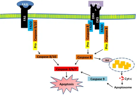
1.10 Apoptosis and Death Receptors
Cell death can be characterized based on its morphological appearance, enzymological
criteria, or immunological characteristics (Kroemer, Galluzzi et al. 2009). Apoptosis is
characterized in two distinct pathways; the death-receptor-mediated extrinsic path and the
Bcl-2-regulated intrinsic path (Hassan, Watari et al. 2014). The extrinsic pathway is initiated by
ligation of an appropriate ligand to a subset of TNF receptor superfamily (TNFRSF) members.
Following ligation, a series of biochemical and morphological changes occur, which are the
Berger et al. 2013). Death receptors are members of the TNFRSF, which include DR4
(TNFRSF10A/TRAIL-R1), DR5 (TNFRSF10B/TRAIL-R2), Fas (CD95/Apo-1), and TNFR1
(Guicciardi and Gores 2009). Ligation of death receptors with cognate death ligands from
anti-tumor immune cells induces apoptotic signals in anti-tumor cells (Grimm, Kim et al. 2010). Death
receptors contain a death domain which recruits the intracellular adaptor molecule Fas-associated
protein with death domain (FADD) and procaspase-8 into the death-inducing signaling complex
(DISC) to initiate apoptosis (Figure 1.6). This pathway is negatively regulated by cellular
FLICE-like inhibitory protein (cFLIP). Activated caspase-8 can directly activate the extrinsic cell
death pathway through activation of caspases-3, -6 and -7 and/or cleave BH3-interacting domain
death agonist (Bid) to trigger mitochondrial intrinsic pathway. Cleavage of Bid by caspase-8
results in mitochondria outer membrane permeabilization, release of cytochrome c, formation of
the apoptosome, and in caspase-9 activation. Both caspase-8 and 9 cleavage cause downstream
activation of the executioner caspases such as caspase-3, -6 and -7 (Scaffidi, Fulda et al. 1998,
MacFarlane 2003).
In our study, the focus is on Fas, DR4, and DR5, which are among the most common
death receptors that CTLs use to kill tumor cells. Fas is expressed in a variety of cell types
including antigen presenting and tumor cells, and is the complementary receptor for Fas-ligand
(FasL), which is expressed on tumor cells, CTLs and NK cells (Ramaswamy, Cleland et al.
2009). The interaction of Fas and FasL plays an essential role in triggering apoptosis (Wajant
2014). During cancer progression, the interaction between Fas and FasL is largely impaired due
to suppression of Fas expression on tumor cells (Zhu, Liu et al. 2005, Pryczynicz,
Guzinska-Ustymowicz et al. 2010). DR4 and DR5 are also essential for driving apoptosis in many types of
apoptosis-Ponti et al. 2004). TRAIL is part of a natural mechanism to kill tumor cells by the immune
system and selectively induces apoptosis in cancer cells with less toxicity towards
healthy/non-cancerous cells (Allen and El-Deiry 2012); however, suppression of DR4/5 by tumor cells leads
to the development of resistance to immune mediated cell killing (Perraud, Akil et al. 2011).
CD8+ T cells and activated NK cells express both TRAIL and its receptor DR5, but both cell
types are resistant to TRAIL-mediated cytotoxicity because these cells express high levels of
c-FLIP and thus physiologically protect NK and CD8+ T cells from apoptotic action (Mirandola,
Ponti et al. 2004).
1.11 Radiation-Induced Gene Expression
Radiation therapy (RT) is commonly used for local tumor control due to its ability to
directly kill tumor cells.RT also induces expression of functionally important molecules in
tumor cells, and thus enhances the ability of immune cells to recognize and eliminate tumor cells
(Ifeadi and Garnett-Benson 2012, Kumari, Cacan et al. 2013). It has also been reported that
sub-lethal doses of irradiation results in upregulation of expression of surface molecules on tumor
cells, which further influences the processes of co-stimulation, adhesion and transvascular
migration of immune cells (Iarilin 1999, Kumari, Cacan et al. 2013, Bernstein, Garnett et al.
2014). Modulation of tumor phenotype by RT enhances anti-tumor T-cell activity in both in vitro
and in vivo models (Bernstein, Garnett et al. 2014, Gameiro, Jammeh et al. 2014). These
findings indicate that RT not only causes DNA damage, but also upregulates expression of
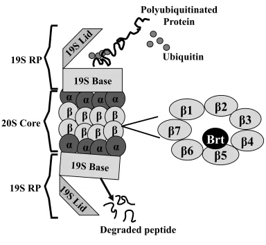
1.12 The 26S Proteasome
The 26S proteasome is a 2.5 MDa multi-protein complex formed by the 19S regulatory
and 20S core subcomponents (Figure 1.7), and it is found in the nucleus and cytoplasm of
eukaryotic cells (Bedford, Paine et al. 2010). The 26S proteasome is the main non-lysosomal
protein degradation machinery and inhibition of the 26S proteasome alters protein turnover and
impacts cellular homeostasis (Chen and Dou 2010). Inhibition of the 26S proteasome also alters
expression of numerous target genes at the transcriptional level by increasing the stability of
transcription factors and/or by regulating epigenetic modifiers (Kinyamu, Jefferson et al. 2008,
Bhat and Greer 2011). It has also been reported that critical components of the 19S particle
interact with chromatin remodeling enzymes and facilitate their recruitment to promoter regions
of some target genes such as MHC-II and class II transactivator (CIITA) promoters, which leads
to activation of histone modifications and an opening of chromatin structure. (Koues, Dudley et
al. 2008, Koues, Dudley et al. 2009). Roles of the 26S proteasome in regulation has been
observed for several transcription factors (Fuchs 2013). For example, inhibition of the 26S
proteasome enhances recruitment of p53 to p21waf1 responsive promoters and some components
of 19S subunit also physically interact with p53 to facilitate its recruitment to p21waf1
responsive promoters (Zhu, Wani et al. 2007). These findings further suggest that the 26S
proteasome can regulate both transcription factors and histone modifying enzymes through its
proteolytic and non-proteolytic functions.
Despite advances in understanding the involvement of 26S proteasome on transcription
regulation of target genes, it is not well understood how proteasome inhibition alters gene
expression. It remains interesting that inhibition of the 26S proteasome promotes its
by stabilizing expression of transcription factors and histone modifying enzymes, and second, by
promoting disassociation of the 19S subunit which then directly interacts with transcription
factors and histone modifying enzymes and recruits them to promoter regions of target genes.
Most of the commercially available proteasome inhibitors directly bind to the 20S catalytic unit
where protein degradation takes place (Shen, Schmitt et al. 2013). Bortezomib inhibits the
chymotrypsin-like activity of the 26S proteasome (Niewerth, Dingjan et al. 2013), is the first
FDA approved 26S proteasome inhibitor, and is being used for the treatment of multiple
myeloma and mantle cell lymphoma (Bross, Kane et al. 2004, Chen, Frezza et al. 2011).
1.13 Summary
The dysregulation of gene expression is a hallmark of cancer (Shay and Roninson 2004,
Sharma, Kelly et al. 2010, Haria and Naora 2013). Dysregulated gene expression has profound
effects on cellular function and is a cause of a majority of human diseases. It is well known that
tumor cells aberrantly activate oncogenic pathways and inhibit or down regulate tumor
suppressor pathways. Cancer arises not only from defects in genetic events but also from
dysregulated epigenetics modifications. Disruption in the balance of DNA methylation or histone
acetylation at specific promoters could lead to aberrant expression of oncogenes or suppression
of tumor suppressor genes. The contribution of histone acetylation and DNA methylation in
dysregulation of gene expression during carcinogenesis is well known, however; involvement of
these epigenetic mechanisms at specific promoters is still quite limited. RGS proteins have
ability to regulate GPCR signaling, thus alteration in the expression of RGS proteins may
regulation RGS proteins in cancer progression. It has been shown that some of the members of
RGS family are dynamically regulated in ovarian cancer cells (Hurst and Hooks 2009, Hooks,
Callihan et al. 2010, Ali, Cacan et al. 2013). A recent study reported that RGS2 expression is
silenced by DNA methylation, which promotes prostate cancer cell growth (Wolff, Xie et al.
2012). These studies suggest that suppression of some RGS proteins could be due to epigenetic
mechanisms. In chapter 2, we show that RGS10 expression is suppressed during ovarian cancer
progression and the suppression of RGS10 is linked to aberrant accumulation of DNMTs and
HDACs. We find that HDAC1 and DNMT1 synergistically suppress RGS10 expression in
chemoresistant ovarian cancer cells. Understanding the regulatory mechanisms that dictate
RGS10 gene expression during ovarian cancer progression is an important step towards
developing better and more specific adjuvants for ovarian cancer.
Of cancers, the most lethal are those that have gained metastatic ability. Metastasized
cancers often down regulate expression of various genes to avoid recognition and response by
the immune system. It is appreciated that epigenetic alterations play critical roles in aberrant
expression of genes which contribute to tumor cell metastasis. Recent evidence indicates one
mechanism utilized by tumor cells to escape recognition and elimination by the immune system
is suppression of cell surface expression of immunogenic genes.
Radiotherapy has been extensively used for cancer therapies including colorectal cancer.
Sub-lethal doses of radiation can modulate gene expression, making tumor cells more susceptible
to T-cell-mediated immune attack (Ifeadi and Garnett-Benson 2012, Kumari, Cacan et al. 2013).
Previous studies and clinical trials have shown that combining radiation with other treatments is
more effective then radiation treatment alone (Finkelstein and Fishman 2012, Finkelstein,
effects of the 26S proteasome inhibitor, bortezomib, alone or in combination with radiotherapy,
on the expression of immunogenic genes in normal colon and in colorectal cancer cell lines. We
examine normal colon and colorectal cancer cell lines for changes in expression of multiple
death receptors (DR4, DR5 and Fas). Our results indicate a combination of 26S proteasome
inhibition and sub-lethal radiation increases the sensitivity of carcinoma cells to apoptosis.
Combination treatment up-regulates cell surface expression of death receptors by increasing their
transcriptional activation. Thus, the combination treatment enhanced sensitivity to killing
through FAS and TRAIL receptors by CD8+ T cells.
In order to better understand radiation induced gene expression, we have investigated
novel regulatory mechanisms of death receptors by sub-lethal radiation in chapter 4. As we
describe above, expression of mammalian genes is regulated at multiple levels, including
transcriptional control. Chapters 4 of this dissertation focuses on the novel roles played by
radiation in regulating transcript expression of death receptors. In this chapter we present novel
ways of regulating transcription of death receptors, where histone acetylation and DNA
Figure 1.1 GPCR-mediated cell survival and acquired chemoresistance.
Agonist binding to GPCR activates G-proteins, which mediate downstream signaling pathways. Akt activation is a potential mechanism for enhanced survival and acquired chemoresistance. RGS proteins inhibit receptor-stimulated Akt activity, and thus suppressed RGS expression in chemoresistant cells indirectly amplifies receptor-stimulated Akt survival signaling to allow cells to escape cisplatin-stimulated cell death.
PI3K
!!
AKT'
Survival'
Chemotherapeu6cs'' (Cispla6n)'
Cycle'arrest'
Caspases'
Apoptosis'
RGS'
γ' β
' γ '
!
'
β '
!
' Gα' GTP'
!
GDP' GTP'
Gα'
GDP' Pi'
???
Agonist'Figure 1.2 Chromatin organization and accessibility.
DNA is packed with histone and non-histone proteins and differential packaging of DNA with these proteins into chromatin determines DNA accessibility during transcription. H2A, H2B, H3 and H4 are four histone proteins, two copies of which form the octameric nucleosome structure along with the linker histone, H1. Histone tails are subject to several modifications that influence the ability of nucleosomes to form stable higher chromatin structures.
Nucleus
Chromatin Fiber
Condensed Chromatin
Histone Octomer
DNA Nucleosome
H3 H2A H2B
H4 H3
met$ met$
Ac$
Figure 1.3 Histone Acetylation
Histone acetylation facilitates gene expression whereas histone deacetylases (HDACs) remove acetyl groups from histones. Acetylation of positively charged lysine residues in histone tails by histone acetyltransferase (HAT) activity influence the ability of histones to neutralize the charge on DNA and thus, reduce the stability of chromatin structure.
Ac#ve,'open'chroma#n' Gene'Expression'
Inac#ve,'closed'chroma#n' Silenced'Genes'
Ac
Ac Ac
Ac Ac Ac
Ac Ac Ac
Ac
Ac Ac
Ac
Ac
Ac
Ac Ac
Ac Ac
Ac Ac
Ac
Ac
Ac
Ac
HATs
Figure 1.4 DNA Methylation
DNA methylation suppresses gene expression and is carried out by DNA methyltransferases (DNMTs) by catalyzing transfer of the methyl groups to cytosine residues in DNA. In
mammalian cells, three active DNMTs have been identified. DNMT3a and DNMT3b facilitate
formation of de novo DNA methylation patterns while DNMT1 is mainly required for
maintenance of the established patterns of DNA methylation. me# me#
me# me# me# me#
me# me#me#
me# me#
Inac(ve,#closed#chroma(n#
Silenced#Genes#
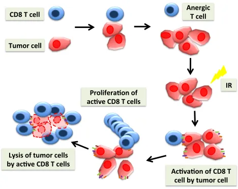
Figure 1.5 Cancer immunotherapy
CD8+ T cells can recognize and eliminate tumor cells. However, dysregulation of surface
receptors on tumor cells results in CD8+ T cell to become anergic and tumor cells continue to
grow. To elicit an effective immune response against tumors, T cells need to recognize tumor antigens presented by MHC molecules in conjunction with appropriate co-stimulation.
Upregulation of surface proteins on tumor cells leads to activation and proliferation of CD8+ T
cells, and activated tumor specific CD8+ T cells can directly contact kill the tumor cells.
CD8$T$cell$
Tumor$cell$
Prolifera1on$of$
ac1ve$CD8$T$cells$
Ac1va1on$of$CD8$T$
cell$by$tumor$cell$$
Lysis$of$tumor$cells$
by$ac1ve$CD8$T$cells$
Anergic$
T$cell$
Figure 1.6 Activation of death receptor mediated apoptosis.
DR4, DR5 and Fas are death receptors and ligation of death receptors with cognate death ligands from anti-tumor immune cells induces apoptotic signals in tumor cells. Activated
caspase-8 can directly activate the extrinsic cell death pathway through activation of caspases-3, -6 and -7 and/or cleave Bid to trigger mitochondrial intrinsic pathway.
Figure 1.7 Structure of the 26S proteasome
The 26S proteasome comprised of core 20S catalytic complex and 19S regulatory complex. The 20S proteasome core has caspase-like, trypsin-like and chymotrypsin-like
activities that are associated with three distinct units: β1, β2, β5, respectively. Proteasome
inhibitor bortezomib (Brt) specifically inhibit the chymotrypsin-like activity at the β5 subunit.
20S Core
19S RP
S7
β4
β5
β6
β2
β1
β3
β
β
19S Base
19S Base
β
β
β
β
β
β
α
α
α
α
α
α
α
α
19S
Li
d
19S
Li
d
19S RP
Degraded peptide
Polyubiquitinated
Protein
Ubiquitin
β7
2 EPIGENETIC REGULATION OF REGULATOR-G PROTEIN SIGNALING 10
(RGS10) PROTEIN AND OVARIAN CANCER CHEMORESISTANCE
Ovarian cancer is one of the deadliest gynecological cancers, with a 60% mortality rate in
patients and a 5-year survival rate of lower than 30% in advanced stage disease (Siegel,
Naishadham et al. 2013). The high mortality rate is due in large part to the development of
resistance to chemotherapeutic drugs (Hooks, Callihan et al. 2010, Liu, Nash et al. 2010). Thus,
understanding the molecular and genetic mechanisms that drive the development of acquired
chemoresistance will enable us to improve current therapeutic agents for ovarian cancer
treatment. G-protein coupled receptors (GPCRs) initiate multiple oncogenic signaling pathways
in cancer cells by activating their associated G-proteins (Cai and Xu 2013, O'Hayre,
Vazquez-Prado et al. 2013). Activation of GPCRs by growth factors such as Lysophosphatidic acid (LPA)
triggers survival signaling pathways that drive resistance to chemotherapeutic drugs such as
cisplatin and taxane (Hurst and Hooks 2009). GPCR activation of G-proteins is opposed by the
activity of regulator of G-protein signaling (RGS) proteins. RGS proteins inhibit G-protein
signaling pathways by directly binding to the activated Gα subunit of G-proteins to accelerate
hydrolysis of GTP into GDP, which returns G-proteins to an inactive state (Berman and Gilman
1998, Zhong and Neubig 2001, Shi, Harrison et al. 2004, Hurst and Hooks 2009). Relevant to
our studies, recent reports indicate that RGS proteins inhibit breast, lung, prostate, and ovarian
cancer cell growth through inhibition of GPCRs signaling pathways (Cao, Qin et al. 2006, Liang,
Bansal et al. 2009, Xie, Wolff et al. 2009, Hooks, Callihan et al. 2010, Ali, Cacan et al. 2013,
RGS10 is among the smallest of the RGS proteins and is highly expressed in a broad
range of cell types (Lu, Gosslau et al. 2008, Garcia-Bernal, Dios-Esponera et al. 2011, Lee,
Chung et al. 2012, Rivero, Gabilondo et al. 2013). RGS10 is an important regulator of cell
survival and chemoresistance (Hooks, Callihan et al. 2010), and RGS10 transcript expression is
significantly suppressed in multiple ovarian cancer cell lines (Ali, Cacan et al. 2013). Thus, the
suppression of RGS10 proteins may contribute to chemoresistance by amplifying
GPCR-mediated cell growth and survival signaling pathways. We have recently shown that suppression
of RGS10 is due in part to DNA hypermethylation and to histone deacetylation, two important
gene-silencing mechanisms which contribute to the progression of many cancers. DNA
methylation is maintained by DNA methyl transferases (DNMTs) (Rhee, Jair et al. 2000) and
histone deacetylation is maintained by histone deacetylases (HDACs) (Ito, P et al. 2000). Often,
these two enzymes coordinately suppress transcriptional activity of genes (Ghoshal, Datta et al.
2002, Cai, Geutjes et al. 2013). Fuks et al. have reported that DNMT1 is associated with histone
deacetylase activity and has the ability to bind HDAC1 (Fuks, Burgers et al. 2000). However, the
molecular mechanisms by which DNA hypermethylation and histone deacetylation suppress
RGS10 and the contribution of these enzymes to acquired chemoresistance remains unknown.
We investigate here the molecular mechanisms of epigenetic regulation of RGS10
expression in ovarian cancer cells and focus on chemosensitive parental A2780 cells and their
derivative cell line, chemoresistant A2780-AD. We identify two important epigenetic regulators,
HDAC1 and DNMT1, which are highly associated with the RGS10 promoter in chemoresistant
ovarian cancer cells. HDAC1 and DNMT1 knock down significantly increases RGS10
expression and cisplatin-stimulated cell death. Our results suggest that HDAC1 and DNMT1
overcoming ovarian cancer chemoresistance.
2.1 RESULTS
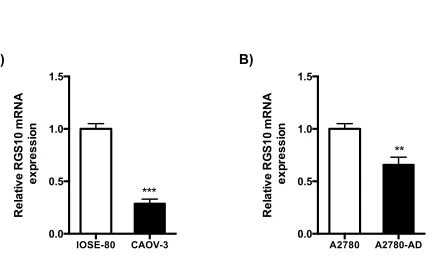
2.1.1 RGS10 Expression is Suppressed in Ovarian Cancer Cells
Previous studies have shown that downregulation of RGS10 expression in ovarian cancer
cell lines with acquired chemoresistance (Hooks, Callihan et al. 2010). To determine if RGS10
transcript expression is also downregulated in ovarian cancer cell lines we compared RGS10
expression in benign IOSE-80 ovarian cells and the ovarian cancer cell line CAOV-3 (Fig 2.1A).
RGS10 transcript expression was significantly lower in CAOV-3 cells compared to IOSE control
cells. Next, we compared RGS10 transcript expression chemosensitive parental A2780 cells to
their derivative chemoresistant A2780-AD cells. Again, we have seen a significant decrease in
RGS10 mRNA expression in chemoresistant cells as compare to parental chemosensitive cells
(Fig 2.1B). Taken together, RGS10 transcript expression is reduced in primary ovarian cancer
cells and the CAOV-3 cancer cell line relative to immortalized ovarian epithelial cells, and in
A2780-AD cells relative to parental cells. These data suggest that RGS10 transcript expression is
decreased as ovarian cancer progress.
2.1.2 Histone Modifications at RGS10 Promoters in Ovarian Cancer Cells
Next, we sought to explore potential mechanisms of regulation of RGS10 expression, we
acetylation at histones associated with the RGS10 promoter in A2780 and A2780-AD cells,
using the GAPDH promoter as a control. Total H3 histone binding was similar at RGS10 and
GAPDH promoters (data not shown). In contrast, acetylated H3 histone levels were significantly
lower at RGS10 promoters in the chemoresistant A2780-AD cells, while similar levels of
acetylated histone H3 were associated with the GAPDH promoter in both cell types (Fig
2.2A-B). Reduced acetylation at Lysine residue 18 in histone 3 (H3K18) is associated with cancer
recurrence and poorer clinical outcome in lung, kidney, and breast cancer patients (Seligson,
Horvath et al. 2005, Seligson, Horvath et al. 2009). To determine if loss of acetylation of this
residue contributed to the loss of histone acetylation in RGS10 promoters in chemoresistant cells,
we performed ChIP assays with H3K18-specific antibodies. We observed a slight but significant
decrease in H3K18 association with the RGS10 promoter in chemoresistant cells as compared to
A2780 parental cells (Fig 2.2C), with no change at the GAPDH control promoter (Fig 2.2D).
Histone acetylation is dynamically regulated in cells by the opposing actions of HATs that add
the acetyl functional group to histones, and HDACs that remove them. Class I HDACs are over
expressed in ovarian cancer tissues and are thought to play a significant role in gene silencing
during ovarian cancer progression (Jin, Pak et al. 2008). We observed a striking increase in
HDAC1 association with RGS10 promoters in A2780-AD cells as compared to parental A2780
cells (Fig 2.3A). This increase reflects a specific recruitment to the RGS10 promoter, as HDAC1
association with GAPDH promoters was unchanged between cell lines (Fig 2.3B), and total
HDAC1 expression levels were not higher in A2780-AD cells (Fig 2.3C).
To confirm these findings in additional cell lines, ChIP assays were carried out in the
chemosensitive ovarian cancer cell line OV2008 and in chemoresistant C13 daughter cells.
significantly lower at RGS10 promoters in the chemoresistant C13 ovarian cancer cells as
compared to chemosensitive OV2008 cells (Fig 2.4B).
We next performed ChIP assays to determine if loss of H3K18 contributes to the loss of
global histone acetylation at RGS10 promoters in chemoresistant C13 cells. A significant
decrease in H3K18 acetylation at RGS10 promoters was observed in chemoresistant C13 cells
(Fig 2.4C), while H3K18 acetylation at the GAPDH promoters in OV2008 and C13 cells
remained unchanged (Fig 2.4F). Together these data suggest the loss of acetylation at RGS10
promoters contributes to the loss of RGS10 expression in two independent cell models of
chemoresistant ovarian cancer.
To determine if histone modifications at RGS10 promoters may account for the
difference in expression in IOSE-80 and CAOV-3 cells, we performed ChIP assays to compare
histone acetylation. Again, the level of acetylated histone H3 levels at the GAPDH promoter
were unchanged between the cell lines (Fig 2.5B), while the level of acetylated histone H3
associated with the RGS10 promoter in CAOV-3 cancer cells was half that observed in IOSE-80
normal ovarian epithelial cells (Fig 2.5A). We also compared the association of HDAC1 with
RGS10 promoters in IOSE-80 and CAOV-3 cells. The level of HDAC1 associated with the
control promoter GAPDH was unchanged between cell lines (Fig 2.5D), but was more than
doubled at RGS10 promoters in the cancer cell line, compared to IOSE-80 cells (Fig 2.5C).
These data show that decreased RGS10 expression in CAOV-3 ovarian cancer cells correlates
with enhanced HADC1 binding and loss of histone acetylation at the RGS10 promoter as
2.1.3 HDAC1 and DNMT1 Suppress RGS10 Expression in Chemoresistant Ovarian Cancer
Cells
We previously demonstrated that HDAC1 proteins bind with significantly increased
frequency to the RGS10 promoter in chemoresistant A2780-AD cells compared to parental
chemosensitive A2780 cells (Ali, Cacan et al. 2013). To investigate molecular roles for HDAC1
in regulating RGS10 expression, a siRNA duplex was utilized to specifically knock down
endogenous HDAC1 expression in A2780-AD cells. siRNA-mediated knockdown of HDAC1
resulted in a more than 3-fold increase in endogenous RGS10 expression as compared to control
siRNA (Fig. 2.6A), suggesting that HDAC1 plays a critical role in regulating RGS10
transcription. In a similar experiment, A2780-AD cells were transfected with HDAC1 to
determine the effects of ectopic expression of HDAC1. Overexpression of HDAC1 dramatically
reduced RGS10 expression in chemoresistant A2780-AD cells (Fig 2.6B). HDAC1 knockdown
increased RGS10 protein expression as well (Fig 2.6C) while HDAC1 overexpression decreased
cellular RGS10 protein level in chemoresistant ovarian cancer cells (Fig 2.6D). Together, these
data indicate that HDAC1 accumulation at RGS10 promoters contributes to suppression of
RGS10 in chemoresistant ovarian cancer cells.
The RGS10 promoter contains a high concentration of CpG dinucleotides making it a
potential target for DNMT maintenance methylation during ovarian cancer progression. We first
showed by western blot analysis that DNMT1 expression levels are similar in both A2780 and
A2780-AD cell lines (Fig 2.7C). To further explore the relevance of DNMT1 in the specific
suppression of RGS10 expression, ChIP assays were carried out in A2780 parental and their
derivative resistant A2780-AD cells. Lysates were immunoprecipitated with control or
total protein abundance, ChIP assays reveal that binding of DNMT1 is significantly increased at
the RGS10 promoter in chemoresistant cells as compared to chemosensitive ovarian cancer cells
(Fig 2.7A). Together these data indicate that accumulation of DNMT1 at the RGS10 promoter
likely contributes to suppression of RGS10 during ovarian cancer chemoresistance.
Accumulation of DNMT1 at the RGS10 promoter led us to determine if that
accumulation affects RGS10 transcript expression level in chemoresistant cells. For this purpose,
A2780-AD cells were transfected with DNMT1 siRNA or control siRNA. RNA was extracted
and generated cDNA was quantified using qRT-PCR with primers and probes specific for the
RGS10 coding region and normalized to housekeeping gene GAPDH expression. The data
reveals that knocking down DNMT1 significantly increases endogenous RGS10 transcript (Fig
2.7B) and protein expression (Fig 2.7D) in A2780-AD cells. This increase suggests that DNMT1
is another important player and functions with HDAC1 to regulate the suppression of RGS10
transcription in chemoresistant A2780-AD cells.
2.1.4 Inhibition of HDAC and DNMT Activity Enhances RGS10 Expression and Decreases
Ovarian Cancer Cell Viability
We next sought to determine if pharmacologic inhibitors of histone deacetylation and
DNA methylation can alter the expression of RGS10 in chemoresistant ovarian cancer cells. The
HDAC inhibitor TSA and DNMT inhibitor 5-Aza-dC were used to inhibit HDACs and DNMTs,
respectively. A2780-AD cells were treated with 500 nM TSA and were incubated for 2 days or
were treated with 20µM 5-Aza-dC and incubated for 3, 5 and 7 days. Total RNA was isolated