Biology Dissertations Department of Biology
4-9-2010
Synthesis, Structure, Function and Biomedical Studies of Nucleic
Synthesis, Structure, Function and Biomedical Studies of Nucleic
Acid Derivatized with Selenium
Acid Derivatized with Selenium
Lina Lin
Georgia State University
Follow this and additional works at: https://scholarworks.gsu.edu/biology_diss
Part of the Biology Commons
Recommended Citation Recommended Citation
Lin, Lina, "Synthesis, Structure, Function and Biomedical Studies of Nucleic Acid Derivatized with Selenium." Dissertation, Georgia State University, 2010.
https://scholarworks.gsu.edu/biology_diss/77
NUCLEIC ACID DERIVATIZED WITH SELENIUM
by
LINA LIN
Under the Direction of Professor Zhen Huang
ABSTRACT
derivatization of nucleic acids has been proven to be a useful strategy for solving the phase problem in nucleic acid X-ray crystallography. Besides the facilitation of nucleic acid crystallography, there is also a wide range of other applications for selenium-derivatized nucleic acids (SeNA).
The investigation presented in this dissertation mainly focuses on the following research subjects
(1) Synthesis and characterization of selenium-derivatized nucleic acids for X-ray crystallography, especially phosphoroselenoate RNAs. They are generated and used for crystallization.
(2) Application of selenium-derivatized RNA for RNA interference. Phosphoroselenoate RNAs are tested for RNAi activities.
(3) Synthesis and characterization of the uridine 5’-triphosphate modified with selenium at position 4.
(4) Facile synthesis and antitumor activities of selenium modified deoxyribonucleosides. MeSe-thymidine nucleosides have shown antitumor activity in cell assays.
. .
NUCLEIC ACID DERIVATIZED WITH SELENIUM
by
LINA LIN
A Dissertation Submitted in Partial Fulfillment of the Requirements for the Degree of
Doctor of Philosophy
in the College of Arts and Sciences Georgia State University
Copyright by Lina Lin
NUCLEIC ACID DERIVATIZED WITH SELENIUM
by
LINA LIN
Committee Chair: Zhen Huang
Committee: Phang C. Tai Irene T. Weber
Electronic Version Approved:
DEDICATION
This thesis is dedicated to my parents Kaiming Lin and Meizhen Xie, who are the initial sources for my creativity and independent thinking. I am deeply indebted to them for their continued love, support and unwavering faith in me.
ACKNOWLEDGEMENTS
TABLE OF CONTENTS
ACKNOWLEDGEMENTS v
LIST OF TABLES x
LIST OF FIGURES xi
LIST OF ABBREVIATIONS xiv
CHAPTER
1 INTRODUCTION 1
1.1 RNA crystallography 1
1.2 Selenium derivatization for nucleic acid crystallography – why we need selenium derivatization for nucleic acids X-ray crystallography and how to introduce selenium into RNA?
3
1.2.1 The phase problem in X-ray crystallography 3 1.2.2 Methods to solve the phase problem 5 1.2.3Selenium derivatization of macromolecules and
MAD phasing
8
1.2.4 The advantages of SeNA for nucleic acids X-ray
crystallography 9
1.2.5 Methods for introducing selenium into RNA 11 1.3 Other applications of Se-derivatized nucleic acids 14 1.3.1 Investigation of nucleotidyl-transfer reactions 14
1.4 Current development of SeNA for X-ray crystallography 24
1.4.1 Selenium modification at sugar 25
1.4.2 Selenium modification at non-bridging phosphate 26
1.4.3 Selenium modification at nucleobase 27
2 PREPARATION OF PHOSPHOROSELENOATE RNA FOR X-RAY
CRYSTALLOGRAPHY 29
2.1 Introduction 29
2.1.1 Brief introduction about the functional RNA molecules in this dissertation
31
2.2 Materials and methods 36
2.2.1 Synthesis, characterization and purification of nucleosides 5’-(alpha-P-seleno) triphosphates
36
2.2.2 DNA template preparation for RNA transcription 40 2.2.3 Phosphoroselenoate modified RNA (PSe-RNA)
transcription with radioactive label
48
2.2.4 PSe-RNA transcription for large scale RNA preparation
49
2.2.5 Purification of PSe-RNA 50
2.2.6 Stability studies of PSe-RNA. 52
2.2.7 Crystallization of PSe-RNA 52
2.3 Results 53
2.3.1 Synthesis, characterization and purification of nucleosides 5’-(alpha-P-seleno) triphosphates
2.3.2 Phosphoroselenoate modified RNA (PSe-RNA) transcription with radioactive label
61
2.3.3 Phosphoroselenoate modified RNA (PSe-RNA) transcription for large scale RNA preparation
64
2.3.4 Purification of PSe-RNA 68
2.3.5 Stability studies of PSe-RNA. 70
2.3.6 Crystallization of PSe-RNA 72
2.4 Discussion 73
3 PHOSPHOROSELENOATE RNA FOR RNA INTERFERENCE
STUDIES
76
3.1 Introduction 76
3.2 Materials and methods 78
3.2.1 siRNA transcription 78
3.2.2 siRNA duplex preparation 79
3.2.3 siRNA duplex Tm study 80
3.2.4 siRNA RNAi activity study 81
3.3 Results 82
3.3.1 Characterization of PSe-siRNA 82
3.3.2 PSe-siRNA interference activity 86
3.4 Discussion 94
4 SYNTHESIS OF 4-Se-URIDINE 5’-TRIPHOSPHATE AND ITS
INCORPORATION INTO RNA
96
4.2 Materials and methods 98
4.2.1 Simple synthetic route for 4-Se-UTP 98
4.2.2 Optimization of 4-Se-UTP in transcription 101 4.2.3 Stability studies of 4-Se-UTP in transcription buffer 102
4.2.4 Kinetic study of 4-Se-UTP enzymatic incorporation
into RNA. 102
4.3 Results 103
4.3.1 Synthesis and characterization of 4-Se-UTP 103 4.3.2 4-Se-UTP stability and transcription studies 105
4.3.3 4-Se-UTP RNA incorporation 113
4.4 Discussion 118
5 FACILE SYNTHESIS AND ANTI-TUMOR CELL ACTIVITY OF Se- CONTAINING NUCLEOSIDES
120
5.1 Introduction 120
5.2 Materials and methods 122
5.2.1 Synthesis of MeSe-nucleosides 122
5.2.2 Anti-Prostate tumor cell evaluation 129
5.3 Results and discussion 130
5.3.1 Synthesis of MeSe-nucleosides 130
5.3.2 Anti-Prostate tumor cell evaluation 132
PUBLICATIONS AND MANUSCRIPTS IN PREPARATION 134
APPENDIX: Natrix (Nucleic acid sparse matrix screen) 135
LIST OF TABLES
Table 1.1: Methods to solve the phase problem for nucleic acid crystal
structures 11
Table 1.2: Methods for introducing selenium into nucleic acid 14
Table 2.1: RNA sequences 36
Table 2.2: Sequences of DNA templates prepared by solid phase DNA synthesizer
43
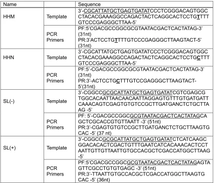
Table 2.3: PCR template sequences and primer sequencesfor plasmid construction
44
Table 2.4: Polymerization primer sequences for P4-P6 47
Table 2.5: PCR template sequences and primer sequences 48
Table 2.6: NTPαSe ESI-MS and yield 54
Table 2.7: 1H-NMR (400 MHz) Chemical Shifts (ppm) of NTPαSe in D2O 55
Table 2.8: 13C-NMR (100 MHz) Chemical Shifts (ppm) of NTPαSe in D2O
55
Table 2.9: 31P-NMR (161.97 MHz) Chemical Shifts (ppm) of NTPαSe in D2O
55
Table 3.1: siRNA sequences 78
Table 3.2: Summary of siRNA MALDI-TOF MS 83
Table 3.3: Summary of siRNA Tm result 83
Table 5.1: Synthesized MeSe-nucleosides (1–3) 130
LIST OF FIGURES
Figure 1.1: Steps for solving a novel macromolecule X-ray crystal
structure. 4
Figure 1.2: T7 RNA polymerase polymerization mechanism 13
Figure 1.3: RNase H cleavage mechanism 16
Figure 1.4: Metal ion mechanism in hammerhead ribozyme catalysis 18
Figure 1.5: Selenomethionine and selenocysteine 21
Figure 1.6: Selenium metabolism pathways 23
Figure 1.7: 2-selenouridine and 5-[(methylamino)methyl]-2- selenouridine (mnm5se2U)
24
Figure 1.8: Atom specific selenium replacement of oxygen in nucleic acids
25
Figure 2.1: (A) NTPαSe structure. (B) Structure of Rp phosphoro- selenoate RNA.
31
Figure 2.2: Secondary structure of wild type hammerhead ribozyme derived from the satellite RNA of tobacco ringspot virus.
32
Figure 2.3: Predicted secondary structure of WNV terminal stem-loop SL (-) and SL (+)
33
Figure 2.4: Secondary structure of P4-P6 domain of group I intron 35
Figure 2.5: Synthesis route of NTPαSe 36
Figure 2.6: Illustration of DNA templates 41
Figure 2.7: HPLC profiles of NTP, NTPαSe diastereomer I and diastereomer II
56
Figure 2.8: ATPαSe Boronate column purification 57
Figure 2.9: GTPαSe Boronate column purification 58
Figure 2.10: CTPαSe Boronate column purification 59
Figure 2.12: Enzymatic incorporation of NTPαSe I and II into HHM RNA using plasmid template
62
Figure 2.13: Enzymatic incorporation of NTPαSe I and II into SL(-) RNA using double strands DNA templates prepared from PCR
62
Figure 2.14: Enzymatic incorporation of NTPαSe I and II into 55.27 RNA using ssDNA templates prepared from solid phase
synthesizer.
62
Figure 2.15: Transcription of a hammerhead ribozyme mutant and wild type using NTP or NTPαSe I
63
Figure 2.16: Transcription of NTPαSe purified by boronate column 63 Figure 2.17: Optimization of large scale UTPαSe RNA transcription
conditions using T7 RNA polymerase in 2-16 hours 65 Figure 2.18: Plasmid template concentration optimization for HHM
transcription condition 65
Figure 2.19: Comparison of normal PCR template and 2’-O-methyl
modified PCR template for P4-P6 RNA transcription by NTP and UTPαSe I
66
Figure 2.20: Concentration test for P4-P6 2’-O-methyl modification PCR DNA template.
67
Figure 2.21: MALDI-TOF of PSe-U hammerhead ribozyme (HHM) 67
Figure 2.22: MALDI-TOF of native hammerhead ribozyme (HHM) 68
Figure 2.23: RNA band of WNV ribosome entry site SL(-) (plasmid template) from NTP and UTPαSe on denature PAGE
69
Figure 2.24: RNA transcribed from single stranded 89.27 ssDNA template and plasmid DNA template HHM (hammerhead ribozyme mutant) and HHN (native hammerhead ribozyme) on denature PAGE
69
Figure 2.25: FPLC anion exchange column purification of PSe-U hammerhead ribozyme
70
Figure 3.1: MALDI-TOF Mass-spectrum of siRNA antisense strand (A) and sense strand (B)
84
Figure 3.2: PSe-siRNA Tm study 86
Figure 3.3: PSe-siRNA RNA knocked down PKM2 87
Figure 3.4: Se-RNA treatment inhibits cell growth 87
Figure 3.5: PSe-siRNA RNA knocked down PKM2 89
Figure 3.6: Se-RNA treatment does not inhibit cell growth 90
Figure 3.7: Western blot analysis of siRNA (A-D) knockdown of PKM2 in SW620 cells
90
Figure 3.8: PKM2 inhibition by siRNA 90
Figure 3.9: The numbers dead cells in siRNA (A-D) knockdown of PKM2 in SW620 cells
92
Figure 3.10: PKM2 inhibition with eight types of siRNAs 93
Figure 3.11: The numbers live cells in eight types of siRNA treatment 93
Figure 3.12: SW240 cell viability in eight types of siRNA 94
Figure 4.1: Synthetic route of 4-Se-UTP 96
Figure 4.2: High resolution MS (ESI) of 4-Se-UTP 103
Figure 4.3: Color comparison of 10 mM UTP and 4-Se-UTP 104
Figure 4.4: 4-Se-UTP UV absorption 104
Figure 4.5: 4-Se-UTP and UTP HPLC profiles. 105
Figure 4.6: 4-Se-UTP stability in transcription buffer 106
Figure 4.7: Mn2+ concentration screening for the best transcription
condition for 4-Se-UTP 115
Figure 4.8: Kinetic curves for incorporation of UTP and 4-Se-UTP into
RNA using 2 mM Mn2+ transcription condition. 116
Figure 5.1: Se-modified nucleosides 121
Figure 5.2: Synthesis of 2’-MeSe-uridine 123
Figure 5.3: Synthesis of 3’-MeSe-thymidine 125
LIST OF ABBREVIATIONS 4-Se-UTP 4-Se-uridine 5’- triphosphate
4-Se-T 4-Se- thymidine
ATPαSe Adenosine 5’-(alpha-P-seleno)triphosphate
BTSe 3H-1,2-benzothaselenol-3-one
CTPαSe Cytidine 5’-(alpha-P-seleno)triphosphate
DMAP 4-Dimethylaminopyridine DMF Dimethylformamide DMTr Dimethoxytrityl
DNA Deoxyribonucleic acid
dsDNA Double stranded DNA
dNTP Deoxyribonucleotide triphosphate
DTT Dithiothreitol
EDTA Ethylenediaminetetraacetic acid
ESI Electrospray ionization
FPLC Fast protein liquid chromatography
GPx Glutathione peroxidase
GSH Glutathione
GTPαSe Guanosine 5’-(alpha-P-seleno)triphosphate
HDV Hepatitis delta virus
HHM Hammerhead ribozyme mutant
HHN Native hammerhead ribozyme
HR-MS High resolution mass spectrum
MAD Multi-wavelength anomalous dispersion
MALDI Matrix-Assisted Laser Desorption/ionization
MeSe Methylselenol
MIR Molecular isomorphorous replacement
mnm5se2U 5-[(methylamino)methyl]-2-selenouridine
MR Molecular replacement
NaBH4 Sodium borohydride
NCR Noncoding region
NMR Nuclear magnetic resonance
NTPαSe 5’-(alpha-P-seleno)triphosphate
PAGE Polyacrylamide gel electrophoresis
PCR Polymerase chain reaction
PEG Poly ethylene glycol
PKM2 Pyruvate kinase type M2
PS Phosphothioate
PSe-RNA Phosphoroselenoate RNA
PTGS Post transcriptional gene silencing
RNA Ribonucleic acid
RNase Ribonuclease
RNAi RNA interference
RISC RNA-induced silencing complex
rRNA Ribosomal RNA
RT Room temperature
SAD Single-wavelength anomalous dispersion
SeCys/Sec Selenocysteine SeMet/SeM Selenomethionine
SeNA Selenium modified nucleic acids
SIR Single isomorphous replacement
SIRAS Single isomorphous replacement with anomalous scattering
siRNA Small interfering RNA
SL Stem-loop
ssDNA Single stranded DNA
TBDMS tert-butyldimethylsilyl
TCA Trichloroacetic acid
TEA Triethylamine
TEAAc Triethylamine-acetic acid buffer
TEABC Triethylamine bicarbonate buffer
TFA Trifluoroacetic acid
THF Tetrahedrofuran
TIBSCl 2,4,6-(Triisopropylbenzene)sulfonyl chloride
TLC Thin layer chromatography
Tm Melting temperature
TMP Trimethyl phosphate.
tRNA Transfer RNA
UV Ultraviolet
UTPαSe Uridine 5’-(alpha-P-seleno)triphosphate
1. INTRODUCTION
1.1 RNA crystallography
In 1953, the DNA double helix model was proposed by Watson and Crick, while at that time scientists still had no clue about the structure and function of the RNA molecule.[1] Until 1974, after the X-ray crystal structure of L-shaped
transfer RNA (tRNA) was determined, the role of RNA in protein biosynthesis started to be revealed.
RNA is an important mediator of gene expression. mRNA transfers genetic information out of the nucleus and serves as a template to guide the synthesis of corresponding proteins. There are also other RNA species playing important roles in the protein synthesis machinery, such as rRNA and tRNA. In biological regulation, researchers previously focused more on protein biology, because protein possesses more diverse structures and biological functions. However, since massive discoveries on non-coding and regulatory RNA molecules, like microRNA [2] and riboswitch,[3] researchers found that nature has very smart and much varied designs for the regulation. Beside regulation of proteins, RNAs, especially mRNAs, are also regulated.[4] These RNA regulations can be performed by both protein and RNA. Furthermore, non-coding RNAs, such as microRNAs, can also regulate proteins expression through RNA-protein interactions.[5] The study of the function and structure of these non-coding and regulatory RNA molecules has become an increasingly active research field.
abilities to catalyze enzymatic reactions.[6] Understanding the biological activities
of RNA molecules also requires the knowledge about the structure and conformational dynamics of these molecules. Another important advance in RNA crystallography afterthe tRNA structure was the crystal structure of several types of ribozymes, such as hammerhead ribozyme (1994),[7] P4-P6 domain of the Tetrahymena group I intron (1996),[8] and hepatitis delta virus (HDV) ribozyme
(1998),[9] which represented a growing interest in the RNA crystallography field. After that, many more crystal structures of functional RNAs were determined, such as ribosomal RNA, [10] hepatitis C virus ribosome entry site [11] and riboswitches.[12] With the assistance of these RNA structures, scientists are able to discover more insights into biological systems.
flexibility of structures; thus it is more difficult to prepare conformationally homogeneous RNA samples. Besides crystallization, the other difficulty in nucleic acid crystallography is how to solve the phase problem.[14] Several methods have been developed for solving the phase problem. [15] The major technical breakthrough for solving the protein phase problem was MAD (multi-wavelength anomalous dispersion) phasing technique with selenomethionine (SeMet) derivatized protein.[16] For nucleic acids, due to the lack of convenient heavy atom derivatization with high stability, the phase problem is still a bottleneck.
Our group pioneered and developed selenium derivatized nucleic acid (SeNA) strategy to solve the phase problem in nucleic acid crystallography, through the MAD phasing technique. What the phase problem is and how selenium-derivatization facilitates the RNA crystallography are explained in detail in the following sections.
1.2 Selenium derivatization for nucleic acid crystallography - why we need selenium derivatization for nucleic acids X-ray crystallography and how to introduce selenium into RNA?
1.2.1 The phase problem in X-ray crystallography
diffraction data is obtained; it can be analyzed and refined to reconstruct the electron density map of the macromolecule.
Figure1.1 Steps for solving a novel macromolecular X-ray crystal structure. Illustration of why selenium derivatization is needed for nucleic acids X-ray crystallography.
In the data analysis step; the reconstruction of the electron density map in order to acquire the electron density at a position (xyz) from the diffraction data requires us to apply Fourier transformation (equation expressed as following) [15].
ρ(xyz ) = 1/V S│Fhkl│exp(iαhkl) exp(-2πihx+ky+lz)
Two components on the right side of the equation, structure-factor amplitude
│Fhkl│ and phase αhkl associated with │Fhkl│, are needed for calculation of the electron density at a position (xyz). The amplitude │Fhkl│ can be measured from the reflection intensity. However, the phase information αhkl is not directly available from the experiment. This problem is called the phase problem of X-ray crystallography.
For two waves with same or similar wavelength, when they are added together, there will be interference. Depending on their relative phases, the interference could be either constructive or destructive. There is either constructive or destructive interference among crystal diffracted X-ray beams. The phase information of diffracted beams is required for determination of the atom position in the structure. In fact, the phase information is very important to produce the right structure. [15] For example, if an electron density map for protein A is desired and is calculated by combining the amplitude of protein A and phase of protein B, a structure like protein B could be generated for protein A, which actually is a wrong structure for protein A.
1.2.2 Methods to solve the phase problem
There are several methods to solve the phase problem in X-ray crystallography: direct methods, molecular replacement, isomorphous replacement and anomalous scattering. [15]
Direct methods [15]
Shake-and-Bake, SHELXD and SHARP, are still using direct methods to find the
substructure of heavy-atom.
Molecular replacement
To carry out the molecular replacement method, a good homolog model for the unknown molecule has to be available. [18] As a rule of thumb, the model
normally has to share at least 25% sequence similarity to that of the unknown molecule, usually much greater similarity is required for success. Patterson method is usually used first to determine the orientation of the model in the new unknown molecule. The oriented model can be then placed into the crystallographic unit cell with the translation function. The phase information can then be extracted from the model.
Isomorphous replacement
Isomorphous derivatives are the derivatives that have same space group and unit cell as native molecule. If one or several heavy-atoms could be introduced into native molecules at specific binding site(s) without changing the crystalline order, such isomorphous heavy-atom derivatives can be used to solve the phase problem of the native molecule. [15, 17] The principle of isomorphous
used to solve the phase of the native molecule.
Soaking the crystal in a solution containing heavy-atom compound can introduce the heavy-atom into the crystal. Another method to introduce the heavy-atom to crystal is growing the crysal with heavy-atom containing molecules[18]. Using one type of heavy-atom derivative in single isomorphous replacement (SIR) alone will acquire two possible solutions to the phase. This is called phase ambiguity. More than one type of heavy-atom derivatives are needed to acquire the only one solution of the phase. This method is called multiple isomorphous replacements (MIR).
Anomalous scattering [15, 17]
isomorphous replacement with anomalous scattering). Generally, at least three different X-ray wavelengths are needed in anomalous scattering to solve the phase problem and this method is called multiple wavelength anomalous diffraction (MAD).
1.2.3 Selenium derivatization of macromolecules and MAD phasing
Molecular replacement is the most convenient method for structure determination, if a good homology model is available, and then there will be no need to obtain a heavy-atom derivative. On the other hand, for new structures, the phase problem has to be solved through heavy-atom methods. Then the question is should we go for isomorphous replacement or anomalous scattering?
With isomorphous replacement, there are several problems. [15] First, different heavy-atom derivatives and native molecule crystals have to be obtained. Second, there could be nonisomorphism among these crystals. Third, all heavy atom positions have to be located and refined. While with MAD technique, it is unable to help on all of the problems of isomorphous replacement. However, it can overcome the nonisomorphism problem since only one type of heavy-atom derivative is needed.
0.9795 Ǻ, which is an ideal wavelength for X-ray diffraction experiments in most synchrotron radiation sources. [15] Secondly, selenium is within the same group as oxygen and sulfur in periodic table, which means there will be insignificant structure perturbation.[20] Thirdly, the building block (amino acid) selenium analog is available, which is selenomethionine (SeMet) with selenium substitution of sulfur in methionine.[21] Fourthly, SeMet can be conveniently incorporated into
proteins by expressing proteins in Met- strain with selenomethionine rich media.[22] Therefore, with advances in the instruments and technical details for MAD, the majority of novel protein structures are now solved by the MAD phasing technique.
1.2.4 The advantages of SeNA for nucleic acids X-ray crystallography
reported by Ferre-D’ Amare and Doudna group.[9, 23] Although the U1A method
has been used to solve two ribozyme structures, it is a considerably labor-intensive approach. Direct and convenient heavy atom derivatization methods for nucleic acids have not been well developed yet.
Without methods to prepare selenium nucleic acid derivatives, bromine (K edge 0.9202 Ǻ) derivatives are usually used for nucleic acid crystallography to solve the phasing problem.[24] Halogen derivatives work fine with B-form DNA. However, in A-form DNA or RNA, halogen derivatives generate significant structural perturbations. Furthermore, due to the chemical properties of halogens, there are limited sites on nucleic acids suitable for halogen modification, usually at the 5-position of pyrimidines. It was also found that the bromine modification was not very stable during X-ray radiation. Debromination can happen with a mild dose of X-ray radiation.
crystals can be obtained overnight. [20]
Nucleic acids are easier to prepare and purify in comparison with proteins. Se-derivatized RNA and DNA can also facilitate solving the phase problem of proteins that bind with nucleic acid. For enzymes in the nucleotidyl transferases superfamily, instead of mutating the protein active site, selenium atomic mutation on DNA or RNA may also be an option to inactivate the enzymes, plus phase information could be obtained without expressing proteins in Met- strain. More detailed information about the relationship of selenium and nucleotidyl transferases is discussed in instruction 1.3.1.
Table 1.1 Methods to solve the phase problem for nucleic acid crystal structures
1.2.5 Methods for introducing selenium into RNA
synthetic incorporation, soaking, and co-crystallization. [28] A comparison of these
methods is shown in Table 1.2. The problem with soaking method is that there is non-specific binding of heavy-atom compounds to nucleic acids, while co-crystallization may also change the structure of nucleic acid due to ion replacement. [28] The benefit of covalent incorporation, like enzymatic incorporation and synthetic incorporation, is that the position of heavy atom is known, which is very important for initial localization of the heavy atom. However, these are no natural building block analogs available to nucleic acids, like selenomethionine (SeMet) to proteins. Artificial heavy-atom derivatized nucleic acids have to be specially prepared.
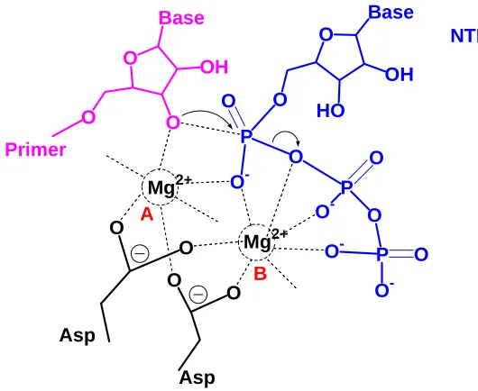
the RNA polymerase is able to take the modified NTP analogs in as substrate. T7 RNA polymerase from bacteriophages is the most commonly used RNA polymerase for in vitro preparation of RNA. The polymerase domain of T7 RNA polymerase is homologous to that of polymerases found in DNA polymerase I family, and shares the catalytic mechanism. [30] The two metal ions catalytic mechanism of T7 RNA polymerase is illustrated in Figure 1.2. T7 RNA polymerase catalysis is an SN2 nucleophilic reaction. In the active site, metal ion
A lowers the pKa of 3’-OH thus increasing its nucleophilicity. Metal ion B helps to bring in NTP, stabilizing the negative charge on the oxygens and facilitates the pyrophosphate release.[31] When preparing modified RNAs by enzymatic synthesis, the mechanism of T7 RNA polymerization should be definitely considered. O Base OH O O O Base OH HO O P O -O O P O O O -P O -O O -O O O
O Mg2+
[image:32.612.169.434.402.619.2]Mg2+ NTP A B Asp Asp Primer
Figure 1.2. T7 RNA polymerase polymerization mechanism
NTP, there was not much difference on the yield of radioactive labeled RNA. In the large scale preparation of RNA with much higher NTP concentrations, there was a difference in the yields for both native RNA and PSe-RNA, especially when dsDNA templates were used. Therefore, the protocol for large scale preparation of PSe-RNA has to be optimized from the native NTP condition. It was suspected that the lower affinity of magnesium to selenium was the reason for the lower yield of PSe-RNA. Therefore, manganese ions were tested in the transcription and indeed improved the transcription yield.
Table 1.2. Methods for introducing selenium into nucleic acid
1.3 Other applications of Se-derivatized nucleic acids 1.3.1 Investigation of nucleotidyl-transfer reactions
events in the beginning of life.
The typical catalytic mechanism of nucleotidyl-transfer reaction, whether by cleavage or ligation, usually involves one or two metal ions. A similar intermediate structure is shared among these nucleotidyl-transferases using two metal ion catalysis, like DNA or RNA polymerase, some nucleases and ribozymes.[32] In the active site(s), metal ions are coordinated by the scissile
phosphate and also nearby amino acid residues (protein) or bases (ribozyme) to promote the nucleophilic attack. Examples of the catalytic mechanism for nucleotidyl-transfer reactions are illustrated in the following sections with discussions later about how selenium modified nucleic acids could be used for investigation of the nucleotidyl-transfer reaction. Moreover, as discussed in 1.2.5, the mechanism of nucleotidyl-transfer reaction must be considered for preparation and application of selenium modified DNA and RNA.
1.3.1.1 Ribonuclease catalytic mechanism and inhibition of RNase by selenium
modification
Ribonuclease catalytic mechanism
RNA induced post transcriptional gene silence mechanism.[34] RNases are also
applied to many analytical purposes, such as RNA sequencing, mapping, and quantitation[35]. Some RNase domains are proposed drug targets in RNA virus infections, such as the RNase H domain in retroviral reverse transcriptase.[36] Advances in the knowledge about the catalysis of RNases provide opportunities for better control of many essential biological processes.
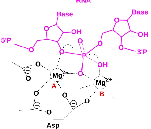
[image:35.612.202.449.243.455.2]O Base OH O O O Base OH O O P O -O OH O O Mg 2+ O O A O O Mg2+ B Asp RNA 5'P 3'P
Figure 1.3. RNase H cleavage mechanism
[36]. The negative charge generated on the oxygen is stabilized by Metal ion A
and this may destabilize the substrate and promote nucleophilic attack. Later, the substrate release may require dissociation of the metal ion B.
Inhibition of RNase by selenium modification
Extensive studies have been carried out on how to control the RNA hydrolysis process, either to accelerate or to delay it. Recently, due to the great desire of extending siRNA lifetime in mammalian cells for therapeutic purposes; studies on inhibiting RNase activities with modified RNA substrate attracted lots of research interests. A variety of chemical modifications have been tested on RNA for RNase resistance and higher base-pairing fidelity.[37]
Our group reported previously that selenium modification at the position of the non-bridging oxygen of RNA phosphate backbone would inhibit the RNase activity.[38] This mechanism is proposed as reduction of magnesium ion affinity to
1.3.1.2 Self-cleavage mechanisms of ribozymes and selenium as an atomic
probe for ribozyme catalytic studies
Ribozyme self-cleavage mechanism
[image:37.612.179.433.378.623.2]The catalytic mechanism of the hammerhead ribozyme is mainly discussed here as an example. Hammerhead ribozymes are small self-cleaving RNAs that first were discovered in a self-cleavage sequence in small RNA satellites of plant viruses [39]. A full-length active hammerhead ribozyme contains three base-paired stems and 15 highly conserved nucleotides.[40] A proposed one metal ion mechanism in hammerhead ribozyme catalysis is shown here (Figure 1.4). The metal ion stabilizes the negative charge generated in the 2’ oxygen in the proposed general acid G-8 to promote nucleophilic in-line attack. [41]
O O O P O O-O N N N N -O
H2N
O O O P O O--O H H O O HO Mg2+ C-1.1 C-17 G-12 G-8
Selenium as an atomic probe for ribozyme studies
In ribozyme studies, it is very important to discover which chemical groups play the main role in structural conformation and which directly participate in chemical catalysis.[42] To answer this question, insertion of chemically modified nucleotides into ribozyme as probe is a very useful strategy. Atomic mutation with elements in the same group is an ideal choice because less perturbation is expected on ribozyme structure. Changes in ribozyme turn-over rate will mainly come from the mutation site.
Originally, there was a strong disagreement on hammerhead ribozyme catalytic mechanism derived from biochemistry studies and the structural data. It was caused by the absence of tertiary interaction of active site in the hammerhead ribozyme structures.[43] Scott and coworkers published a Schistosma hammerhead ribozyme structure in 2006, which basically solved
most of the structure-function conflicts and organized the majority of the biochemical data.[43] However, in Scott’s structure, no metal ion was found in the active site.[40]
Mg2+, the activity was rescued.[38] Selenium on the phosphate backbone was
considered to produce no significant perturbation in the structures, if the phosphate was exposed to solvent.[27] The inhibition of cleavage activity was caused by the atomic mutation. This is an example of how selenium modified nucleotides can be used as an atomic mutation to probe the function of chemical groups for ribozymes.
1.3.2 Selenium and diseases
Selenium is an unusual trace element. Discovered in 1818, and named after the goodness of the moon, this element was only known for its toxicity and as a possible carcinogen for a long period of time.[46] The essentiality of
selenium for life rather than a hazard was primarily recognized in the late 1950s, which was almost one and a half centuries after the identification of selenium element. The systematically documented diseases caused by selenium deficiency in humans were rare, except for the well known severe heart failure in Keshan disease.[47] However, there is evidence from both laboratory and observational studies suggesting that selenium may affect human health from diverse aspects, such as immune responses, cancer, HIV infection, cardiomyopathy, rheumatoid arthritis, asthma and Alzheimer’s disease.[48]
prostate cancer risk was reduced by 63% for selenium enriched yeast. [49] In
contrast, the more recent and larger scale clinical trial result for Selenium and Vitamin E Cancer Prevention Trial (SELECT), funded by the National Cancer Institute (NCI) and the National Institutes of Health (NIH) showed that selenium and vitamin E supplements, either alone or in combination, did not prevent prostate cancer.[50] These results seem suggest that selenium may only benefit
people who have low serum selenium level. Although the result from SELECT was a huge disappointment, as a trace element, the essentiality of selenium to life was still indisputable.
The biological effects of selenium on life are exerted through a variety of selenium-containing molecules in cells:
HSe OH
NH2 O
OH NH2
O Se
SeMet SeCys Figure 1.5. Selenomethionine and Selenocysteine
In protein, selenium is inserted in two forms, selenomethionine (SeM, SeMet) and selenocysteine (Sec, SeCys) (Figure 1.5). [51] No physiological function differences were observed between proteins with selenomethionine or methionine. [21] Selenoenzymes, the majority of which are involved in catalyzing redox reactions, are generally referred as enzymes with Sec in the active sites for specific functions. In the human genomes, there are 25 genes encoding for
peroxidase (GPx), which catalyzes the reduction of hydrogen peroxide or phospholipid peroxide to water or corresponding alcohols using glutathione (GSH).[53] GPxs constitute an important part of the cell antioxidant defense system and have also been related with oxidative stress recovery, reduction of UV-induced DNA damage, protection against neurodegenerative diseases and cancer prevention. [54]
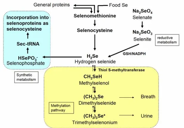
Selenium levels in the body are mainly determined by the dietary selenium content. The major natural selenium species in selenium rich plants are selenate, SeMet, SeCys, Se-methyl-selenocysteine (MeSeCys) and g-glutamyl-Semethyl-Selenocysteine (GluMeSeCys). [55] In general selenium metabolic pathways, selenium containing amino acids from food, such as SeMet and SeCys are metabolized to hydrogen selenide (H2Se) through enzymatic reactions (Figure
1.6).[56] Selenomethionine could also be incorporated into ordinary proteins through the normal protein synthesis pathway in the place of methionine. Inorganic selenite (SeO32–) and selenate (SeO42-) are also reduced to H2Se
through several metabolic steps involving glutathione and NADPH-dependent reductases.[57-58] H2Se is an intermediate among several selenium pathways,
such as selenium reductive pathway, synthetic metabolism and methylation metabolism. H2Se can be incorporated into phosphate by selenophosphate
synthetase.[59] The selenophosphate (H3PO3Se) is used to synthesize sec-tRNA,
which conducts insertion of selenocysteine into selenoproteins through the UGA code. H2Se could also be methylated by thiol S-methyltransferase to mono-, di-
Nevertheless, as first proposed by Ip and coworkers, methylselenol (MeSe, CH3SeH) was considered to be the most active metabolite of selenium
[image:42.612.142.451.184.399.2]compounds in selenium chemoprevention.[60] There were many researches on the chemprevention activities of MeSe.
Figure 1.6. Selenium metabolism pathways
Compared with the most extensively studied selenoproteins and small selenium metabolites, it is not well-known that selenium also naturally occurs in bases of tRNA, usually in the wobble base position, which may have some special functions. [61] However, detailed studies about the natural Se-derivatized RNAs are rather limited. Selenium was found in tRNAs, mainly in the forms of 2-selenouridine and 5-[(methylamino)methyl]-2-2-selenouridine (mnm5se2U), as shown in Figure 1.7. These seleno-tRNAs are usually found in species such as Escherichia coli, Clostridium sticklandii, Methanococcus vannielii, and
It is reasonable that the essentiality of selenium for life could be the combined effects from selenoproteins, seleno-nucleic acids and selenium metabolites in body, which increases the complexity of the evaluation of selenium biological activities. NH O Se N O OH OH HO NH O Se N O OH OH HO N H
Figure 1.7. 2-selenouridine and 5-[(methylamino)methyl]-2-selenouridine (mnm5se2U)
In our lab, many selenium-containing nucleosides, nucleotides and nucleic acids have been made. Considering the possible therapeutic activity of selenium compounds, these nucleosides, nucleotides and nucleic acids have been sent for primary anticancer, antimicrobial screenings. Some of them indeed shown biological activities.[68] Further investigation on selenium-containing molecules for health will definitely offer more information about this type of molecule.
1.4 Current development of SeNA for X-ray crystallography
Figure 1.8. Atom specific selenium replacement of oxygen in nucleic acids
1.4.1 Selenium modification at sugar
Selenium substitution of oxygen at 5’ of A, C, G, T and U as methylselenol, was the first attempt of derivatizing nucleic acid with selenium for X-ray crystal structure studies. [69] This modification was designed for terminal selenium
derivatization of nucleic acid. Selenium containing phosphoramidites were prepared and selenium was introduced into nucleic acids through a solid phase synthesizer. The successful synthesis of selenium containing oligonucleotides proved that it was possible to incorporate selenium into nucleic acid through solid phase synthesizer.
Selenium was also introduced to 2’-position of nucleic acid as methylselenol group through solid phase synthesis.[20, 26, 70-73] This modification later turned out to be a great success, even more than expected. Selenium modified DNA grew crystals in many buffer conditions of the Hampton kit
overnight. [20] The comparison results showed that DNA derivatized with
selenium at 2’-position could crystallize at broader buffer condition and had better crystallizability than native or bromine modified DNA. A high resolution selenium derivatized small DNA duplex structure (1.28 Å) was solved by MAD with 2’-methylseleno modification. Selenium derivatization was also present in the minor groove of DNA and did not affect minor groove hydration significantly.
Micura’s group in Austria has developed strategies for solid phase synthesis of RNA with 2’-methylseleno modifications.[74-76] The shorter RNA strands prepared by solid phase synthesis with site specific 2’-methylseleno modification were linked together to form longer RNA (up to 100 nt) by T4 RNA ligase.[75] A ligated 49 nt RNA with 6 selenium atoms was crystallized in the
same conditions as native and the crystals diffracted at about 3 Å resolution. It was stated that, with site specific insertion, 2’-methylseleno modification did not significantly change the global folding of RNA. As with what happened in DNA, a crystallization screening showed that 2’-methylseleno modified RNA was able to crystallize at broader buffer conditions than the nonmodified counterpart. Crystal packing analysis suggested that selenium atoms may contribute to bring RNA molecules closer to each other. [76]
1.4.2 Selenium modification at non-bridging phosphate
under crystallization conditions for months. It was reported that selenium modification close to the 5’ end was more stable than inside positions. There were only minimal differences between PSe-DNA structure and the native DNA structure. The Mg2+ ion coordination was identical to that in original native DNA structure. The PSe-DNA generated from solid phase synthesis method was a mixture of Rp and Sp diastereomers for phosphorous chiral center and needed further purification. Diastereomeric pure PSe-DNA could be made from enzymatic synthesis [77-78]. Our group reported a synthesis method for all nucleoside 5’-(α-P-seleno)triphosphates and also incorporation of one of the two NTPαSe diastereomers into RNA [38]. RNase resistance was observed for PSe-RNA.
1.4.3 Selenium modification at nucleobase
Derivatives of DNA at the 4-position of thymidine (4-Se-T) have been prepared through both solid phase synthesis and enzymatic incorporation. [79-80] Crystallization was attempted for solid phase synthesized 4-Se-DNA. A selenium mediated hydrogen bond was observed in the 4-Se-DNA duplex with distance of 2.87 Å.[80] The UV-melting experiment showed that the 4-selenium modification did not significantly affect the melting point of a 9mer DNA comparing with the native DNA duplex, which indicated that there was not much perturbation on base pairing.
In order to investigate whether a C-H (or CH3) group can form a hydrogen
the 5-methyl group of thymidine.[81] A DNA duplex crystal was obtained with
selenium at 5-methyl position of thymidine from solid phase synthesis In the crystal structure, the selenium linker extended the methyl group toward the phosphate and the distance between the methyl and phosphate was only 2.93 Å, which is within the normal hydrogen bond distance. The possible interaction between methyl and phosphate was suspected to be involved in DNA duplex unwinding by reducing the energy barrier.
2 PREPARATION OF PHOSPHOROSELENOATE RNA FOR X-RAY CRYSTALLOGRAPHY
2.1 Introduction
The explosive discoveries on RNA in recent years, such as small regulatory RNA [2, 83] and the riboswitch, [3] lead to a great demand for structural insights of RNA and RNA related molecules. Compared with protein crystallography, RNA crystallography has some unique problems (as discussed in the introduction). One major problem is lack of a well developed heavy atom derivatization method to solve the phase problem.[13] In the case of novel protein structure, the phase problem is usually solved by selenomethionine (SeMet) derivative via MAD phasing technique.[16, 19] Halogen derivatives have been
traditionally used for nucleic acid heavy atom derivatization. However, halogen derivatives usually produce crystal structures that are different from native structures and are unstable during X-ray radiation.[24] Since 1998, Huang’s
research group has pioneered the development of selenium derivatization of nucleic acids for X-ray crystallography. [69] It was found that selenium derivatives are much more stable than halogen derivatives under X-ray radiation and less structural perturbation has been observed for the selenium derivatives.[20, 25]
Amare and Doudna groups.[9, 23] Although this U1A method has been used to
solve two ribozyme structures, it is a labor-intensive approach. For direct covalent incorporation of a heavy atom into nucleic acids, there are two methods - enzymatic (RNA polymerase) and synthetic (solid phase synthesis).[29] A drawback of solid phase synthesis for large-scale RNA preparation is that the length of RNA is limited (<50 nt). To solve this problem, a T4 RNA ligase method for RNA 2’-methylseleno modification was developed by Micura’s group to produce longer selenium derivatized RNA.[75] For most laboratories that are not equipped with a solid phase synthesizer and not familiar with organic synthesis, enzymatic incorporation is a much more accessible method. A large quantity of long RNA molecules can be obtained using enzymatic synthesis by RNA polymerase. In addition to assisting in RNA crystallography, phosphoroselenoate RNA (PSe-RNA) was also reported to be RNase resistant,[38] as such it serves as a potential application for RNA interference therapeutics.
To incorporate selenium into RNA through RNA polymerase, the 5’-(alpha-P-seleno) triphosphates (NTPαSe) have to be prepared and must be capable of being accepted as substrate by RNA polymerase. We have reported previously a method to prepare NTPαSe with protective groups on the sugar moiety, [38, 78] and their incorporation into RNA by T7 RNA polymerase.
protective groups, which greatly reduced efforts for purification and increased the yield of NTPαSe and PSe-RNA. All of the nucleoside 5’-(α-P-seleno)triphosphate diastereomers synthesized by the new method were characterized by NMR, MS and HPLC and tested for RNA incorporation on multiple types of DNA templates. Different DNA template preparation methods (single strand, plasmid and PCR) for RNA crystallization are also described in detail to standardize the protocol. All of the NTPαSe were purified by RP-HPLC or boronate column and purified NTPαSes were tested for several RNA incorporations through different types of DNA template. The PSe-RNA was purified and the selenium derivatization was confirmed by MALDI-TOF.
O OH OH O Base P O P -O O P -O O-O O O O OH O O Base O OH O O Base P Se-O 3'-Oligonucleotide 5'-Oligonucleotide
Figure 2.1. (A) NTPαSe structure. (B) Structure of Rp phosphoroselenoate RNA.
2.1.1 Brief introduction about the functional RNA molecules in this dissertation Hammerhead ribozyme
hammerhead ribozyme has of three base-paired stems and a conserved core of 15 nucleotides (shown in grey in Figure 2.2). The catalytic mechanism of the hammerhead ribozyme has been discussed in detail in the Introduction 1.3.1.2 Self-cleavage mechanisms of ribozymes and selenium as an atomic probe for
ribozyme catalytic studies. Basically, the nucleophilic attack starts from the
[image:51.612.206.399.295.489.2]2’-hydroxyl of a nucleotide in the conserved core to its adjacent phosphodiester bond, and generates a 2’, 3’-cyclic phosphate and a free 5’-hydroxyl end.[85]
Figure 2.2. Secondary structure of wild type hammerhead ribozyme derived from the satellite RNA of tobacco ringspot virus. Conserved bases are labeled in grey
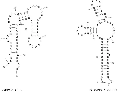
West Nile virus (WNV) terminal stem-loop (SL)
complementary negative strand of this region, which also formed a SL structure (SL(-)) (Figure 2.3.A), could serve as a promoter for replication of the positive strand. The existence of the terminal secondary structure in the complementary negative strand was confirmed by RNase digestion experiment. [88] These SL structures are probably cell protein binding sites. Analysis of the sequence and structure of the WNV genome can allow potential applications in the treatment and diagnosis of West Nile Virus in humans and animals.
[image:52.612.99.482.262.560.2]
A. WNV 3’ SL(-) B. WNV 5’ SL (+)
Figure 2.3. Predicted secondary structure of WNV terminal stem-loop SL (-) and SL (+)
3’ 5’
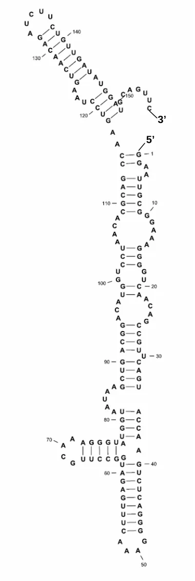
P4-P6 domain of group I intron
Figure 2.4. Secondary structure of P4-P6 domain of group I intron
3’
Table 2.1. RNA sequences
Name Sequences
HHM 5’-GGGAGCCCUGUCACCGGAUGUGCUUUCCGGUCUGA UGAGUCCGUGAGGAC AAAACAGGGCUCCCGAAUU-3’
HHN 5’-GGGAGCCCUGUCACCGGAUGUGCUUUCCGGUCUGA UGAGUCCGUGAGGACGAAACAGGGCUCCCGAAUU-3’
SL(-)
5’-CAGCUCGCACCGUGUUAAUUGUUGUUAAUCCUCACA AACACUACUAAGUUUGUCAGCUCACACAGGCGAACUACU -3’
SL(+) 5’- AGUAGUUCGCCUGUGUGAGCUGACAAACUUAGUAGU GUUUGAGAGGAUUAACAACAAUUAACACGGTGCGAGCU G -3’
89.24 5’-GGGAGCCCUGUCACCGGAUGUGCUUUCCGGUCUGA UGAGUCCGUGAGGAC AAAACAGGGCUCCCGAAUU-3’ 55.27 5’-GGCAACCUGAUGAGGCCGAAAGGCCGAAACGUACA-3’ 44.1 5’-G GCA ACCGGAUGAGGCCGA AAGGC-3’
P4-P6 5’GGAAUUGCGGGAAAGGGGUCAACAGCCGUUCAGUACC AAGUCUCAGGGGAAACUUUGAGAUGGCCUUGCAAAGGG UAUGGUAAUAAGCUGACGGACAUGGUCCUAACACGCAG CCAAGUCCUAAGUCAACAGAUCUUCUGUUGAUAUGGAU GCAGUUC-3’
2.2 Materials and methods
2.2.1 Synthesis, characterization and purification of nucleosides 5’-(alpha-P- seleno) triphosphates O HO Base OH OH O O O P Cl O P -O O -O
P O -O -O + DMF/TBA O O -O D M F /T B A O O OHOH Base Se S O 1)Dioxane/TEA
2) H2O
O O OHOH Base P O P O P O O O -O O -P O P O P O O -O O -O P O Se -O -P O P O O -O O
[image:55.612.128.485.469.680.2]2.2.1.1 Synthesis of NTPαSe
The following procedure was performed under argon gas with stirring and all liquids were dried and purged with argon before use:
Tributylammonium pyrophosphate (426 mg, 0.9 mmol, 2 eq., flask 1), individual nucleoside (varied mass, 0.45 mmol, 1 eq., flask 2), and 3H-1, 2-benzothaselenol-3-one (BTSe, flask 3) (195 mg, 0.9 mmol, 2 eq., flask 3),[92-93]
30 min then 14k rpm 30 min at 4 oC. The pellet was redissolved in small amount
of water and the concentration was checked by UV at 260 nm.
2.2.1.2 HPLC purification of nucleosides 5’-(alpha-P- seleno) triphosphates:
The synthesized NTPαSes were purified by reverse phase HPLC (RP-HPLC). NTPαSe was eluted using a combination of two solvents at varying proportions: buffer A (20mM TEAAc in water) and buffer B (20mM TEAAc, 50% water, 50% acetonitrile). Analytic HPLC program for ATPαSe, CTPαSe and GTPαSe was 20 min 12.5% acetonitrile with 20 mM TEA·Ac buffer (pH7) at 1 mL/min. Analytic HPLC program for UTPαSe was 20 min 20% ethanol with 20 mM TEA·Ac buffer (pH7) at 1 mL/min. Each diastereomer was collected seperately. Fractions were lyophylizated and one additional ethanol precipitation (as described in 2.2.1.1) was performed for desalting before NMR data collection or transcription.
2.2.1.3 Boronate column chromatography of nucleosides 5’-(alpha-P-seleno)
triphosphates
Wash buffer: 1 M TEABC buffer was prepared by bubbling CO2 through
a solution with 28.2 ml triethylamine in 140 ml H2O at 4 oC until pH drops to 9.5
and then bring the solution volume up to 200 ml. The pH was tested each time before the buffer was used. The prepared TEABC buffer was stored at 4 oC.
buffer.
Procedure: 0.2 g of boronate affigel (Biorad) was hydrated in 5 mL of TE buffer (10 mM Tris HCl pH 8.0, 1 mM EDTA) and packed in 1 X 4 cm column. The column was equilibrated using 5 mL of 1 M TEABC pH 9.5 at 4 oC. An ethanol precipitation was done to 200 µL NTPαSe (20 mM) sample using 600 µL ethanol, 20 µL 3 M NaCl and 20 µL 1M DTT before the NTPαSe was loaded on column . The NTPαSe was then redissolved in 1 mL, 1M TEABC buffer, applied to the column and incubated for 15 mins at 4 oC. Afterwards, washing buffer (1 M TEABC, pH 9.5, 4 oC) was added. Flow through was collected by 0.5 mL per tube. Fractions were collected until the maximum absorbance value dropped below 0.01 as determined by UV. Maximum absorbance values are as follows: ATPαSe-259nm, UTPαSe-262 nm, GTPαSe-252 nm, CTPαSe-271 nm. Elution was done using the elution buffer (0.05% acetic acid pH 4.5) and fractions were collected by 0.5 mL per tube. The tubes were labelled as E1, E2, and so on depending on the fraction collected. All working buffers were kept on ice during the procedure. DNA quantification was done by UV spectrometer and the absorbance values noted and concentrations calculated. Analytical HPLC analysis was performed to determine the quality of purified NTPαSe.
adjusted to 20 mM, which was used as 10X solution in transcription reactions. The HPLC programs for analyzing NTPαSe eluted from the boronate column: NTPαSe was eluted using a combination of two solvents at varying proportions: buffer A (20 mM TEAAc in water) and buffer B (20 mM TEAAc, 50% water, 50% ethanol). The HPLC program for UTPαSe: 20 min 12.5% acetonitrile with 20 mM TEA·Ac buffer (pH 7) eluted at 1 mL/min. HPLC program for ATPαSe, and GTPαSe: 20 min 22.5% ethanol with 20 mM TEA·Ac buffer (pH 7) eluted at 1 mL/min. CTPαSe was 20 min 20% ethanol with 20 mM TEA·Ac buffer (pH 7) eluted at 1 mL/min.
2.2.2 DNA template preparation for RNA transcription
To transcribe RNA, beside the preparation of NTPαSe substrates, the DNA template must be prepared. Three types of DNA templates, plasmid, PCR and synthetic DNA templates were used to test for NTPαSe transcription. Based on the target RNA requirement, different DNA templates can be chosen for transcription.
For the PCR dsDNA template (Figure 2.6B) preparation, the template for PCR could be plasmid, or synthetic DNA. Advantages of PCR dsDNA template for RNA transcription are that there is neither a specific sequence requirement nor length limit for the target RNA molecule. After the PCR reaction and spin column purification, the PCR product can be directly used for RNA transcription.
Figure 2.6. Illustration of DNA templates. A: plasmid (pUC19, Amp+) inserted region for RNA transcription; B: PCR dsDNA template; C: synthetic ssDNA template.
Another choice for DNA template is synthetic template (Figure 2.6C). In general, although T7 RNA polymerase works better on dsDNA template, considering the expense and preparation time, ssDNA is a good option if it is possible. The ssDNA template is usually prepared as illustrated in Figure 2.6C, a full length ssDNA template strand and a 21mer promoter strand. There is no
Example of plasmid (pUC19, Amp+) inserted region for RNA transcription A. Plasmid dsDNA
B. PCR dsDNA
RNA gene
T7 RNAP promotor
RNA gene EcorR I
T7 RNAP promotor NgoMIV
C. ssDNA template
T7 RNAP promotor
sequence requirement for the template but there is length limit for synthetic template (<100 nt). Due to the length limit, synthetic ssDNA template can not be used directly for these long RNAs. However, synthetic ssDNA template is a very convenient and economical way for template preparation of short RNAs, like siRNA.
2.2.2.1. Purification of ssDNA template synthesized by solid phase DNA
synthesizer
in this dissertation were also purified by this method.
Table 2.2 Sequences of DNA templates prepared by solid phase DNA synthesizer.
Name Sequence
21.1 5’-GCGTAATACGACTCACTATAG-3’
55.27 3’-CGCATTATGCTGAGTGATATCCGTTGGACTACTCCGGC TTTCCGGCTTTGCATGT-5’
89.24 3’-CGCATTATGCTGAGTGATATCCCTCGGGACAGTGGCCT ACACGAAAGGCCAGACTACTCAGGCACTCCTGTTTTGTCCCGAGGGC TTAA-5’
2.2.2.2 Plasmid construction for RNA transcription template
a. PCR of inserted fragment (New England Biolabs PCR Kit):
Characteristic PCR reactions (100 μL) were carried out using 1 μL plasmid template - pWNV (80 ng/mL) or 8 μL ssDNA (10 nM) from solid phase synthesizer, 10 μL each of forward and reverse primers (10 μM), 10 μL 10 X PCR standard buffer with Mg2+, 0.4 μL Taq DNA polymerase (5 u/μL), 2 μL 10 mM dNTP, and the proper amount of water to make a final volume of 100 µL. PCR started at 94 oC for 2 min, then repeated for 35 following cycles 94 oC (30 sec), 55 oC (30 sec, temperature varied for different sequences. Basically, it was 5 oC
below Tm of primers) and 72 oC (30 sec) and the final extension at 72 oC for 2 min. The PCR product was purified by Qiagen MinElute PCR purification kit using the manufacturer’s suggested protocol. The product was eluted with 20 μL of water and checked on a 2% agarose gel.
b. Restriction endonuclease digestion of PCR product:
A typical 20 μL double digestion reaction was performed as follows: 6
NEB4 buffer (New England Biolabs restriction endonuclease Buffer 4) and desired amount of water to make final volume 20 µL. The reaction mixture was kept at 37 °C for 2 hours. The product was purified by Qiagen MinElute PCR purification kit using the manufacturer suggested protocol. The product was eluted with 20 μL of water and checked on a 2% agarose gel.
Table 2.3. PCR template sequences and primer sequences for plasmid construction
Name Sequence
HHM Template
3’-CGCATTATGCTGAGTGATATCCCTCGGGACAGTGGC CTACACGAAAGGCCAGACTACTCAGGCACTCCTGTTTT GTCCCGAGGGCTTAA-5’
PCR Primers
PF:5’CGACGCCGGCGCGTAATACGACTCACTATAG-3’ (31nt)
PR:3’ACTCCTGTTTTGTCCCGAGGGCTTAAGTACT-5’ (31nt)
HHN Template 3’-CGCATTATGCTGAGTGATATCCCTCGGGACAGTGGC CTACACGAAAGGCCAGACTACTCAGGCACTCCTGCTTT GTCCCGAGGGCTTAA-5’
PCR Primers
PF:5’-CGACGCCGGCGCGTAATACGACTCACTATAG-3’ (31nt)
PR:3’-ACTCCTGC TTTGTCCGAGGGCTTAAGTACT-5’(31nt)
SL(-) Template
3’-CGGCCGCGCATTATGCTGAGTGATATCGTCGAGCG TGGCACAATTAACAACAATTAGGAGTGTTTGTGATGATT CAAACAGTCGAGTGTGTCCGCTTGATGANCTCTGCTTA AG -5’ PCR Primers PF: 5’-CGACGCCGGCGCGTAATACGACTCACTATAGCA GCTCGCACCGTGTTAATT -3’ (51nt)
PR:3’-CGAGTGTGTCCGCTTGATGANCTCTGCTTAAGTG CAC -5’ (37 nt)
SL(+) Template 3’-CGGCCGCGCATTATGCTGAGTGATATCTCATCAAGC GGACACACTCGACTGTTTGAATCATCACAAACACTCCT AATTGTTGTTAATTGTGCCACGCTCGACCATGGCTTAAG -5’ PCR Primers PF:5’CGACGCCGGCGCGTAATACGACTCACTATAGAGTA GTTCGCCTGTGTGAGC -3’ (51nt)
c. Plasmid restriction endonuclease digestion and purification:
The obtained pUC19 based plasmids (pSAM) were double digested by EcoR I and NgoMIV as follows: 7 μL purified pSAM (1.4 μg/μL), 1 μL EcoR I (20 u/ μL) and 1 μL NgoMIV(10 u/μL), 2 μL 10x NEB4 (New England Biolabs restriction endonuclease Buffer 4) and proper amount of water to make a final volume 20 µL. The reaction mixture was kept at 37 °C, 2 h and purified on a 0.6% Agarose gel. Linear plasmid bands, visualized by UV photo illuminator were cut and purified by Qiagen gel extraction kit using the manufacturer suggested protocol. The product was eluted by 20 μL of water.
d. Ligation:
Purified, restriction endonuclease digested PCR product and linearized pHHM were ligated with T4 DNA ligase under the following conditions: 1 µL 10X T4 DNA ligase buffer, 1 µL T4 DNA ligase, 2 µL linearized pHHM, 2 µL PCR product and 2 µL water were mixed and kept in 16 oC for 1 hours. The reaction was stopped by inactivating T4 ligase with heating to 65 oC for 10 min.
e. Transformation (Invitrogen Top10 cell):
f. Large scale plasmid purification and digestion for RNA transcription
The clone with correctly sequenced plasmid was used for large scale plasmid purification by Qiagen Plasmid Maxi kit. Before transcription, the plasmid was linearized by EcoR I as follows: 25 μL plasmid (1.5 μg/μL), 3 μL EcoR I (20 u/ μL), 10 μL NEB buffer 4 and proper amount of water to make final volume 100 µL. The reaction mixture was kept at 37 °C 4 h, and then 65 °C for 20 min to inactivate EcoR I. The product was checked on a 0.6% Agarose gel. The linear plasmid was isolated by NaCl/ethanol precipitation from digestion buffer. 20 μL of water was added to redissolve the plasmid. The plasmid solution was further desalted by 3K Nanosep centrifugal devices (Pall) at 10K rpm for 10 min. The desalting procedure was repeated once by adding 20 μL more water and centrifuging again. Then the plasmid was recovered by 20 μL of RNase free water
2.2.2.3 dsDNA Template prepared by PCR
a. Klenow extension for making full length long DNA template:
Due to the size limit for the oligonucleotide that could be obtained from solid phase synthesizer, two fragments of P4-P6 DNA template (a 98mer and a 99mer) were synthesized by solid phase DNA synthesizer for Klenow DNA polymerization to get full length template.
concentration of 10mM, 0.5 μL of 10X Klenow buffer, 0.2 μL of Klenow DNA Polymerase (5 U/ μL) and desired amount of water to make a final volume of 5
μL. This reaction was incubated at 37 oC for an hour and checked via Agarose gel electrophoresis. The Klenow extended full length double stranded product was purified by 12% denature PAGE.
Table 2.4. Polymerization primer sequences for P4-P6
Name Sequence
P4-P6
Polymerization primer 1 (99 nt)
5’-GCGTAATACGACTCACTATAGGGAATTGCGGGA AAGGGGTCAACAGCCGTTCAGTACCAAGTCTCAG GGGAAACTTTGAGATGGCCTTGCAAAGGGTAT-3’
Polymerization primer 2 (98 nt)
5’-GAACTGCATCCATATCAACAGAAGATCTGTTGAC TTAGGACTTGGCTGCGTGTTAGGACCATGTCCGT CAGCTTATTACCATACCCTTTGCAAGGCCA-3’
b. PCR (New England Biolabs PCR Kit):
Characteristic PCR reactions (100 μL) were carried out using 1 μL plasmid template - pWNV (80 ng/mL) or 8 μL ssDNA (10 nM) from solid phase synthesizer, 10 μL each of forward and reverse primers (10 μM), 10 μL 10 X PCR standard buffer with Mg2+, 0.4 μL Taq DNA polymerase (5 u/μL), 2 μL 10 mM
dNTP, and desired amount of water to make a final volume of 100 µL. PCR started at 94 oC for 2 min, then repeated 35 following cycles 94 oC (30 sec), 55
oC (30 sec, temperature varied for different sequences). Basically, it was 5 oC
modification at the last two nucleotides of 5’ of DNA increased the fidelity of RNA transcription.[94] Therefore, two types of primers were tested. One set was just a commonly used native primers; the other set has one primer with 2’-O-methyl modification at the last two nucleotides of 5’, which was the end of transcription.
Table 2.5. PCR template sequences and primer sequences
Name Sequence
SL(-) Template 3’-GCGCATTATGCTGAGTGATATCGTCGAGCGTGGC ACAATTAACAACAATTAGGAGTGTTTGTGATGATTCA AACAGTCGAGTGTGTCCGCTTGATGA -5’
PCR
Primers P2: 3’CGAGTGTGTCCGCTTGATGmAm5’ (21nt) 21.15’-GCGTAATACGACTCACTATAG-3’ (21nt)
SL(+) Template 3’-GCGCATTATGCTGAGTGATATCTCATCAAGCGGAC ACACTCGACTGTTTGAATCATCACAAACACTCCTAAT TGTTGTTAATTGTGCCACGCTCGAC -5’
PCR
Primers P2: 3’TTAATTGTGCCACGCTCGAmCm5’ (21nt) 21.15’-GCGTAATACGACTCACTATAG-3’ (21nt)
P4-P6 Template
3’-CGCATTATGCTGAGTGATATCCCTTAACGCCCTTT CCCCAGTTGTCGGCAAGTCATGGTTCAGAGTCCCCT TTGAAACTCTACCGGAACGTTTCCCATACCATTATTC GACTGCCTGTACCAGGATTGTGCGTCGGTTCAGGAT TCAGTTGTCTAGAAGACAACTATACCTACGTCAAG-5’
P4-P6 PCR Primers P2: 5’-GmAmACTGCATCCATATCA ACAG-3’ (21nt) 21.15’-GCGTAATACGACTCACTATAG-3’ (21nt)
2.2.3 Phosphoroselenoate modified RNA (PSe-RNA) transcription with radioactive label
1 h at 37 oC. Each time one type of 10 mM NTPαSe was used to substitute one
type of normal 10 mM NTP solution in protocol (for example, to ATPαSe I tube, ATPαSe I, CTP, GTP and UTP were added). NTP sample was labeled both by ATP [α-32P] and CTP [α-32P]. Different types of templates were tested in the transcription reaction. A typical 10 µL reaction was performed under the final condition of 1 µM ssDNA template and top promoter strand or 50 ng/µL plasmid or 6 ng/µL dsDNA PCR template, 0.5 mM NTP or NTPαSe, 10 mM DTT, 1 µL of 10 X Epicentre T7 RNA transcription buffer, 1 µL of Epicentre T7 RNA polymerase and desired amount of RNase-free water to add volume up to 10 µL. ATPαSe and GTPαSe samples were labeled by CTP[α-32P], and CTPαSe and UTPαSe samples were labeled by ATP[α-32P]. The transcription reaction was
quenched by adding equal volume of loading dye containing EDTA (100 mM). The transcription result was determined by 15% denature PAGE gel by exposure to film.
2.2.4 PSe-RNA transcription for large scale RNA preparation Hammerhead ribozyme as an example:
Mutant hammerhead ribozyme (HHM) transcripts (500 µL) were prepared according to the protocol of the manufacturer (Epicentre T7 flash transcription kit). The reaction was also includeing 0.1 u/μL phosphatase and 2 mM MnCl2 and the reaction was incubated for 2 h at 37 oC. 100 mM NTPαSe
and top promoter strand, or 150 ng/µL plasmid, or 50 ng/µL dsDNA PCR template, 9 mM ATP, 9 mM CTP, 9 mM GTP and 9 mM UTP (when one type of NTPαSe was used, the corresponding NTP was not added to the transcription mixture), 10 mM DTT, 0.008 unit pyrophosphatase, 2 mM MnCl2, 1 µL of 10 X
Epicentre T7 RNA transcription buffer, 1 µL of Epicentre T7 RNA polymerase and proper amount of RNase-free water to add volume up to 10 µL. After transcription incubation, 10 µL (10 u) Epicentre DNase I was added at 37 oC for 15 min to remove DNA template. After 37 oC incubation was finished, an NaCl/ethanol precipitation was performed (14K RPM, 10 min) to remove enzymes, short RNA fragments and NTP. RNA pellet was redissolved in 200 µL water.
2.2.5 Purification of PSe-RNA 2.2.5.1 Gel purification of PSe-RNA
band was cut from the gel, transferred to a clean tube and crushed by a RNase-free spatula (All of the solutions used after this step, including RNase-RNase-free water, ethanol, 3M NaCl and so on, were filtered by 0.2 µm nylon membrane filter. For PSe-RNA purification, 20 mM DTT was added to all RNase-free water used in the purification steps to dissolve the PSe-RNA). RNase-free water (2 gel volume) was added to the crushed gel placed in a clean tube for soaking. After soaking for 4 h at 4 oC with rotation, the gel soaking solution was centrifuged (3K rpm, 10 min, at 4 oC) to remove gel fragments. The supernatant was transferred to a fresh tube and RNase-free water (one more gel volume) was added to the gel for washing. The solution was centrifuged again (3K rpm, 10 min, at 4 oC) and the supernatant was combined with the previous supernatant. This washing step was repeated two more times. All of the supernatant was collected and filtered through 0.2 μm nylon syringe filter to a high speed centrifuge tube. NaCl/ethanol precipitation was then performed to pellet the RNA out of the solution. A typical ethanol precipitation started with adding 1/10 volume of 3 M NaCl, 3 volume of ethanol and 10 mM DTT and mixed well. The mixture was left in -80 oC for 30 min and centrifuged at 14K rpm (4 oC) for 30 min. The RNA pellet was redissolved in water and desalted by 10K Nanosep centrifugal device (Pall) at 8K rpm for 10 min.
2.2.5.2 FPLC purification of PSe-RNA
![Figure 1.4. Metal ion mechanism in hammerhead ribozyme catalysis [41].](https://thumb-us.123doks.com/thumbv2/123dok_us/9129865.987589/37.612.179.433.378.623/figure-metal-ion-mechanism-hammerhead-ribozyme-catalysis.webp)