RESEARCH ARTICLE
The
Drosophila
Hox gene
Ultrabithorax
controls appendage
shape by regulating extracellular matrix dynamics
JoséM. De las Heras1,*,‡, Celia Garcıa-Corté ́s1, David Foronda1, JoséCarlos Pastor-Pareja2, L. S. Shashidhara3and Ernesto Sánchez-Herrero1,‡
ABSTRACT
Although the specific form of an organ is frequently important for its function, the mechanisms underlying organ shape are largely unknown. In Drosophila, the wings and halteres, homologous appendages of the second and third thoracic segments, respectively, bear different forms: wings are flat, whereas halteres are globular, and yet both characteristic shapes are essential for a normal flight. The Hox gene Ultrabithorax (Ubx) governs the difference between wing and haltere development, but how Ubx function in the appendages prevents or allows flat or globular shapes is unknown. Here, we show that Ubx downregulates Matrix metalloproteinase 1 (Mmp1) expression in the haltere pouch at early pupal stage, which in turn prevents the rapid clearance of Collagen IV compared with the wing disc. This difference is instrumental in determining cell shape changes, expansion of the disc and apposition of dorsal and ventral layers, all of these phenotypic traits being characteristic of wing pouch development. Our results suggest thatUbxregulates organ shape by controlling Mmp1 expression, and the extent and timing of extracellular matrix degradation.
KEY WORDS:Drosophila, Extracellular matrix, Hox genes, Matrix metalloproteinases, Ultrabithorax, Organ shape
INTRODUCTION
Animal species are characterized by particular morphologies that distinguish them from other animals. The form of distinct organs is the result of evolutionary forces that determine how genetic pathways are implemented to regulate size and shape. How the shape of a structure is determined is an interesting biological problem, as the form of an organ is frequently related to its function.
Drosophila melanogaster is an organism well suited for this
analysis. The knowledge of genetic pathways and cell properties in the fruitfly (and other organisms) has allowed the identification of some mechanisms needed to confer shape (Gilmour et al., 2017). These have been analyzed mostly in adult derivatives of imaginal discs: groups of cells that grow during the larval period and
differentiate in pupal stages (Cohen, 1993). A well-studied example is the development of the wing. This appendage derives from a region of the wing disc known as the wing pouch, and undergoes a complex series of morphogenetic processes leading to the adult shape (Baena-López et al., 2005; Etournay et al., 2015; Guirao et al., 2015; Diaz de la Loza and Thompson, 2017).
The mechanisms that establish differences in shape between organs are perhaps more amenable to be investigated when comparing homologous structures, such as the two Drosophila dorsal thoracic appendages, wings and halteres. Wings, which are derived from wing imaginal discs, emanate from the second thoracic segment and are considerably larger than halteres, which are small appendages that arise from the third thoracic segment and are derived from haltere imaginal discs. Difference in size, however, is not the only obvious distinction between wings and halteres: wings are flat, whereas halteres have end-knob morphology, with a distal part (capitellum) that is globular in nature. Importantly, this structural difference has been shown to be crucial for a stable flight (Dickinson, 1999). At the end of the larval period, wing and haltere discs differ in size but are morphologically similar, both at the cellular and disc levels. It is thus clear that the two structures acquire characteristic flat (wing) and globular (haltere) shape during the subsequent pupal development.
At the start of metamorphosis, the wing disc everts through its peripodial membrane. The pouch region expands to form the wing blade, dorsal and ventral surfaces appose, and the constituent epithelial cells change from columnar to cuboidal. These events are completed by 8 h after puparium formation (APF). Ventral and dorsal surfaces separate and appose again by 18-20 h APF. Different processes of morphogenesis and differentiation take place afterwards, ending up with the formation of the adult wing (Fristrom and Fristrom, 1975, 1993; Fristrom, 1976; Aldaz et al., 2010; Díaz de la Loza and Thompson, 2017). Development of the pupal haltere disc has not been described in as much detail. Nevertheless, it has been reported that the morphology of epithelial cells of the haltere disc starts to diverge from that of the wing disc cells at 32-36 h APF, and that dorsal and ventral surfaces do not appose in the distal haltere disc, with the pouch remaining hollow throughout development (Roch and Akam, 2000).
The Hox gene Ultrabithorax (Ubx) is the master regulator of haltere identity (Lewis, 1963).Ubxis present in the haltere disc, but not in the wing disc, and Ubxfunction is necessary to generate halteres. Furthermore, ectopic expression ofUbxin the wing disc is sufficient to transform wings into halteres (Lewis, 1963; Cabrera et al., 1985; White and Akam, 1985). Therefore, the distal globular form of halteres depends on Ubx activity. Although this distinct cellular architecture emerges during pupal development (Roch and Akam, 2000), the precise identity of Ubx targets that control this morphogenetic process is currently unknown. It is also unclear when Ubx regulates haltere identity by preventing wing
Received 5 December 2017; Accepted 17 May 2018
1Centro de Biologıa Molecular Severo Ochoa, Universidad Autó ́noma de Madrid,
Nicolás Cabrera 1, Cantoblanco, Madrid 28049, Spain.2School of Life Sciences,
Tsinghua University, Beijing 100084, China.3Indian Institute of Science Education
and Research Pune, Dr Homi Bhabha Road, Pune, Maharashtra 411008, India. *Present address: Institut Curie, PSL Research University, CNRS UMR 3215, INSERM U934, 75248 Paris, France.
‡Authors for correspondence ( jose.de-las-heras-chanes@curie.fr; esherrero@cbm.csic.es)
J.M.D.H., 0001-5421-8169; D.F., 0003-2598-7852; E.S.-H., 0000-0001-5688-2303
DEVEL
O
development in the haltere primordia, although Hox input appears to be continuously needed until the beginning of differentiation (Morata and García-Bellido, 1976; Roch and Akam, 2000).
To better understand howUbxis generating haltere morphology, we have undertaken a detailed analysis of the early pupal development of both wing and haltere discs, and analyzed the role ofUbxin generating the morphogenetic distinctions between the two structures. We report here thatUbxengineers haltere morphogenesis in the pre-pupal stage by inhibiting three important phenotypic traits diagnostic of wing development: changes in cell shape, control of adhesion competence between cell layers and regulation of organ expansion. Ubx achieves this by downregulating Matrix
metalloproteinase 1 (Mmp1) expression in the haltere disc, thus
delaying elimination of the basal extracellular matrix (ECM). Our results suggest thatUbx prevents haltere discs from acquiring the flat wing morphology, at least in part, by controlling the elimination of ECM in the first hours of pupal development.
RESULTS
The different shape between wings (flat; Fig. 1B) and halteres (end-knob structures; Fig. 1D) is acquired during the pupal stage, as larval wing and haltere discs have similar shapes (Fig. 1A,C). We therefore analyzed the morphogenetic events leading up to the generation of the adult structures after the larval stage.
Ubxprevents the haltere pouch from acquiring wing pouch-like morphology and cellular architecture
TheDrosophilawing blade structure results from the apposition of
the basal side of two epithelial monolayers. Previous studies have shown that ventral and dorsal surfaces of the wing disc align in the first hours of pupariation (Fristrom et al., 1993, 1994; Fristrom and Fristrom, 1993; Aldaz et al., 2010). Therefore, we decided to compare at this stage the development of the distal part of the wing and haltere discs, the wing and haltere pouches, using phalloidin staining to visualize F-actin. In most of our experiments we have focused on analyzing frontal sections of the distal wing or haltere discs in order to observe the basal ECM distribution in the lumen and to illustrate
differences in morphology and gene expression. At about 3 h APF there is incipient apposition of dorsal and ventral layers in the wing pouch, except in its central region, the presumptive L3 pre-vein (Fristrom et al., 1994) (Fig. 1E). Three hours later, after disc eversion, the tissue expands and the region of apposition increases (Fig. 1F). At 9 h APF, the dorsal and ventral layers are tightly apposed (Fristrom and Fristrom, 1993; Fristrom et al., 1994; Blair, 2007) (Fig. 1G).
A different pattern is observed in the haltere disc. At about 3 h APF dorsal and ventral surfaces do not start to align, and a large lumen is observed in the distal part, corresponding to the presumptive capitellum (Fig. 1H). Three hours later (6 h APF), epithelial expansion is absent and so is dorso-ventral apposition (Fig. 1I). Similar observations with respect to the absence of dorso-ventral apposition have previously been described at late pupal stages (Roch and Akam, 2000). At 9 h APF, the tight apposition and zippering that takes place in the wing pouch are not observed in the haltere pouch (Fig. 1J).
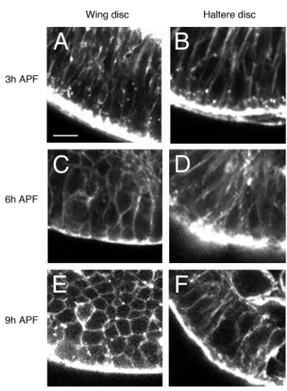
Close inspection of the discs at different time points indicated a change in cell shape in the wing disc at late stages. In transverse sections of the distal wing pouch, at 3 h APF, the epithelium shows a columnar pseudostratified disposition (Fig. 2A); however, the cells of the epithelium show a more isodiametric shape at 6 h APF and, more clearly, at 9 h APF, as the wing disc expands (Fig. 2C,E). At 3 h APF, the haltere disc cells show an elongated shape, similar to that of the wing disc at the same stage (Fig. 2B). However, in contrast to the wing disc, cells in the haltere primordium do not change shape at 6 h or 9 h APF, thus keeping a columnar epithelial organization (Fig. 2D,F).
Therefore, when comparing prepupal development of distal wing and haltere discs, three main differences can be observed: (1) there is apposition of dorsal and ventral layers in the wing pouch but not in the haltere pouch; (2) the expansion observed in the wing pouch is absent in the corresponding region of the haltere disc; and (3) wing disc cells, but not haltere disc cells, become more isodiametric as the disc expands and tightly apposes ventral and dorsal surfaces.
[image:2.612.50.338.485.740.2]As the presence of Ubx is a crucial molecular distinguisher between haltere and wing discs, we decided to analyze prepupal disc
Fig. 1. Wing and haltere discs acquire different shapes in early pupa.(A) Third instar wing disc. (B) Adult wing. h, haltere. (C) Third instar haltere disc. Note the similar shape of both discs. (D) Haltere. Its globular shape differs from the flat wing. (E-J) Optical frontal sections of the distal part of wing (E-G) and haltere (H-J) discs at 3 h (E,H) (n=35), 6 h (F,I) (n=20) and 9 h (G,J) (n=15) APF, stained with phalloidin. The wing disc apposes dorsal and ventral surfaces, and extends as the pupa develops (F,G), whereas the haltere disc remains unexpanded, and dorsal and ventral surfaces do not appose (H-J). Scale bars: 25μm.
DEVEL
O
morphology in Haltere-mimic (Hm) mutants, in which Ubx is derepressed in the wing disc and wings are transformed into halteres (Lewis, 1982; Fig. S1A). In Hm animals, the wing disc fails to display the characteristic expansion or the apposition of dorsal and ventral surfaces, and resembles a haltere disc (Fig. S1B). Then, we decided to focus on how Ubx activity suppresses disc elongation, surface apposition and cell shape changes in the haltere disc.
Components of the basal ECM persist longer in the prepupal haltere pouch than in the wing pouch
Some of the differences observed between the two disc structures appear to be related to basal adhesion, suggesting a possible role of basal lamina proteins in differentiating haltere and wing disc development. In fact, ECM composition and remodeling has been shown to be a key element in defining organ morphology (Hynes, 2009; Haigo and Bilder, 2011; Pastor-Pareja and Xu, 2011; Crest et al., 2017). Furthermore, the analysis of genes regulated byUbxin the pupa has uncovered a small group of genes coding for proteins that affect ECM turnover (Pavlopoulos and Akam, 2011). We therefore wondered whether regulation of the ECM could prove to be crucially different between the two appendages. We decided to analyze ECM elements and their regulators in early pupal wing and haltere discs.
There are four main basement membrane components in
Drosophila: Collagen IV (the most abundant one), Laminin,
Perlecan and Nidogen (LeBleu et al., 2007). We decided to analyze the distribution of two of these components, Collagen IV and Perlecan, for which there are well-characterized GFP protein
traps (Morin et al., 2001) inserted in the genesviking(vkg), coding for one Collagen IV α chain (Yasothornsrikul et al., 1997), and
terribly reduced optic lobes(trol), coding for Perlecan (Voigt et al.,
2002; Park et al., 2003). We have followed Vkg-GFP and Trol-GFP dynamics in fixed andex-vivoimaginal discs.
At 3 h APF, there is an accumulation of Vkg-GFP basally, which is mainly associated with the lumen ( presumptive L3 vein), in the distal region of the wing disc (Murray et al., 1995) (Fig. 3A,A′). Three hours later, when the discs have everted, Vkg-GFP is hardly detected in the lumen of the wing disc (Fig. 3C,C′) and its presence disappears completely by 9 h APF (Fig. 3E,E′). In the 3 h APF haltere disc, Vkg-GFP accumulation is associated exclusively with the lumen and the levels are considerably higher than in the wing disc (Fig. 3B,B′; see also Fig. 4A,B). At 6 h APF, and in contrast to what is observed in the wing disc, there are still high levels of Vkg-GFP in the basal side of the haltere pouch cells (Fig. 3D,D′). The Vkg-GFP signal is almost completely lost from the basement membrane at 9 h APF, when Collagen IV can be seen associated to hemocytes inside the lumen (Fig. 3F,F′).
To further assess the dynamics of the ECM remodeling, we decided to employ time-lapse imaging. In ex vivo cultures of prepupal wing discs we observed that Vkg-GFP is lost in the wing pouch before the complete retraction of the peripodial membrane, which takes place before 6 h of prepupal development (Movie 1; Fig. S2). In contrast, high amounts of Vkg-GFP are detected in the pouch of haltere discs even after the eversion of the disc (Movie 2; Fig. S3). The expression of a Trol-GFP protein trap follows similar dynamics to Vkg-GFP in both discs: the Trol-GFP signal in the wing pouch is lost during retraction of the peripodial membrane (Movie 3; Fig. S4), whereas GFP expression is still observed in 6 h APF haltere discs, after the eversion concludes (Movie 4; Fig. S5). The correlation of Trol-GFP and Vkg-GFP expression is expected as vkgis required for Perlecan deposition in the basement membranes of imaginal discs (Pastor-Pareja and Xu, 2011). Taken together, our results indicate that the elimination of basal ECM components in the haltere disc of the prepupa is delayed when compared with the wing disc of the same age.
UbxdownregulatesMmp1expression and delays Vkg degradation in the prepupal haltere pouch
The analysis presented in the preceding sections suggests thatUbx ( present in the haltere disc and absent in the wing disc) delays Vkg and Perlecan degradation in the prepupal haltere pouch when compared with the wing pouch. These ECM components are synthesized in the fat body, secreted into the hemolymph and incorporated into the basement membrane of the discs (Pastor-Pareja and Xu, 2011). Thus, it is unlikely thatUbxregulatesvkgor troltranscription in the haltere pouch in a disc-autonomous manner. Because Matrix metalloproteinases (Mmps) and Tissue inhibitor of metalloproteinases (Timp) control ECM turnover (Page-McCaw, 2008), we hypothesized thatUbxmight regulate ECM remodeling indirectly by controlling the expression of these proteins. There are two genes coding for metalloproteinases inDrosophila,Mmp1and Mmp2. Although it was initially thought that Mmp1 codes for a secreted protein and Mmp2 codes for a protein tethered to the membrane (Llano et al., 2000, 2002; Page-McCaw et al., 2003; Page-McCaw, 2008), both membrane-anchored and secreted isoforms of both proteins have recently been reported (LaFever et al., 2017).
Metalloproteinases are inhibited by Timp, and the Drosophila genome harbors a single gene coding for Timp (Godenschwege et al., 2000; Wei et al., 2003; Page-McCaw, 2008). In the fly, Timp
Fig. 2. Changes in cell shape occur in the prepupal wing disc but not in the haltere disc.(A-F) Optical transverse sections of the distal wing (A,C,E) and haltere (B,D,F) discs at 3 h (A,B) (n=7), 6 h (C,D) (n=4) and 9 h (E,F) (n=6) APF, stained with phalloidin. The distal part of the appendage is at the bottom. At 3 h APF, cells of the distal wing region show the columnar shape of the third instar imaginal disc (A), but show a more isodiametric shape at 6 h and 9 h APF (C,E) as the disc extends. By contrast, haltere pouch cells maintain the elongated shape at 3 h, 6 h and 9 h APF (B,D,F). Scale bar: 5μm.
DEVEL
O
[image:3.612.72.274.52.327.2]abrogates Mmp1 and Mmp2 activity completely (Page-McCaw et al., 2003). Metalloproteinases remodel tissue by degrading the Collagen IV-containing basement membrane (Srivastava et al., 2007) and, interestingly, Mmp1, Mmp2 and Timp have been identified as targets ofUbx(Pavlopoulos and Akam, 2011):Mmp1 is downregulated byUbxin pupal stage whereasMmp2andTimp are upregulated byUbxin late larval and pupal stages, respectively. All these data prompted us to analyze whether or not the delay in the elimination of Collagen IV in the prepupal haltere disc, with respect to the wing disc, might result from Ubx-mediated regulation of metalloproteinase activity.
To test this, we first analyzed Mmp1and Mmp2 expression in wing and haltere prepupal discs.Mmp2expression can be detected with an Mmp2-GFP line (Deady et al., 2015). The GFP signal is non-uniformly distributed in both wing and haltere prepupal discs, and there appears to be higher expression levels in groups of cells of the wing pouch than in the corresponding region of the haltere pouch; however, these cells are mainly in the peripheral region of the disc (Fig. S6A,B). To carefully compare the relative levels of Mmp2-GFP levels in the two discs, we looked at the GFP signal in Mmp2-GFPpbxmutants, in which the posterior compartment of the haltere disc is transformed into the corresponding region of the wing disc. As shown in Fig. S6C,C′, inpbxhaltere discs there appears to be only a somewhat higher Mmp2-GFP expression in the dorsal, but not the ventral, cells contacting the lumen, in the mutant (wing-like) compartment.
A significant difference is readily observed, by contrast, when studying Mmp1 expression with an anti-Mmp1 antibody: the wing pouch lumen shows notably higher levels of Mmp1 protein than the haltere pouch at the same stage (Fig. 4A,B). When comparing simultaneously Vkg-GFP and Mmp1 expression in both discs at 3 h
APF we noticed that, in the wing pouch, the higher levels of Mmp1 were correlated with lower Vkg-GFP signal, and the reverse was true for the distal haltere disc (Fig. 4A,B). Quantification of such levels in eight wing and haltere disc pairs showed that the wing disc lumen accumulated about twice as much Mmp1 as did the haltere disc lumen, and that the level of Vkg-GFP in the former is∼40% lower than in the latter, after normalization (Fig. 4C). We have also observed consistent results with high-throughput techniques identifying genes that are regulated by Ubx and differentially expressed between wing and haltere discs of third instar larvae. Although ChIP-seq suggests that bothMmp1andMmp2are direct targets ofUbx, RNA-seq data suggest that only transcripts ofMmp1 (one isoform), and not of Mmp2, are differentially expressed (S. Khan and L.S.S., unpublished observations).
We have also analyzedTimp expression in prepupal wing and haltere discs byin situ hybridization (ISH) because no antibody against Timp or Timp protein trap is available. Timp RNA is distributed in small, discrete patches in the 3 h APF wild-type wing disc; none of these is close to the lumen (Fig. 4D). In the haltere disc, by contrast, there appear to be two patches ofTimp -expressing cells abutting the lumen (Fig. 4E). However, as we could not detect protein expression, the possible difference in distribution of Timp protein between the two discs in the lumen, and therefore possible Mmp1 inactivation, could not be properly assessed.
The differences in Mmp1 and Vkg-GFP expression between wing and haltere discs suggestUbx-mediated control. If so,‘gain’or
[image:4.612.48.345.52.383.2]‘loss’of Ubx activity ought to result in reciprocal changes in the expression of these genes in both wing and haltere discs. To test this in the mutant conditions, in this and the following experiments we selected 3 h and 6 h APF as the best time-points to show the different Mmp1 and Vkg-GFP signals between wing and haltere
Fig. 3. Vkg-GFP decays more rapidly in prepupal wing discs than in haltere discs.Optical frontal sections of Vkg-GFP distal wing (A,A′,C,C′,E,E′) and haltere (B,B′,D,D′,F,F′) discs of 3 h (A-B′) (n=11), 6 h (C-D′) (n=6) and 9 h (E-F′) (n=6) APF, showing GFP signal and stained with phalloidin. In the wing disc, Vkg-GFP signal is observed in the basal membrane at 3 h APF (A, arrowhead in A′), decays at 6 h APF (C, arrowhead in C′) and no traces are detected at 9 h APF (E,E′). By contrast, in the haltere pouch, Vkg-GFP remains at high levels at 3 h (B, arrowhead in B′) and 6 h APF (D, arrowhead in D′), and decays later on, showing signal in only hemocytes (F, arrow in F′). Scale bars: 25μm.
DEVEL
O
discs. When Ubx levels were compromised by employing an UbxRNAi construct driven by thescalloped(sd)-Gal4 line, which directs expression in the appendage region of both wing and haltere discs (Klein et al., 1997), we observed a significant increase in Mmp1 accumulation (Fig. 4F, compare with 4B), and reduction of Vkg-GFP (compare Fig. 4G with Fig. 3D), in the distal haltere disc. Conversely, ifUbxis expressed ectopically in the wing disc (Hm mutants), Mmp1 expression is reduced by∼50% (Fig. 4H, compare with 4A) and Vkg-GFP signal is significantly increased (compare
Fig. 4I with Fig. 3C). These results, for the 3 h APF discs, are quantified and compared with the wild-type levels in Fig. 4C.
Mmp1andTimpregulate Vkg-GFP expression and organ shape
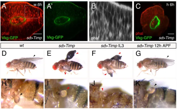
We have shown thatMmp1 expression and Vkg-GFP abundance appear inversely correlated in wing and haltere discs, and also that decreasing levels of Vkg-GFP coincide with significant morphological changes in the wing disc. We resolved to analyze whether changes in Mmp1 activity were responsible for the differences in Vkg-GFP presence and morphology between the two discs, with a further analysis of the possible impact on adult morphology. At 6 h APF, overexpression ofTimpin the wing pouch prevents the elimination of Vkg-GFP in the basal side of the cells; in addition, there is neither apposition of tissue layers nor expansion of the tissue (Fig. 5A,A′). The cells exhibit an elongated shape, resembling the wild-type 6 h APF haltere cells and not the wild-type wing disc cells of the same stage (compare Fig. 5B with Fig. 2C,D). In this mutant condition, therefore, the distal wing disc adopts several characteristics of the haltere disc. In the haltere pouch,Timp overexpression does not elicit significant changes at 3 h or 6 h APF, although the morphology in frontal sections appears rounder than in the wild type (Fig. 5C).
Next, we studied the possible effect in adult appendage development of expressing Timp with sd-Gal4. As this driver is inserted on the X chromosome, and therefore dosage compensated, there is high male lethality in this and other mutant combinations. We therefore focused our study on females heterozygous for the Gal4 insertion. IncreasingTimpexpression in the wing pouch led to animals with abnormal wings, shorter than in the wild type, and in which dorsal and ventral surfaces were not apposed (Fig. 5E, compare with the wild type in 5D). The halteres in most of these animals (in 28/39 females) presented a more globular capitellum than in the wild type, with a square-like shape (compare Fig. 5I with 5H, the wild type). A very similar phenotype in wings and halteres is observed ifTimp expression is induced only in middle-late third instar larvae (insd-Gal4tub-Gal80tsUAS-Timpflies shifted from
17°C to 29°C; Fig. 5F,J). By contrast, if these flies are shifted to 29° C at 12 h APF, the flies are wild type, suggesting that regulation of Mmp activity is important only prior to this time-point (Fig. 5G,K). We performed the reciprocal set of experiments by increasing Mmp1expression. The induction of highMmp1levels with thesd -Gal4 line resulted in high larval lethality, so we used the -Gal4/ Gal80ts system and temperature-shift experiments with 24 h
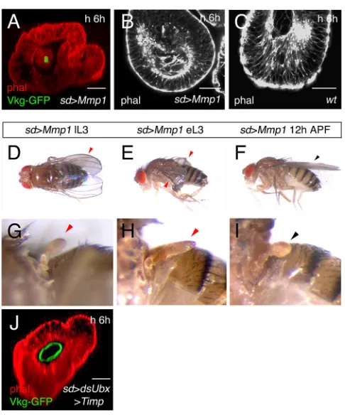
resolution to reduce the lethality rate. When we expressedMmp1 in the haltere pouch in middle-late third instar larvae, we observed elimination of most Vkg-GFP expression at 6 h APF, but no dorso-ventral apposition (Fig. 6A). The epithelium is no longer pseudostratified, resembling more the epithelium of the 6 h APF wild-type wing disc than that of the 6 h APF wild-type haltere disc (Fig. 6B, compare with 6C, the wild-type haltere disc). The wing disc of the prepupa overexpressingMmp1does not differ much from the wild type (not shown). Adult flies emerging after this treatment presented reduced or crumpled (but flat) wings, whereas others showed adumpyphenotype (Fig. 6D). Halteres in nearly 50% of the flies (8/18) have an elongated and flattened capitellum (compare Fig. 6G with the wild type in Fig. 5H). Interestingly, if Mmp1 expression is induced earlier, at the early-middle third larval instar stage, the capitellum of the halteres was, in most cases (7/11), significantly flatter and more elongated than in the wild type (Fig. 6H). The wings also presented the crumpled phenotype observed when the temperature shift was made at middle-late third
Fig. 4.Ubxregulates Mmp1 and Vkg levels in the haltere pouch.
(A,B) Optical frontal sections showing that at 3 h APF, the amount of Mmp1 (red) is higher, but that of Vkg-GFP (green) is lower, in the wing disc (A) (n=8) compared with in the haltere disc (B) (n=8). Phalloidin is shown in white. (C) Quantification of Mmp1 and Vkg-GFP levels in the 3 h APF wing (wild type andHm) and haltere (wild type andsd-Gal4 UAS-dsUbx DfUbx109) discs.
The expression in the wing disc has been normalized to 1 (arbitrary units). Data are mean±s.d. *P<0.01. (D,E) ISH with aTimpprobe in 3 h APF wing (D) (n=30) and haltere (E) (n=27) imaginal discs, showing RNA expression close to the haltere pouch but not the wing pouch (arrows). (F,G) Optical frontal sections of a 3 h (F) and 6 h (G) APFsd-Gal4 UAS-dsUbx DfUbx109haltere
disc, showing increased Mmp1 expression (F) (n=7) and lower Vkg-GFP signal (G) (n=6) with respect to the wild type. Note also the apposition of dorsal and ventral surfaces. (H) In 3 h APFHmwing discs, the expression of Mmp1 is drastically reduced (some remaining signal is indicated by an arrowhead) (n=7). (I) Optical frontal section of the distal part of anHm/Vkg-GFP prepupal wing disc at 6 h APF, showing that the ectopic expression ofUbxprevents Vkg degradation (compare with the wild type in 3C) (n=6). Scale bars: 21μm.
DEVEL
O
instar larvae (Fig. 6E). As seen when overexpressingTimp, forcing Mmp1 expression at 12 h APF onwards is immaterial to wing development and most halteres also develop normally (Fig. 6F,I).
Finally, we analyzed whether the changes in Vkg-GFP expression observed when Ubx expression is modified were mediated by the regulation of Mmps. In pupae in which we simultaneously reduceUbxexpression and Mmp activity (sd-Gal4
UAS-dsUbxUAS-Timp) the haltere discs show, at 6 h APF, the ring
of Vkg-GFP expression and the lack of expansion and dorso-ventral apposition that are characteristic of the wild-type haltere, and not of theUbx-mutant haltere (Fig. 6J, compare withsd-Gal4 UAS-dsUbx haltere disc in Fig. 4G). This strongly suggests thatUbxregulates Vkg-GFP expression and morphology through the control of Mmps. We also analyzed whether changes inUbxexpression during the third larval instar stage cause phenotypes related to those observed when modifying Mmp1. If Ubx is downregulated in the early-middle third larval stage, the halteres are transformed into small wings, most of which are flat (17/27) (compare Fig. 7B with the control haltere in 7A). Some of these winglets have, for unknown reasons, dark patches. By contrast, ifUbxexpression is reduced at the late third larval stage, the halteres are enlarged but globular (Fig. 7C). These data suggest that the size of the transformation to wing correlate to a certain extent with appendage shape: bigger transformations make flatter organs whereas small transformations produce a globular shape.
Taken together, our results indicate that: (1) the levels of Mmp1 in the wing pouch lumen are significantly higher than in the corresponding haltere lumen at the first 3 h of prepupal development; (2) this higher expression negatively correlates with GFP levels in the basal membrane of the wing disc, and Vkg-GFP persists longer in the distal haltere disc than in the distal wing disc; (3)Ubx, which is expressed only in the haltere disc, maintains Vkg-GFP levels in the haltere pouch for longer, most likely by directly downregulatingMmp1expression; and (4) changes in Mmp
activity impact pupal disc shape and adult appendage morphology– halteres elongate and get flatter if Mmp1 expression is increased, and wings shorten and get inflated if Mmp activity is reduced.
DISCUSSION
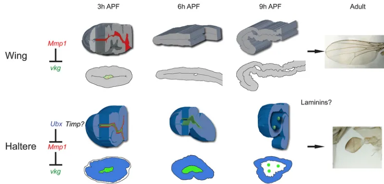
The homeotic gene Ubx has served as a paradigm of cell fate determination mediated via transcriptional regulation.Ubxis one of the few well-characterized master control genes, as its activity is necessary and sufficient for haltere fate specification. Although a number of transcriptional targets ofUbxhave been identified using different molecular strategies, how individual targets contribute to the molecular and biochemical composition of the haltere structure remains unclear. Equally elusive are the components of the morphogenetic pathways that influence the development of the specific structural and morphological characteristics of the halteres. As Ubx activity is necessary to suppress wing fate, comparative analysis employing molecular and biochemical criteria between the two structures has proven to be highly instructive. Here, we have addressed howUbxcontrols organ shape to prevent the flat shape of the wings and develop the globular form of the halteres. Our results show that the presence ofUbxin the haltere disc directs its shape by controlling the ECM dynamics in the prepupal haltere pouch. Importantly, our data reveal that the molecular targetMmp1assists Ubxin achieving this task. This regulation prevents the expansion and apposition observed in the wing pouch and consequently, instead of a flat wing-like structure, a globular-shaped haltere emerges. A summary of our results is shown in Fig. 8.
Ubxand regulation of ECM component expression
[image:6.612.152.461.57.248.2]Previous studies have reported Collagen IV expression (and other components of the ECM) in the pupal wing disc (Fristrom et al., 1993; Murray et al., 1995). By using GFP protein trap lines, and in fixed tissue, we have shown that Perlecan and Collagen IV are eliminated from the wing pouch at 6 h APF because of high levels of
Fig. 5. Reducing Mmp activity modifies prepupal wing and haltere disc shape, and adult appendage morphology.(A,A′) Optical frontal section of a 6 h APF distal wing disc overexpressingTimp(sd-Gal4 Vkg-GFP UAS-Timp): Vkg-GFP is not degraded, the disc shows a globular shape, and there is no elongation or apposition of dorsal and ventral surfaces (n=7). (B) Boxed area in A. The cells present an elongated shape, similar to wild-type haltere (but not wing) cells at this stage. (C) Optical frontal section of a 6 h APFsd-Gal4 UAS-Timpdistal haltere disc, showing a round shape (n=4). (D) Wild-type fly, showing flat wings. (E) Insd-Gal4 UAS-Timpanimals, grown at 29°C, the wings are shorter and do not appose their dorsal and ventral surfaces (36/39). (F) A similar phenotype is observed insd-Gal4tub-Gal80tsUAS-Timpflies, shifted from 17°C to 29°C at late third larval instar (lL3) (34/36). (G) If the shift is made at 12 h APF, the
flies have normal wings (n=8). (H) Detail of a wild-type haltere. (I) Insd-Gal4 UAS-Timpflies grown at 29°C most of the halteres are globe shaped (28/39). (J) Asd-Gal4 UAS-Timp tub-Gal80tsfly shifted from 17°C to 29°C at about the middle-late third larval instar: the capitellum is more globular than in the wild type
(30/36). (K) When pupae from this genotype are shifted from 17°C to 29°C at 12 h APF, the capitellum is wild type (n=8). Black arrowheads indicate normal development; red arrowheads indicate abnormal development. Scale bars: 21μm.
DEVEL
O
Mmp1 activity, but persist at this stage in the haltere pouch. A microarray analysis of genes regulated by Ubx in the pupal haltere disc also identified Mmp1 as aUbxtarget, although the precise spatial and temporal expression of this gene was not described (Pavlopoulos and Akam, 2011). Our analysis shows thatUbxdownregulatesMmp1expression in the haltere pouch at 3 h and 6 h APF, and that Mmp1 degrades Collagen IV. By contrast, contribution of Mmp2 to this regulation, based on its distribution, is probably negligible. We have also expressed an Mmp2RNAi line in wing and haltere discs with thesd-Gal4 driver but found no phenotype (not shown). Our data thus argue that the ability of Ubxto attenuate Mmp1 activity is crucial for haltere disc morphogenesis.
There is a correlation between elimination ofvkgin wing discs at 6 h APF and pouch cells acquiring a more isodiametric shape. Our results are consistent with previous studies showing that the basement membrane constricts cells to give the columnar shape to the disc cells, and that upon degradation of ECM there is transition from a columnar pseudostratified epithelium to a cuboidal epithelium and a flattening of the tissue (Domínguez-Jiménez et al., 2007; Pastor-Pareja and Xu, 2011). This is what we observed in the wild-type pupal wing disc. Therefore, the rapid clearance of the ECM in the wing disc would lead to two events: first, the tissue expands as the shape of the cells changes from columnar to cuboidal; and second, the dorsal and ventral epithelia appose. If we inactivate Mmp1 and prevent Vkg degradation by expressingUbxor Timp, there is no dorso-ventral apposition and no expansion, thus resembling haltere disc development. Supporting our conclusions, previous experiments have reported that dorso-ventral adhesion is sensitive to ectopicUbxonly during the larval and prepupal stages (Pavlopoulos and Akam, 2011). This latter result also suggests that the second separation and adhesion of wing surfaces, which take place after prepupa (Fristrom et al., 1993), may not be as relevant forUbx control of dorso-ventral adhesion as the gene regulation we have described at 3-6 h APF. Our data, therefore, strongly suggest that elimination of the ECM in the basal side of the wing pouch cells is indispensable for the flattening and expansion of the tissue and development of the wing. By preventing Mmp activity, and so preserving ECM integrity, ectopic Ubx makes the prepupal wing pouch acquire a haltere pouch-like shape. In the wild-type haltere disc, by reducingMmp1expressionUbxdelays basal ECM degradation, the transition from columnar to cuboidal epithelium, the early disc expansion, and the apposition of dorsal and ventral layers.
[image:7.612.53.296.53.345.2]Even though the presence of ECM in the basal membrane, which is regulated byUbx, is enough to prevent a wing shape, the absence of this regulation in the prepupa may not be sufficient to give a wing-like form to the haltere disc. If we force Mmp1 expression in the haltere pouch, Vkg-GFP signal is not observed at 6 h APF and the epithelium is no longer pseudostratified, as in the wild-type wing pouch. However, ventral and dorsal layers do not make contact and there is no expansion of the tissue. We also noted that in the 9 h APF wild-type haltere pouch there is no Collagen IV in the basement membrane (and we do not know the mechanism that degrades it), but the shape of the cells and the absence of dorso-ventral apposition is different from the 9 h APF wing pouch. Apart from Mmp1, other genes are likely to have an impact in distinguishing wing and haltere morphology.
Fig. 6. IncreasingMmp1expression changes prepupal wing and haltere disc shape, and adult wing and haltere morphology.(A) In asd-Gal4 UAS-Mmp1Vkg-GFPtub-Gal80tshaltere pouch at 6 h APF, after induction of Mmp1at the middle third instar, Vkg-GFP expression is not detected in the basement membrane (compare with the wild type in Fig. 3D) (n=7). (B,C) In optical transverse sections of these discs (distal part at the bottom) an increase in lumen size is observed, and the epithelium is cuboidal (B), rather than columnar pseudostratified, as in the wild type (C; see also Fig. 2D) (n=4). (D) In
sd-Gal4 UAS-Mmp1 tub-Gal80tsflies shifted from 17°C to 29°C at about
middle-late third instar larvae (sd>Mmp1lL3) (n=18), several of the flies show a
dumpyphenotype. About half of these flies (8/18) have halteres that show elongation and flattening of the capitellum (G; the wild type in Fig. 5H). (E) If the temperature shift is made at early-middle of the third larval instar (sd>Mmp1
eL3), the adults show crumpled wings (n=11). (H) The halteres of most of these flies (7/11) are elongated and flattened, very noticeably in some specimens. (F,I) If the temperature shift is made at 12 h APF, the adults show wild-type wings and halteres (n=15). (J) Frontal section of a 6 h APFsd-Gal4
UAS-dsUbxUAS-Timp tub-Gal80tsprepupa after a change from 17°C to 29°C at
early third larval stage. There is a ring of Vkg-GFP expression at the basal side of cells abutting the lumen, and the disc has a haltere-like morphology (no dorso-ventral apposition or expansion), much more similar tosd-Gal4 UAS-Timp
wing discs than tosd-Gal4 UAS-dsUbx Df109haltere discs (compare with Fig. 5A and Fig. 4G) (n=10). Black arrowheads indicate normal development; red arrowheads indicate abnormal development. Scale bars: 21μm.
Fig. 7. Effect of downregulation ofUbxon size and shape.(A) Control haltere of ansd-Gal4 UAS-dsUbx Df109 tub-Gal80tsfly grown at 17°C. The
capitellum is slightly enlarged because of the haploinsufficiency forUbx
(Df109). (B) If larvae of the same genotype are shifted from 17°C to 29°C at about early third instar (sd>UbxieLIII), the halteres are transformed into small wings, most of them flat (17/27). (C) By contrast, if larvae of the same genotype undergo this shift at about late third instar (sd>UbxilLIII), the halteres are enlarged, but are much smaller than the wings of the early shift and globular in shape (40/42). Black arrow indicates normal development; red arrows indicate abnormal development.
DEVEL
O
[image:7.612.323.552.57.137.2]For example,blistered (Fristrom et al., 1994) and laminin genes (Henchcliffe et al., 1993; Martin et al., 1999) are required to maintain a flat wing blade, and mutations in genes such asdumpy,
piopioorminiature, which encode apical matrix proteins, also cause
abnormal wing development (Carlson, 1959; Prout et al., 1997; Wilkin et al., 2000; Jazwiń ́ska et al., 2003; Roch et al., 2003; Bökel et al., 2005; Ray et al., 2015). All these genes have been shown to be regulated by Ubxat different developmental stages (Roch et al., 1998; Slattery et al., 2011; Pavlopoulos and Akam, 2011). However, we have analyzed Laminin A expression (Sarov et al., 2016) and found no major difference between wing and haltere prepupal discs at 3-9 h APF (not shown). The apposition of dorsal and ventral surfaces probably needs the cooperation of mechanisms regulating apical and basal ECM proteins, and the wayUbxmight control them to distinguish haltere from wing shape remains unexplored.
Timp and Mmp activity, and organ shape
The differences in ECM dynamics affect adult organ shape: changes in Mmp1 activity impact pupal disc shape and adult dorsal appendage morphology. The interpretation of the results is somehow complicated because of the high lethality observed when expressing Mmp1at certain developmental stages. However, we find a broad correlation between Mmp1 activity and organ shape: if Mmp1 expression is increased, halteres are longer and flatter, and wings are shorter and crumpled; ifMmp1activity is reduced, the halteres are more round, and wings are shorter and more globular. Timing of major gene requirement forMmp1expression does not seem to be the same for wing and haltere discs. ElevatedTimpexpression in the wing disc at late larval stages, or throughout development, prevents apposition of dorsal and ventral layers and expansion of the pouch, and the adults have inflated and abnormal wings. AlthoughMmp1expression in the haltere disc is low, high levels ofTimpresult in halteres with a slightly more globular form, suggesting that further reduction of Mmp1 and Mmp2 activity impacts haltere shape. Interestingly, the effects observed in both appendages occur when Timp is expressed in middle-late third instar larvae onwards, but not after 12 h APF. Therefore, the time when Timp expression affects appendage morphology includes the prepupal stage, when Mmp activity regulates the presence of Vkg-GFP.
In contrast, whenMmp1expression is forced in the haltere disc we see elongation and flattening of the haltere, but this diminishes if the expression is induced in late third instar larvae, suggesting Mmp1might affect haltere shape mainly, but not exclusively, at the early-middle third larval stage. It seems, therefore, thatUbxmight control other pathways to maintain the globular haltere shape, even if Vkg is absent in prepupal stages. Supporting this, it has been demonstrated that simultaneous activation of the Akt pathway and downregulation ofexpanded(or activation of Yorkie) in the haltere disc also results in flattened halteres (Singh et al., 2015). These mechanisms might contribute to maintain the globular shape of the haltere pouch throughout the rest of the pupal development to form the adult appendage.
The control of size and shape byUbx
[image:8.612.106.500.56.245.2]The formation of an organ requires the coordination of mechanisms that regulate size, shape and differentiation.Ubxregulates haltere disc size by controlling the Decapentaplegic and Hippo pathways (Crickmore and Mann, 2006; de Navas et al., 2006; Makhijani et al., 2007; Singh et al., 2015). Most of these observations are based on Ubx-mediated regulation of patterning events in the third instar larval stage. Our results reported here suggest thatUbxmay, to some extent, regulate haltere shape early in third instar larvae and that elimination of the Hox protein immediately before pupariation may change cell fate but not organ shape. A delayed effect of Ubx activity was also reported in the control of haltere cell differentiation (Roch and Akam, 2000). We also noticed that timing of Ubx expression, shape and size, might correlate in appendage development. The early downregulation of Ubx makes large appendages that are flat (small wings), but late downregulation produces smaller appendages that are globular. Although early overexpression ofMmp1makes halteres slightly bigger and flatter than those of the wild type, the conversion to a completely flat appendage might require its reaching a critical size. Cell differentiation, by contrast, might be changed byUbxexpression in pupal stages (Roch and Akam, 2000), suggesting differentiation might be uncoupled from size and shape. Further studies are required to understand how Ubx regulates gene expression to coordinate size, shape and differentiation in haltere development.
Fig. 8. Model of Ubx activity and the control of appendage shape.Simplified scheme of the early development of wing and haltere pupal discs, and the role ofUbxin the haltere disc. In the wing pouch,Mmp1expression at 3 h APF degrades Vkg and allows wing development. In the haltere pouch,Ubx
downregulatesMmp1expression and therefore Vkg is not degraded. As a result, the expansion of the disc and apposition of dorsal and ventral surfaces, which are observed in the wing pouch, are prevented. There is probably also Timp protein expression in the haltere disc, and a role for laminins and other ECM components in determining shape.
DEVEL
O
MATERIALS AND METHODS
Genetics
The following mutations were used:pbx1(Lewis, 1963),DfUbx109(Lewis, 1978) andHm(Lewis, 1982). The Gal4/UAS system (Brand and Perrimon, 1993) was used to drive or inactivate gene expression with the following Gal4 and UAS lines:sd-Gal4 (Calleja et al., 1996), UAS-dsUbx(Monier et al., 2005), and UAS-Mmp1and UAS-Timp(Page-McCaw et al., 2003). Other lines used wereMmp2-GFP (Deady et al., 2015),vkg-GFP (vkgG454) andtrol-GFP
(trolGFPZCL1700) (Morin et al., 2001), and DE-cad-Tomato (Huang et al.,
2009). To activate or inactivate genes at precise times, we used the Gal4/Gal80ts
system (McGuire et al., 2003). The white pupa stage was taken as 0 h APF.
Immunochemistry
Staining of imaginal discs was performed as described previously (Roch and Akam, 2000), with slight modifications. The experiments comparing wing and haltere discs were carried out with the same experimental conditions of fixation, staining and acquisition using a confocal microscope. We used a mouse anti-Mmp1 monoclonal antibody cocktail (Page-McCaw et al., 2003; Developmental Studies Hybridoma Bank, 3A6B4, 3B8D12 and 5H7B11, 1:50), and TRITC-phalloidin (Sigma Aldrich, 1:200), Phalloidin-Atto 488 and Phalloidin-Atto 647N (Life Technologies, 1:200) to detect F-actin. Secondary antibodies were anti-mouse Alexa Fluor 448 and 555 (ThermoFisher Scientific, A-21202 and A-31570, respectively, 1:100). Images were taken with LSM710 vertical and LSM710 inverted multiphoton microscopes (Zeiss).
In situhybridization
ISH was carried out as described in Wolff (2000). The probes were generated using the cDNA GH26186 from the collection of expression sequence tags (ESTs) of the BerkeleyDrosophilaGenome Project, with RNA polymerase T7 and SP6 promoter sequences in its ends. The transcription was carried out using the RNA polymerases T7 or SP6 (Roche) for 2 h.
Time-lapse movies
Time-lapse movies were captured ofex vivoimaginal discs as described in Aldaz et al. (2010) and Guarner et al. (2014), with small modifications. The time of development observed in the movies does not exactly agree with the time of development in pupae. We have taken the retraction of the peripodial membrane, which occurs at around 4 h APF, and the disc eversion as reference time points to analyze the presence of Vkg-GFP and Trol-GFP.
Measurements and statistical analysis
To quantify Mmp1 and Vkg-GFP levels in wing and haltere discs, we took pairs of discs from the same pupa and measured the intensity in 3 h APF wing and haltere segmented lumens using a user-made macro for phalloidin channel in FIJI. An RStudio macro was generated to normalize and generate a wing/ haltere ratio, which was later implemented in Excel (Microsoft) to generate the average graphs. Analysis for both Mmp1 and Vkg-GFP was performed on six to eight discs. Images were taken using LSM710 vertical and LSM710 inverted multiphoton microscopes. A two-tailed Mann–Whitney–Wilcoxon test was used to compare wild-type wing and haltere samples, wild-type and
Hmwings, and wild-type andsd-Gal4 UAS-UbxRNAi halteres.
Acknowledgements
We thank Antonio Tarruell for collaborating in the study of the adult phenotypes; M. Akam, Maria Dolores Martin-Bermudo, theDrosophilaBloomington Stock Center, the ViennaDrosophilaRNAi Centre, and the Developmental Studies Hybridoma Bank (DSHB) at the University of Iowa for providing stocks and antibodies; and the confocal microscopy service at the CBMSO for excellent service. We thank members of E.S.-H.’s and L.S.S.’s labs for help and comments, and Y. Bellaïche, J. Casanova and G. Deshpande for comments on the manuscript.
Competing interests
The authors declare no competing or financial interests.
Author contributions
Conceptualization: J.M.D.H., E.S.-H.; Formal analysis: J.M.D.H., C.G.-C., D.F., J.C.P.-P., L.S.S., E.S.-H.; Investigation: J.M.D.H., C.G.-C., D.F.; Writing - original
draft: E.S.-H.; Writing - review & editing: J.M.D.H., C.G.-C., D.F., J.C.P.-P., L.S.S., E.S.-H.; Funding acquisition: L.S.S., E.S.-H.
Funding
This work was supported by the Ministerio de Ciencia e Innovación (BFU2011-26075 to E.S.-H.); the Ministerio de Economıa, Industria y Competitividad, Gobiernó de España (BFU2014-51989-P to E.S.-H.); by a Spanish-Indian Program, the Department of Scientific and Industrial Research, Ministry of Science and Technology (DST/INT/Spain/P-12/2009 to L.S.S.) and the Ministerio de Ciencia e Innovación (ACI2009-0857 to E.S.-H.); and by the Fundación Ramón Areces (institutional grant) to E.S.-H.
Data availability
Macros for the quantification of Mmp1 and Vkg in wing and haltere discs have been deposited in Figshare under accession number 10.6084/m9.figshare.6615218.
Supplementary information
Supplementary information available online at
http://dev.biologists.org/lookup/doi/10.1242/dev.161844.supplemental
References
Aldaz, S., Escudero, L. M. and Freeman, M.(2010). Live imaging of Drosophila imaginal disc development.Proc. Natl. Acad. Sci. USA107, 14217-14222. Baena-López, L. A., Baonza, A. and Garcı́a-Bellido, A.(2005). The orientation of
cell divisions determines the shape of Drosophila organs.Curr. Biol.15, 1640-1644. Blair, S. S. (2007). Wing vein patterning in Drosophila and the analysis of
intercellular signaling.Annu. Rev. Cell Dev. Biol.23, 293-319.
Bökel, C., Prokop, A. and Brown, N. H.(2005). Papillote and Piopio: Drosophila ZP-domain proteins required for cell adhesion to the apical extracellular matrix and microtubule organization.J. Cell Sci.118, 633-642.
Brand, A. H. and Perrimon, N.(1993). Targeted gene expression as a means of altering cell fates and generating dominant phenotypes.Development118, 401-415. Cabrera, C. V., Botas, J. and Garcı́a-Bellido, A.(1985). Distribution of Ultrabithorax
proteins in mutants of Drosophila bithorax complex.Nature318, 569-571. Calleja, M., Moreno, E., Pelaz, S. and Morata, G.(1996). Visualization of gene
expression in living adultDrosophila.Science274, 252-255.
Carlson, E. A.(1959). Allelism, complementation, and pseudoallelism at the dumpy locus in Drosophila melanogaster.Genetics44, 347-373.
Cohen, S. M.(1993). Imaginal disc development. InThe Development of Drosophila melanogaster(ed. M. Bate and A. Martinez-Arias), pp. 747-841. Plainview, New York: Cold Spring Harbor Laboratory Press.
Crest, J., Diz-Muñoz, A., Chen, D. Y., Fletcher, D. A. and Bilder, D.(2017). Organ sculpting by patterned extracellular matrix stiffness.Elife6, e24958.
Crickmore, M. A. and Mann, R. S.(2006). Hox control of organ size by regulation of morphogen production and mobility.Science313, 63-68.
Deady, L. D., Shen, W., Mosure, S. A., Spradling, A. C. and Sun, J.(2015). Matrix metalloproteinase 2 is required for ovulation and corpus luteum formation in Drosophila.PLoS Genet.11, e1004989.
de Navas, L. F., Garaulet, D. L., and Sanchez-Herrero, E. (2006). The Ultrabithorax Hox gene of Drosophila controls haltere size by regulating the Dpp pathway.Development133, 4495-4506.
Diaz de la Loza, M. C. and Thompson, B. J.(2017). Forces shaping the Drosophila wing.Mech. Dev.144, 23-32.
Dickinson, M. H.(1999). Haltere-mediated equilibrium reflexes of the fruit fly,
Drosophila melanogaster.Philos. Trans R. Soc. Lond. B Biol. Sci.354, 903-916. Domı́nguez-Jiménez, P., Brown, N. H. and Martin-Bermudo, M. D. (2007). Integrin-ECM interactions regulate the changes in cell shape driving the morphogenesis of the Drosophila wing epithelium.J. Cell Sci.120, 1061-1071. Etournay, R., Popović, M., Merkel, M., Nandi, A., Blasse, C., Aigouy, B., Brandl, H.,
Myers, G., Salbreux, G., Jülicher, F. et al.(2015). Interplay of cell dynamics and epithelial tension during morphogenesis of the Drosophila pupal wing.Elife4, e07090. Fristrom, D.(1976). The mechanism of evagination of imaginal discs of Drosophila
melanogaster. III. Evidence for cell rearrangement.Dev. Biol.54, 163-171. Fristrom, D. and Fristrom, J. W.(1975). The mechanism of evagination of imaginal
discs of Drosophila melanogaster. 1. General considerations.Dev. Biol.43, 1-23. Fristrom, D. and Fristrom, J. W.(1993). The metamorphic development of the adult epidermis. InThe Development of Drosophila melanogaster(ed. M. Bate and A. Martinez-Arias), pp. 843-897. Plainview, New York: Cold Spring Harbor Laboratory Press.
Fristrom, D., Wilcox, M. and Fristrom, J.(1993). The distribution of PS integrins, laminin A and F-actin during key stages in Drosophila wing development.
Development117, 509-523.
Fristrom, D., Gotwals, P., Eaton, S., Kornberg, T. B., Sturtevant, M., Bier, E. and Fristrom, J. W.(1994). blistered: a gene required for vein/intervein formation in wings of Drosophila.Development120, 2661-2671.
Gilmour, D., Rembold, M. and Leptin, M. (2017). From morphogens to morphogenesis and back.Nature541, 311-320.
DEVEL
O
Godenschwege, T. A., Pohar, N., Buchner, S. and Buchner, E.(2000). Inflated wings, tissue autolysis and early death in tissue inhibitor of metalloproteinases mutants of Drosophila.Eur. J. Cell Biol.79, 495-501.
Guarner, A., Manjón, C., Edwards, K., Steller, H., Suzanne, M. and Sá nchez-Herrero, E.(2014). The zinc finger homeodomain-2 gene of Drosophila controls Notch targets and regulates apoptosis in the tarsal segments.Dev. Biol.385, 350-365. Guirao, B., Rigaud, S. U., Bosveld, F., Bailles, A., López-Gay, J., Ishihara, S., Sugimura, K., Graner, F. and Bellaïche, Y. (2015). Unified quantitative characterization of epithelial tissue development.Elife4, e08519.
Haigo, S. L. and Bilder, D.(2011). Global tissue revolutions in a morphogenetic movement controlling elongation.Science331, 1071-1074.
Henchcliffe, C., Garcia-Alonso, L., Tang, J. and Goodman, C. S.(1993). Genetic analysis of laminin A reveals diverse functions during morphogenesis in Drosophila.Development118, 325-337.
Huang, J., Zhou, W., Dong, W., Watson, A. M. and Hong, Y.(2009). Directed, efficient, and versatile modifications of the Drosophila genome by genomic engineering.Proc. Natl. Acad. Sci. USA106, 8284-8289.
Hynes, R. O.(2009). The extracellular matrix: not just pretty fibrils.Science326, 1216-1219.
Jaźwińska, A., Ribeiro, C. and Affolter, M.(2003). Epithelial tube morphogenesis during Drosophila tracheal development requires Piopio, a luminal ZP protein.
Nat. Cell Biol.5, 895-901.
Klein, T., Brennan, K. and Martı́nez-Arias, A.(1997). An intrinsic dominant negative activity of serrate that is modulated during wing development in Drosophila.Dev. Biol.189, 123-134.
LaFever, K. S., Wang, X., Page-McCaw, P., Bhave, G. and Page-McCaw, A. (2017). Both Drosophila matrix metalloproteinases have released and membrane-tethered forms but have different substrates.Sci. Rep.7, 44560.
LeBleu, V. S., Macdonald, B. and Kalluri, R.(2007). Structure and function of basement membranes.Exp. Biol. Med.232, 1121-1129.
Lewis, E. B.(1963). Genes and developmental pathways.Am. Zool.3, 33-56. Lewis, E. B.(1978). A gene complex controlling segmentation in Drosophila.Nature
276, 565-570.
Lewis, E. B.(1982). Control of body segment differentiation in Drosophila by the bithorax gene complex. InEmbryonic Development, Part A: Genetic Aspects(ed. M.M. Burger and R. Weber), pp. 269-288. New York, USA: Alan R. Liss. Llano, E., Pendás, A. M., Aza-Blanc, P., Kornberg, T. B. and López-Otı́n, C.
(2000). Dm1-MMP, a matrix metalloproteinase from Drosophila with a potential role in extracellular matrix remodeling during neural development.J. Biol. Chem.
275, 35978-35985.
Llano, E., Adam, G., Pendás, A. M., Quesada, V., Sánchez, L. M., Santamaria, I., Noselli, S. and López-Otı́n, C.(2002). Structural and enzymatic characterization of Drosophila Dm2-MMP, a membrane-bound matrix metalloproteinase with tissue-specific expression.J. Biol. Chem.277, 23321-23329.
Makhijani, K., Kalyani, C., Srividya, T. and Shashidhara, L. S.(2007). Modulation of Decapentaplegic gradient during haltere specification in Drosophila.Dev. Biol.
302, 243-255.
Martin, D., Zusman, S., Li, X., Williams, E. L., Khare, N., DaRocha, S., Chiquet-Ehrismann, R. and Baumgartner, S.(1999).wing blister, a newdrosophila
Laminin a chain required for cell adhesion and migration during embryonic and imaginal development.J. Cell Biol.145, 191-201.
McGuire, S. E., Le, P. T., Osborn, A. J., Matsumoto, K. and Davis, R. L.(2003). Spatiotemporal rescue of memory dysfunction in Drosophila. Science 302, 1765-1768.
Monier, B., Astier, M., Sémériva, M. and Perrin, L.(2005). Steroid-dependent modification of Hox function drives myocyte reprogramming in the Drosophila heart.Development132, 5283-5293.
Morata, G. and Garcia-Bellido, A.(1976). Developmental analysis of some mutants of the bithorax system of Drosophila.Wilehm Roux Arch Dev. Biol.179, 125-143. Morin, X., Daneman, R., Zavortink, M. and Chia, W. A.(2001). A protein trap
strategy to detect GFP-tagged proteins expressed from their endogenous loci in Drosophila.Proc. Natl. Acad. Sci. USA98, 15050-15055.
Murray, M. A., Fessler, L. I. and Palka, J. (1995). Changing distributions of extracellular matrix components during early wing morphogenesis in Drosophila.
Dev. Biol.168, 150-165.
Page-McCaw, A.(2008). Remodeling the model organism: matrix metalloproteinase functions in invertebrates.Semin. Cell Dev. Biol.19, 14-23.
Page-McCaw, A., Serano, J., Santé, J. M. and Rubin, G. M.(2003). Drosophila matrix metalloproteinases are required for tissue remodeling, but not embryonic development.Dev. Cell4, 221-233.
Park, Y., Rangel, C., Reynolds, M. M., Caldwell, M. C., Johns, M., Nayak, M., Welsh, C. J., McDermott, S. and Datta, S. (2003). Drosophila perlecan modulates FGF and hedgehog signals to activate neural stem cell division.Dev. Biol.253, 247-257.
Pastor-Pareja, J. C. and Xu, T.(2011). Shaping cells and organs in Drosophila through the opposing effects of fat body-secreted Collagen IV and Perlecan.Dev. Cell21, 245-256.
Pavlopoulos, A. and Akam, M.(2011). Hox gene Ultrabithorax regulates distinct sets of target genes at successive stages of Drosophila haltere morphogenesis.
Proc. Natl. Acad. Sci. USA108, 2855-2860.
Prout, M., Damania, Z., Soong, J., Fristrom, D. and Fristrom, J. W.(1997). Autosomal mutations affecting adhesion between wing surfaces in Drosophila melanogaster.Genetics146, 275-285.
Ray, R. P., Matamoro-Vidal, A., Ribeiro, P. S., Tapon, N., Houle, D., Salazar-Ciudad, I. and Thompson, B. J.(2015). Patterned anchorage to the apical extracellular matrix defines tissue shape in the developing appendages of drosophila.Dev. Cell34, 310-322.
Roch, F. and Akam, M.(2000). Ultrabithorax and the control of cell morphology in Drosophila halteres.Development127, 97-107.
Roch, F., Baonza, A., Martı́n-Blanco, E. and Garcı́a-Bellido, A.(1998). Genetic interactions and cell behaviour in blistered mutants during proliferation and differentiation of the Drosophila wing.Development125, 1823-1832.
Roch, F., Alonso, C. R. and Akam, M.(2003). Drosophila miniature and dusky encode ZP proteins required for cytoskeletal reorganisation during wing morphogenesis.J. Cell Sci.116, 1199-1207.
Sarov, M., Barz, C., Jambor, H., Hein, M. Y., Schmied, C., Suchold, D., Stender, B., Janosch, S., Vinay Vikas, K. J., Krishnan, R. T. et al.(2016). A genome-wide resource for the analysis of protein localisation in Drosophila.Elife
25, e12068.
Singh, S., Sánchez-Herrero, E. and Shashidhara, L. S.(2015). Critical role for Fat/ Hippo and IIS/Akt pathways downstream of Ultrabithorax during haltere specification in Drosophila.Mech. Dev.138, 198-209.
Slattery, M., Ma, L., Négre, N., White, K. P. and Mann, R. S.(2011). Genome-wide tissue-specific occupancy of the Hox protein Ultrabithorax and Hox cofactor Homothorax in Drosophila.PLoS ONE6, e14686.
Srivastava, A., Pastor-Pareja, J. C., Igaki, T., Pagliarini, R. and Xu, T.(2007). Basement membrane remodeling is essential for Drosophila disc eversion and tumor invasion.Proc. Natl. Acad. Sci. USA104, 2721-2726.
Voigt, A., Pflanz, R., Schäfer, U. and Jäckle, H.(2002). Perlecan participates in proliferation activation of quiescent Drosophila neuroblasts. Dev. Dyn. 224, 403-412.
Wei, S., Xie, Z., Filenova, E. and Brew, K.(2003). Drosophila TIMP is a potent inhibitor of MMPs and TACE: similarities in structure and function to TIMP-3.
Biochemistry42, 12200-12207.
White, R. A. H. and Akam, M.(1985).Contrabithoraxmutations cause inappropriate expression ofUltrabithoraxproducts inDrosophila.Nature318, 567-569. Wilkin, M. B., Becker, M. N., Mulvey, D., Phan, I., Chao, A., Cooper, K., Chung,
H.-J., Campbell, I. D., Baron, M. and MacIntyre, R.(2000). Drosophila dumpy is a gigantic extracellular protein required to maintain tension at epidermal-cuticle attachment sites.Curr. Biol.18, 559-567.
Wolff, T.(2000). Histological techniques for the Drosophila eye. Part I: Larva and Pupa. InDrosophila Protocols(ed. W. Sullivan, M. Aahburner and R. S. Hawley), pp. 210-227. Plainview, New York: Cold Spring Harbor Laboratory Press. Yasothornsrikul, S., Davis, W. J., Cramer, G., Kimbrell, D. A. and Dearolf, C. R.
(1997). viking: identification and characterization of a second type IV collagen in Drosophila.Gene198, 17-25.