IJPSR (2018), Volume 9, Issue 12 (Research Article)
Received on 06 April, 2018; received in revised form, 06 June, 2018; accepted, 12 June, 2018; published 01 December, 2018
ANTIMICROBIAL AND ANTIPLASMODIAL ACTIVITY DISPLAYED BY ACTINOMYCETES ISOLATED FROM SOILS FROM KAVANGO AND HARDAP REGIONS IN NAMIBIA
Albertina Mariina Ndinelao Iikasha * 1, Florence Dushimemariya 2, Maria Mbewe 2, Henrique Eiman 2 and Davis Ropafadzo Mumbengegwi 3
Department of Anatomy 1, Department of Biological Sciences 2, Multidisciplinary Research Center 3, University of Namibia, 340 Mandume Ndemufayo Avenue Pioneers Park, Windhoek, Namibia.
ABSTRACT: Plasmodium falciparum and multidrug resistant bacteria such as Mycobacterium avium, Staphylococcus aureus and Escherichia coli are among the top causes of morbidity and mortality especially in developing countries. These pathogens are responsible for the reported antibiotic resistance incidents globally. Due to the increase in antibiotic resistance worldwide, there is an urgent need for new bioactive compounds. This study aimed at determining the antiplasmodial and antimicrobial activity of secondary metabolites produced by soil actinomycetes. Soil samples were collected from Kavango and Hardap regions and cultured on various media to isolate and identify actinomycetes. Secondary metabolites produced were extracted using different solvents. The antimicrobial activity of different isolated secondary metabolites was determined against M. avium, S. aureus and E. coli. Antiplasmodial activity of four bioactive fractions with potent antibacterial activity was further determined against a chloroquine resistant (D10) P. falciparum strain at various concentrations. Bioactive compounds from two isolates displayed activity against M. avium, whilst another two showed activity against E. coli. None of the secondary metabolites displayed activity against S. aureus. A concentration dependent reduction in parasitaemia for P. falciparum was observed across all four fractions both after 24 and 48 h. These results display a vast potential antibacterial and antiplasmodial activity and should be considered for future drug development.
INTRODUCTION: Infectious and parasitic diseases account for 9.5 million deaths reported annually on a global scale 1. Most reported deaths are caused by multidrug resistant pathogens such as
Mycobacterium avium, Staphylococus aureus,
Escherichia coli, Vibrio cholera, Salmonella typhi
and Shingella dysentery which cause more deaths
especially in immune- compromised patients 2, 3.
QUICK RESPONSE CODE
DOI:
10.13040/IJPSR.0975-8232.9(12).5191-96
Article can be accessed online on: www.ijpsr.com
DOI link: http://dx.doi.org/10.13040/IJPSR.0975-8232.9(12).5191-96
Apart from these diseases, malaria which is transmitted through the bite of infected Anopheles (female) mosquitoes is also a public health concern especially in developing countries 4. Globally, an estimated 3.3 billion people in 97 countries and territories are at risk of being infected with malaria
4
. There are four parasitic species that cause malaria in humans. Among the four species that cause malaria, Plasmodium falciparum and
Plasmodium vivax are the most common of the
malaria-causing parasites, while Plasmodium
falciparum is the most deadly 4. These parasites
invade the human red blood cells and subsequently also infect the liver cells and also invade brain tissue in severe malaria.
Keywords:
Actinomycetes, Antimicrobial, Antiplasmodial,
Secondary metabolites, Kavango, Hardap Correspondence to Author:
Albertina Mariina Ndinelao Iikasha
Department of Anatomy, Hage G. Geingob Campus,
University of Namibia, 340 Mandume Ndemufayo Avenue Pioneers Park, Windhoek, Namibia.
Over the years, antimalarial drugs such as quinine, chloroquine and primaquine and antibacterial drugs such as penicillin, fluoroquinolones and cephalosporins, vancomycin and teicoplanin have been developed, that interfere with the growth and reproduction of the pathogens. However, the pathogens have developed resistance to the drugs, hence there is a persistent need to develop safe and effective medicine to be used in treating malaria and other infectious diseases 3, 5, 6.
Namibia has diverse habitats and soils that is composed of unique microorganism including Actinomycetes, which can be used as a source of new antibiotics based on their ability to effectively produce secondary metabolites. Most members of the genus have saprophytic, free-living lifestyles, and contend for resources with several other organisms in oligotrophic environments and they grow as hyphae, which frequently branch to form an intricate vegetative mycelium 7.
Actinomycetes are known to produce secondary metabolites during the stationary phase of their life cycle, with antibacterial antitumor, antifungal, antiviral, anti-parasitic activities 8, 9, 10. The discovery of new antimalarial and antibacterial drugs is a major motive of research on infectious diseases. Most researches on actinomycetes in Namibia have only gone as far as characterization of the microbes as well as the secondary metabolites they produce; antiplasmodial and antibacterial activity of these actinomycetes is rarely researched. Hence, the possibility of discovering novel antibiotics extracted from actinomycetes from soil sampled in Namibia is greater, considering its unique and diverse climate. This study was conducted to evaluation the antimicrobial and antiplasmodial effects of secondary metabolites extracted from Actinomyctes from Kavango and Hardap regions in Namibia.
MATERIALS AND METHODS:
Sample Collection: All the soil samples used were randomly collected on January 2015 and grouped into 2 groups based on the region of collection. Group 1 soil samples were collected from a farm 40km east of Rehoboth in Hardap region and group 2 soil samples were collected along the Kavango River in Kavango region.
Isolation of Actinomycetes: A serial dilution was carried out on a mixture of 1g of soil and 9 ml of distilled water until a 10-6 dilution factor was achieved. Approximately 0.1 ml of the undiluted, 10-1 and 10-3 dilution factors was inoculated onto prepared agar plates of Yeast extract/Malt extract agar and Kuster‟s agar, the plates were then incubated at 30 ºC for 11 days. Single Actinomycetes colonies were selected and sub-cultured into separate respective agar plates for 11 days 11.
Primary Antibiotic Screening: The selected Actinomyces were stab-inoculated in the center of Yeast extract/Malt extract agar and Kuster‟s agar plates. The plates were then incubated at 30 ºC for 7 days, three bacterial strains (Escherichia coli,
Mycobacterium avium and Staphylococcus aureus)
were used for screening. All three organisms were grown in Luria Broth for 24 h and 48 h respectively. The optical density of each organism was determined at 600 nm and the values obtained used to determine the amount of bacterial culture to be transferred into test tubes containing 6ml of sloppy agar. The formula used for the volume determination was as follows:
E. coli: 4 = OD600 × µL
M. avium, S. aureus: 160 = OD600 × µL
The inoculated Sloppy agar was then poured over the stab-inoculated media around the colony and the plates were incubated at 37 ºC over-night for E. coli and 36 h for M. avium and S. aureus.
Scale up of the Actinomycetes Cell Mass for Secondary Metabolites Production: In order to increase cell mass, the Actinomycetes colonies on plates were inoculated into 50 ml conical flasks containing 50 ml of Nutrient broth and incubated at 37 ºC on a shake flask incubator for 7 days. Following this, a gram-staining procedure was carried out to validate the purity of the culture. Stock culture was prepared by mixing 700 µL of culture with 300 µL of a mixture of 50% glycerol and 50% deionized water into an eppendorf tube and stored at -20 ºC for future research.
liters of Nutrient broth and incubated in a 37 ºC shaking incubator for 14 days. Filtration was carried out to separate the cells from the broth. The broth was then divided into 3 equal volume which were placed into individual bottles. An equal amounts of chloroform and toluene were poured into each of two bottles of aliquots respectively.
The remaining was kept frozen until required. The residual cells were place into falcon tubes and mixed with 10 ml of methanol. The organic-solvent mixtures were subjected to continuous shaking for 2 days to extract the antibiotics into the solvent. Following this extraction, the solvents were separated from the broth and placed under a fume hood until all the solvent evaporated. The antibiotic remaining after evaporation, was re-dissolved in 2 ml of the respective solvent, and stored at -20 ºC until required.
Antibacterial Screening of the Extracted Secondary Metabolites: Five µl of each of the antibiotic-solvent solutions was dotted onto 3 TLC plates and left to dry (solvent evaporated completely). Each plate was dabbed with one of the test organisms grown in Luria Broth for 2 days for
S. aureus and M. avium and 24 h for E. coli using
cotton wool. The plates were placed on a wet paper towel and subsequently placed into a plastic container, which was sealed and placed into a 37 ºC incubator for 24 h. After overnight incubation, the plates were dabbed with MTT solution and placed in an incubator for 20 min to allow the MTT to react with the respiring cells (the reaction with respiring cells was indicated by the appearance of a purple color), after which each antibiotic „dot‟ was analyzed for the presence of an inhibition zone (clear “halo”) 12
.
Anti-malarial Bioassay of the Extracted Secondary Metabolites: The anti-plasmodial assay was carried out according to methods implemented by Nafuka & Mumbengegwi 13. P. falciparum cells were thawed, maintained in media and the parasitemia was determined by preparing blood smears on microscope slides. The parasites were maintained in RPMI 1640 supplemented with L-glutamine, NaOH, D-glucose, HEPES, gentamycin and serum was used for culture maintenance. Red blood cells obtained from O+ donors were added to reach required hematocrit.
To conduct the anti-plasmodial assay, four extracts were chosen based on their observed activity against M. avium and other bacterial species, at 10, 50 and 100 µg/ml, n=5. The assay was conducted in 96 well plates, for two time points, 24 and 48 h, with parasite culture at 2% parasitaemia. Wells treated with Chloroquine served as positive control, while wells receiving media only acted as negative control.
To determine the effect of the treatment after 24 and 48 h, pseudo-smears were analyzed. This was done by missing the contents 3/5 of the replicates of each category in an eppendorf tube. The parasitaemia was then determined for each smear by microscopy.
Statistical Analysis: All experiments were repeated three times for each experiment. The percentage parasite-growth inhibition was calculated, as follows:
{Parasitaemia (Negative control) – Parasitaemia Treatment/ Parasitaemia (Negative control)} × 100
The results obtained in parasitaemia were analyzed using the GenStats® Discovery computer software. The test for normality was carried out initially for each data set in order to determine the type (parametric or non-parametric) of statistical analysis to be implemented. One-way Anova was carried out to determine the significance between the treatments and the negative control at 24 hours and 48 h and the MANOVA test was used for the difference in parasitaemia between 24 and 48 h of exposure.
RESULTS AND DISCUSSION:
Actinomycetes Isolation and Antibacterial Activity: The soil samples cultured on yeast/malt extract and Kuster‟s agar yielded three distinct colonies each as shownin Table 1. The isolated colonies were classified as actinomyctes based on their morphological appearance, concave or convex colonies with a powdery consistency which stuck firmly to agar surface 14.
TABLE 1: ISOLATED ACTINOMYCETE COLONIES ON DIFFERENT MEDIA
Media Number of Colonies Designation
Yeast extract/Malt extract (YEME) agar
3 1A, 1B, 1D
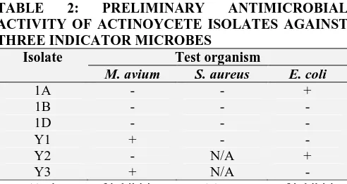
The actinomycetes isolates were screened against three bacterial strains, M. avium, S. aureus and E. coli. The susceptibility of these strains is presented in Table 2. Antibacterial activity was only observed against M. avium by isolates Y1 and Y3. Isolates 1A and Y3 also showed preliminary antibacterial activity against E. coli. Secondary metabolites are produced in the stationery phase of microbial growth, which was evidenced by the length of the period required for growth. Bunch and Harris (2013) 15 also reported of actinomycetes isolate 3725 that was susceptible against E. coli and
Bacillus subtilis. However, no zones of inhibition
were observed on all six isolates against S. aureus.
Table 2 shows the findings on preliminary antimicrobial activity of different isolates against
M. avium, S. aureus and E. coli. These findings are
contrary to those by Chaudhary et al., (2013) 16 who reported on 31 actimonycetes isolates with activity against S. aureus and 11 other infectious pathogens. Furthermore, Streptomyces sp. VITBRK2 are also reported todisplay potent antibacterial activity against Multi-drug resistant S.
aureus and Enterococcus strains 17. The observed
activity was due to the presence of indolo compounds in Streptomyces sp. VITBRK2. It is probable that that none of the six actinomycetes from Namibian soil had the ability to produce indole compounds which are responsible for
anti-Staphylococcus activity. The zones of inhibition
observed in the antibacterial preliminary screen suggested the presence of secondary metabolites produced by these isolated actinomycetes colonies.
TABLE 2: PRELIMINARY ANTIMICROBIAL
ACTIVITY OF ACTINOYCETE ISOLATES AGAINST THREE INDICATOR MICROBES
Isolate Test organism
M. avium S. aureus E. coli
1A - - +
1B - - -
1D - - -
Y1 + - -
Y2 - N/A +
Y3 + N/A -
Key: (-) absence of inhibition zone, (+) presence of inhibition zone, N/A - not applicable
Secondary metabolites produced by actinomycetes were fractionated using three solvents of varying polarities: toluene, chloroform and methanol. These fractions were then screened in a secondary antimicrobial susceptibility screen against M.
avium, S. aureus and E. coli. Table 3 shows the
results for secondary antimicrobial activity against three indicator microbes by three fractions of the actinomycete‟s secondary metabolites. Secondary metabolites eluted in toluene appeared not to have antibacterial activity. The most active faction was eluted in chloroform, followed by Methanol as depicted in Table 3.
TABLE 3: CONFIRMATORY SECONDARY ANTIMICROBIAL ACTIVITY AGAINST THREE INDICATOR MICROBES BY THREE FRACTIONS OF THE ACTINOMYCETE’S SECONDARY METABOLITES
Test Organism M. avium S. aureus E. coli
Solvent Tol Ch Met Tol Ch Met Tol Ch Met
Isolate
SO4 - + - + + - + + -
SO5 - + - - + + - + -
SO6 - + - - + - - + -
SO7 - + - - + - - - -
SO10 - - - - + - - + -
SO13 - + - - + + - + -
1A - - - -
1B - - - -
1D - - - -
Y1 - - - -
Y2 - - - -
Y3 - - - -
However, Rajan and Kannabiran (2014) 17 used ethyl acetate to characterize the metabolites and identify the antibiotics. Isolate 1A displayed antibacterial activity against E. coli in the primary screen but not in the secondary confirmatory
[image:4.612.317.563.120.251.2]The lack of activity in the secondary screen may be because the secondary metabolites produced did not elute into any of the solvent systems used. It is also possible that the secondary metabolites produced were not effective against the indicator microbes. The observation of more than one active antibacterial fraction from one isolate indicates that more than one antibacterial compound is produced.
Antiplasmodial Activity: Four fractions were chosen for antiplasmodial activity based on their broad spectrum activity, and their growth inhibitory activity against M. avium and S. aureus as depicted in Table 4. Antiplasmodial activity of the fractions in Table 4 against chloroquine sensitive plasmodial parasites (D10) is shown in Fig. 1 and 2 and is expressed as percentage parasitemia. All fractions exhibited antiplasmodial activity although reduction in parasitemia was not significantly
different between treatment concentrations (P=0.05). This may be a result of extract saturation at active sites. There was a significant difference in parasitemia between 24 and 48 h time points (P<0.001) indicating that fractions were more active on mature stages of the parasites (schizonts). This observation could also mean that a longer period of exposure to the fractions is required to reduce the level of parasites significantly 19.
TABLE 4: ACTINOMYCETE SECONDARY
META-BOLITES CHOSEN FOR SCREENING AGAINST
PLASMODIA BASED ON THEIR SECONDARY
CONFIRMATORY ANTIMICROBIAL ACTIVITY
Extract Property
SO4 Broad spectrum of activity SO5 Strongest activity against M. avium SO7 Activity against M. avium SO13 Largest inhibition zone against S.
aureus by methanol extract
Dahari et al., 2016 20 reported on acetone crude extracts of H11809 and FH025 that each displayed active anti-plasmodial activity with IC50 values of
0.57 ± 0.09 and 1.28 ± 0.11 µg/mL, respectively. Extracts with IC50 ≤ 50 μg/mL are regarded as
active whereas those with values higher than 50 μg/mL are considered inactive 20
. However, for this study, since there was no significant difference in reduction in parasitaemia between the concentrations of each treatment, this could mean that the efficacy of the treatments was concentration independent (time dependent). Hence, the rate and extent of microorganism killing remain unchanged regardless of antimicrobial concentration.
Therefore IC50 (the concentration of an extract that
inhibits the growth of 50% of the parasites; a measure of the strength of the extract) could not be calculated.
The growth inhibitory activity of these Actinomycetes derived fractions against test microorganisms, bacteria and plasmodia, indicates that the fractions have a range of secondary metabolites which exert their bioactive activities through more than one mode of action.
CONCLUSION: Based on the findings from this study, soils collected from Kavango and Hardap regions displayed potent antimicrobial activity against M. avium and S. aureus as well as anti- plasmodial activity against chloroquine resistant (D10) P. falciparum. Hence, further studies should be conducted to fully fraction and isolate compounds with anti-bacterial and anti- plasmodic activity from the Kavango and Hardap soils in Namibia. This can help to address the increasing antibiotic resistance and the emerging infectious diseases.
FIG. 1: REDUCTION IN PARASITEMIA AFTER TREATMENT WITH ACTINOMYCETES SECONDARY
[image:5.612.70.558.185.449.2]METABOLITESFOR 24 h
ACKNOWLEDGEMENT: University of Namibia for providing the facilities to conduct the study.
CONFLICT OF INTEREST: No conflict of interest.
REFERENCES:
1. World health Organization. Health in 2015: From MDGs to SDGs. Infectious diseases 2015; 1-204.
2. Lim C, Takahashi E and Hongsuwan M: Epidemiology and burden of multidrug-resistant bacterial infection in a developing country. Abdool Karim Q, ed. eLife 2016; 5: e18082:1-18.
3. Asit KC: Multi-Drug Resistant Genes in Bacteria and 21st century problems associated with antibiotic therapy. BioTechnology: An Indian Journal 2016; 12(12): 114. 4. World health organization. World malaria report 2014;
1-178.
5. Harten RM, Willems RJL, Martin NI and Hendrickx APA:
Multidrug-Resistant Enterococcal Infections: New
Compounds, Novel Antimicrobial Therapies? Trends in Microbiology 2017; 25(6): 467-479.
6. Fair RJ and Tor Y: Antibiotics and Bacterial Resistance in the 21st Century. Perspectives in Medicinal Chemistry 2014; 6: 25-64.
7. Cruz-Morales P, Vijgenboom E and Barona-Gómez F: The
Genome Sequence of Streptomyces lividans 66 reveals a novel tRNA-dependent peptide biosynthetic system within a metal-related genomic. Island.Geo Bio Evol 2013; 5(6): 1165-1175.
8. Chaudhary HS: Diversity and versatility of actinomycetes and its role in antibiotic production. Journal of Applied Pharmaceutical Science 2013; 3(8): 83-94.
9. Manikkam R, Venugopal G, Ramasamy B and Kuma V: Effect of critical medium components and culture conditions on antitubercular pigment production from novel Streptomyces sp D25 isolated from Thar Desert, Rajasthan. Journal of Applied Pharmaceutical Science 2015; 5(06): 015-019.
10. Loganathan K, Kumar G, Kirthi AV, Rao KV and Rahuman AA: Entomopathogenic marine actinomycetes as potential and low-cost biocontrol agents against blood-sucking arthropods. Parasitol Res 2013; 112(11): 39-519.
11. Mohseni M, Norouzi H, Hamedi J and Roohi A: Screening of Antibacterial Producing Actinomycetes from Sediments of the Caspian Sea. International Journal of Molecular and Cellular Medicine 2013; 2(2): 64-71.
12. Maataoui H, Iraqui M, Jihani S, Ibnsouda S and Haggou A: Isolation, characterization and antimicrobial activity of a Streptomyces strain isolated from deteriorated wood. African Journal of Microbiology Research 2014; 8(11): 1178-118.
13. Nafuka SN and Mumbengegwi DR: Phytochemical
analysis and in-vitro anti-plasmodial activity of selected ethnomedicinal plants used to treat malaria associated symptoms in Northern Namibia. Int. Sci. Technol. J. Namibia 2013; 2: 78-93.
14. Janardhan A, Kumar AP, Saigopal BVDR and Narasimha
G: Production of Bioactive Compounds by Actinomycetes and Their Antioxidant Properties. Biotechnology Research International 2014; 1-8.
15. Bunch AW and Harris RE: The Manipulation of Micro-organisms for the Production of Secondary Metabolites. Biotechnology and Genetic Engineering Reviews 2013; 4(1): 117-144.
16. Chaudhary HS, Yadav J and Gopalan N: Antibacterial activity of actinomycetes isolated from different soil samples of Sheopur (A city of central India). Adv Pharm Technol Res 2013; 4(2): 118-123.
17. Rajan BM and Kannabiran K: Extraction and identification of antibacterial secondary metabolites from marine
Streptomyces sp. VITBRK2. Int J Mol Cell Med 2014; 3(3): 130-137.
18. Abdelmohsen UR, Grkovic T, Balasubramanian S, Kamel
MS, Quinn RJ and Hentschel U: Elicitation of secondary metabolism in actinomycetes. Biotechnology Advances 2015; 33(6): 798-811.
19. Sánchez-Valdéz FJ, Padilla A, Wang W, Orr D and Tarleto R: Spontaneous dormancy protects Trypanosoma cruzi
during extended drug exposure.eLife 2018; 7: e34039. DOI: https://doi.org/10.7554/eLife.34039.
20. Dahari DE, Salleh RM, Mahmud F, Chin LP, Embi N and
Sidek HM: Anti-malarial activities of two soil acti-nomycete isolates from Sabah via inhibition of glycogen synthase sinase 3β. Tropical Life Sciences Research 2016; 27(2): 53-71.
All © 2013 are reserved by International Journal of Pharmaceutical Sciences and Research. This Journal licensed under a Creative Commons Attribution-NonCommercial-ShareAlike 3.0 Unported License. This article can be downloaded to ANDROID OS based mobile. Scan QR Code using Code/Bar Scanner from your mobile. (Scanners are available on Google Playstore)
How to cite this article: