IJPSR (2017), Volume 8, Issue 9 (Research Article)
Received on 03 February, 2017; received in revised form, 24 April, 2017; accepted, 27 May, 2017; published 01 September, 2017
FORMULATION AND IN VITRO CHARACTERIZATION OF ANTICANCER DRUG
LOADEDSOLIDLIPIDNANOPARTICLES
Uma Maheshwari Bandi *, K. Philip, D. Basker Reddy, A. Swaroopa, L. Prabakaran and G. Parthasarathy
R.R. College of Pharmacy, Guddadahalli, Bangalore - 550 098, Karnataka, India.
ABSTRACT: The aim of the study was to prepare Temozolomide loaded solid lipid nanoparticles and evaluating loaded solid lipid nanoparticles. Temozolomide loaded solid lipid nanoparticles were prepared by micro emulsification method using different lipid (stearic acid) concentration keeping drug concentration unchanged, poloxamer 188 and cetyl alcohol as surfactant and co-surfactant. The prepared SLN were characterized for surface morphology by SEM analysis, drug loading and % entrapment efficiency, FTIR, zeta potential and in vitro diffusion studies. The lowest and highest % Entrapment Efficiency was found to be 85.85% and 97.45% for F1 to F5 batches respectively. Release studies were done by using saline phosphate buffer pH 7.4 using dialysis bag diffusion method. Zeta potential was found to be - 20.3 mV and particle size was found to be 119.34. For 8 hrs in vitro drug release found to be 79.16%, 74.45%, 71.42%, 61.36%, and 55.13% respectively. The release data was analysed by different kinetic models and found that the formulations best fit model is peppas and the drug release mechanism was Fickian diffusion (n-value 0.3808).
INTRODUCTION: Cancer chemotherapy is now established value and highly specialized field and complete re-emission should be the goal of this therapy in which the drugs are often used in maximum tolerated doses. Intensive regimens used earlier yield better results. Cancers were treated
with one drug at a time 1. The main problem of
cancer therapy is not the lack of efficient drugs, but that these drugs are very difficult to concentrate in the tumour tissue without leading to toxic effects
on neighbouring organs or tissues 2. So now days
going for targeted drug delivery systems i.e.
nanoparticles. The uncontrolled and often rapid proliferation of cells can lead to either a benign or
malignant tumor 2, 3.
QUICK RESPONSE CODE
DOI:
10.13040/IJPSR.0975-8232.8(9).3808-12
Article can be accessed online on: www.ijpsr.com
DOI link: http://dx.doi.org/10.13040/IJPSR.0975-8232.8 (9).3808-12
Nanoparticles are solid colloidal particles ranging in size from 1 to 1000 nm and composed of
macromolecular materials 4. Solid lipid
nanoparticles were defined as oil in water emulsion for parenteral nutrition, but the liquid lipid (oil) of
the emulsion has been replaced by a solid lipid i.e.
yielding solid lipid nanoparticles.
Solid Lipid Nanoparticles Provide the Following Advantages:
Control and target drug release
Improves the stability of pharmaceuticals
High and enhanced drug content when
compared to other carriers
Feasibility of carrying both lipophilic and
hydrophilic drugs
Water based technology
Easy to scale-up and sterilize
Good biocompatibility
Low toxicity
SLNs particularly those in the range of 120-200
nm are not taken up readily by the cells of the
Keywords:
Temozolomide, Solid lipid nanoparticles,
In vitro drug release kinetics
Correspondence to Author: Uma Maheshwari Bandi
M. Pharm
D/O Saideshwar Rao Bandi Chinthakani Village and Mandal, Khammam -507208, Telangana, India.
reticulo-endothelial system and thus bypass
liver and spleen filtration 4, 5.
The main advantage of the study is to provide drug delivery by oral route to the targeted site. SLN are colloidal lipid systems, which have been proposed for several administration routes, such as parenteral, oral and topical. These solid lipid nanopaticles were prepared by different methods as following
High shear homogenization
Hot homogenization
Cold homogenization
Ultrasonication/high speed homogenization
Solvent emulsification/evaporation
Micro emulsion based SLNs preparations
SLNs preparation by using supercritical fluid
Spray drying method
Double emulsion method
Hot homogenization followed by ultrasonication
6- 8
.
Temozolomide: It is an oral alkylating agent which can be used for the treatment of Grade IV astrocytoma an aggressive brain tumor, also known as glioblastoma multiforme as well as Melanoma, a
form of skin cancer. It is having protein binding
about 15% and having 100% bioavailability, metabolism is spontaneously hydrolyzed at physiologic pH to the active species, 3methyl -(triazen-1-yl) imidazole-4-carboxomide (MTIC) and to Temozolomide acid metabolite and half life
is 1.8 hours 9, 10. The objective of the present
research was to study the preparation of loaded solid lipid nanoparticles of Temozolomide by
micro-emulsification technique and to study the effects of composition of lipid materials and surfactant mixture on the particle size, zeta potential, drug entrapment efficiency, FTIR, SEM and in vitro drug release.
MATERIALS AND METHODS:
Materials: Temozolomide, stearic acid from SD fine chem. Limited, Mumbai, poloxamer 188 as a git sample from MSN Laboratories, hyd and cetyl alcohol as gift sample from trident pharmaceuticals, Pondicherry and all other ingredients were analytical grade.
Methods:
Preformulation Study: The drug-excipient interaction studies were performed by suitably mixing the drug with the chosen additives and stored the mixture for 4 weeks. The drug excipient mixture was evaluated periodically for their physical properties like TLC, FTIR and physical observations and compared with the drug raw
material observations. Calibration curve of the drug
was estimated in saline phosphate buffer pH 7.4 and the suitably diluted solutions were analyzed by spectro photometrically using double beam UV spectro photometer at 266.8 nm. The estimation was performed in triplicate and the regression co-efficient was derived to ensure the linearity of the calibration curve.
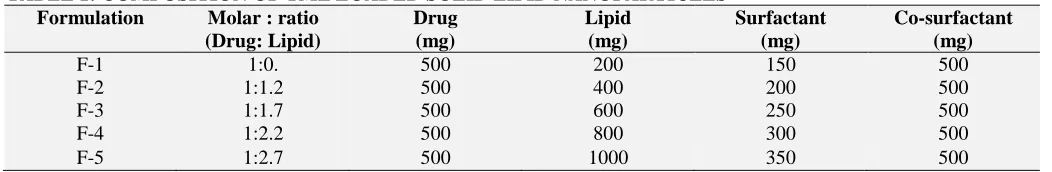
[image:2.612.45.571.558.644.2]Preparation of Solid Lipid Nanoparticles: SLN were prepared from a warm w/o/w micro emulsion containing Stearic acid as lipid, poloxamer 188 as surfactant, and cetyl alcohol as co-surfactant.
TABLE 1: COMPOSITION OF TMZ LOADED SOLID LIPID NANOPARTICLES Formulation Molar : ratio
(Drug: Lipid)
Drug (mg)
Lipid (mg)
Surfactant (mg)
Co-surfactant (mg)
F-1 1:0. 500 200 150 500
F-2 1:1.2 500 400 200 500
F-3 1:1.7 500 600 250 500
F-4 1:2.2 500 800 300 500
F-5 1:2.7 500 1000 350 500
Temozolomide SLN was prepared by micro emulsification technique. Stearic acid was first heated to 75 °C to ensure total melting and then Temozolomide was dispersed in it. Double-distilled water was heated separately (at least 5 °C above the melting point of the lipid). Poloxamer 188 and cetyl alcohol were added to the hot water under
Characterization of Solid Lipid Nanoparticles: Fourier Transform Infra-Red Spectroscopy (FT-IR) Analysis: Drug and lipid compatibility was studied by infrared spectroscopy using Avatar
Thermo Nicolet FT-IR spectrophotometer. The
spectra was recorded in the wavelength region of
400-4000 cm-1 for drug, lipid and Temozolomide,
after mixing respective samples with dried KBr powder and compressing to a disc by a hydraulic press at 5 t compression.
Determination of Drug Loading and % Entrapment Efficiency: Equivalent weight of prepared nanoparticles was dissolved in a mixture of methanol and chloroform (5:5). Require dilutions were performed with the same solvent
mixture and analyzed in a UV-Visible
spectrophotometer at 266.8 nm against the blank.
Amount of drug present in nanoparticles = concentration of dug × dilution factor
Particle Size, Surface Morphology and Zeta Potential: The surface morphology (roundness, smoothness, and formation of aggregates) and particle size were studied by scanning electron microscopy (SEM). Zeta potential of the best formulation was determined by zeta metre.
Evaluation of In vitro Drug Release: In vitro drug release was evaluated by using a dialysis bag
diffusion technique. Equivalent weight of
nanoparticles were packed in dialysis bag and placed in basket immersed in 900 ml of saline phosphate buffer pH 7.4 as dissolution medium at 37 °C paddle rotating at 75 rpm. An aliquote amount of 5 ml sample was drawn in a time
interval of 30, 60, 90,120, 180, 240, 360 and 480 min and replaced with fresh dissolution medium. Dilutions to be done if necessary and absorbance to be recorded at 266.8 nm.
In vitro Drug Release Kinetics: In order to investigate the mechanism of release, the release
data were analysed with the following
mathematical models: zero-order kinetic (Eq. 3), first-order kinetic (Eq. 4) and Higuchi kinetic (Eq. 5)
The following plots were made: Qt versus t
(zero-order kinetic model), log (Q0–Qt) versus t
(first-order kinetic model) and Qt versus t1/2 (Higuchi
model), where Qt is the percent of drug released at
time t, Q0 is the initial amount of drug present in
the lipid nanoparticles and K0, K1 and KH are the
rate constants of the equations 3, 4 and 5, respectively.
korsmeyer-peppas model and Hixson crowell model were applied to interpret the drug release kinetics from the formulations. Based on the
highest regression values (r2) for correlation
coefficients for formulations, the best fit model was decided.
RESULTS AND DISCUSSION:
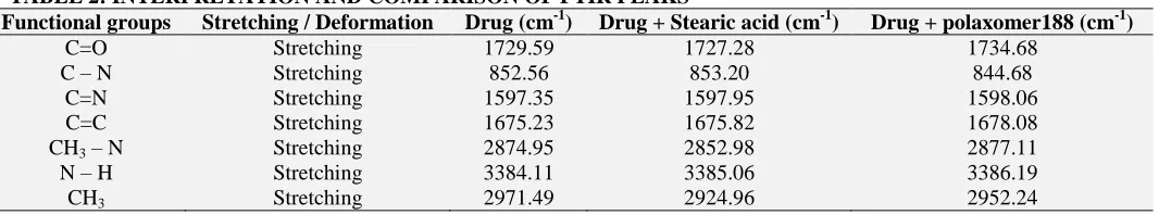
Drug Polymer Interaction by FT - IR Analysis:
Identification of Temozolomide was done by FTIR. Drug excipient interaction studies showed that there is no any physicochemical interaction between the drug and the selected additives of
[image:3.612.44.577.642.741.2]formulations. The results were shown in Table 2.
TABLE 2: INTERPRETATION AND COMPARISON OF FTIR PEAKS
Functional groups Stretching / Deformation Drug (cm-1) Drug + Stearic acid (cm-1) Drug + polaxomer188 (cm-1)
C=O Stretching 1729.59 1727.28 1734.68
C – N Stretching 852.56 853.20 844.68
C=N Stretching 1597.35 1597.95 1598.06
C=C Stretching 1675.23 1675.82 1678.08
CH3 – N Stretching 2874.95 2852.98 2877.11
N – H Stretching 3384.11 3385.06 3386.19
Drug Loading and % Entrapment Efficiency:
Many different drugs had been incorporated in SLNs. The prerequisite to obtain a sufficient loading capacity was a sufficiently high solubility of the drug in the lipid melt. Relative higher drug EE % was one of the major advantages of SLNs. The drug content in five batches of Temozolomide solid lipid nanoparticles was studied. The amount of drug present in TMZ solid lipid nanoparticles
was determined in each batch. Table 3 shows the
results of the drug entrapment efficiency in each of these formulations. It was observed that the entrapment efficiency increased with the increase in concentration of lipid in the formulations. The maximum entrapment was found in F-5 of 97.45% and lowest entrapment in F-1 of 85.85%.
TABLE 3: DRUG LOADING AND % ENTRAPMENT EFFICIENCY OF TMZ LOADED SLN
Formulation Drug Loadind
% Entrapment Efficiency
F-1 50.5% 85.85%
F-2 40.90% 90%
F-3 33.33% 91.05%
F-4 29.62% 94.8%
F-5 26.33% 97.45%
Particle Size, Surface Morphology and Zeta Potential: Average particle size of TMZ loaded solid lipid nanoparticles was found to be 119.34 nm.
FIG. 1: PARTICLE SIZE DISTRIBUTION OF TMZ LOADED SLN OF F-2
Surface morphology was done by using instrument JEOL-JSM-T330A. Magnification was done at 7500- 20000X for taking photographs. Shown in
Fig. 2.
From surface morphology studies it was revealed that the nanoparticles were spherical in shape. Surface charge analysis of the TMZ loaded solid lipid nanoparticles was done by Malvern Zetasizer
and zeta potential was found to be -20.3mV which
indicates they are slightly stable. The Fig. 3 was
[image:4.612.315.566.106.244.2]shown below.
[image:4.612.313.564.266.395.2]FIG. 2: SCANNING ELECTRON MICROSCOPY OF F-2
FIG. 3: ZETA POTENTIAL OF TMZ LOADED SLN OF F-2
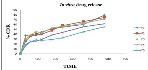
In vitro Drug Release Studies: In vitro release of all batches of Temozolomide loaded SLN (F1-F5) were carried out in saline phosphate buffer pH 7.4 and the formulation showed the release 55.13%, 61.36%, 71.42%, 74.45% and 79.16% respectively at the end of 8 hrs. Among all the formulations F2 showed good controlled release and able to release entire amount in 12 hrs. Therefore it may be considered as the optimized formulation. The dissolution profiles of all the formulations were
shown in Fig. 4.
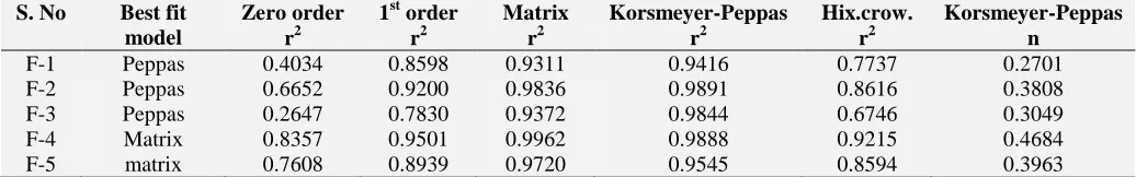
[image:4.612.48.300.465.594.2] [image:4.612.312.566.594.715.2]In vitro Drug Release Kinetic Studies: Data
obtained from in vitro release studies of prepared
TMZ loaded solid lipid nanoparticles were fitted to various kinetic models such as zero order, first order, Higuchi model, Hixson Crowell cube root and korsmeyer-Peppas model. The formulation F2 showed best fit in korsmeyer-Peppas model release
and with the r2 value 0.9891 and n = 0.3808 which
obeys Fickian diffusion. The consolidated
dissolution data analysis of all the formulations is
shown in Table 4 and drug release model of TMZ -
SLN F2 is shown in Fig. 5.
[image:5.612.314.564.55.187.2]FIG. 5: CUMULATIVE PERCENTAGE RELEASE PROFILE WITH MODEL FITTING OF FORMULATION F-2
TABLE 4: KINETIC VALUES OBTAINED FROM IN VITRO RELEASE DATA OF DIFFERENT TEMOZOLOMIDE SOLID LIPID NANOPARTICLES
S. No Best fit model
Zero order 1st order Matrix Korsmeyer-Peppas Hix.crow. Korsmeyer-Peppas
r2 r2 r2 r2 r2 n
F-1 Peppas 0.4034 0.8598 0.9311 0.9416 0.7737 0.2701 F-2 Peppas 0.6652 0.9200 0.9836 0.9891 0.8616 0.3808 F-3 Peppas 0.2647 0.7830 0.9372 0.9844 0.6746 0.3049 F-4 Matrix 0.8357 0.9501 0.9962 0.9888 0.9215 0.4684 F-5 matrix 0.7608 0.8939 0.9720 0.9545 0.8594 0.3963
ACKNOWLEDGEMENT: Nil.
CONFLICT OF INTEREST: There is no conflict
of interest.
REFERENCES:
1. Tripathi K.D: A Text book of Essentials of medical pharmacology. Jaypee Brothers Medical Publishers Pvt. Ltd., Sixth Edition 2008.
2. Cancer cited 2012 June 15. Available from: URL: http://en.wikipedia.org/wiki/cancer.
3. Malcolm R Alison. Cancer. Encyclopedia of life sciences. 2011: 1-8.
4. Pragati S, Kuldeep S, Ashok S and Satheesh M: Solid Lipid Nanoparticles: A Promising Drug Delivery Technology. International Journal of Pharmaceutical sciences and Nano technology 2009; 2(2): 509-516. 5. Mukherjee S, Ray S and Thakur R.S: Solid Lipid
Nanoparticles: A Modern Approaching in Drug Delivery
System. Indian journal of pharmaceutical sciences 2009; 71(4):349-58.
6. Krishna Sailaja A, Amareshwar P and Chakravarty P: Formulation of solid lipid nanoparticles and their application. Current Pharmaceutical Research 2011; 1(2): 197 - 203.
7. Vidyavathi Maravajhala, Sandya Papishetty and Sarika Bandlapalli: Nanotechnology in development of drug delivery system. International Journal of Pharmaceutical Sciences Research 2012; 3(1): 84-96.
8. Vishvajit A. Kamble, Deepali M. Jagdale and Vilasrao J. Kadam: Solid lipid nanoparticles as drug delivery system. International Journal of Pharm and Bio Sciences 2010; 1(3): 1-9.
9. Temozolomide cited 2012 may 28. Available from: URL: http://en.wikipedia.org/wiki/Temozolomide.
10. Malcolm J.M. Darkes, Greg L. Plosker and Blair Jarvis: Temozolomide: A Review of its Use in the Treatment of Malignant Gliomas, Malignant Melanoma and Other Advanced Cancers. American Journal of Cancer 2002; 1(1):55-80.
All © 2013 are reserved by International Journal of Pharmaceutical Sciences and Research. This Journal licensed under a Creative Commons Attribution-NonCommercial-ShareAlike 3.0 Unported License.
This article can be downloaded to ANDROID OS based mobile. Scan QR Code using Code/Bar Scanner from your mobile. (Scanners are available on Google Playstore)
How to cite this article:
[image:5.612.47.565.254.335.2]