ORIGINAL RESEARCH ARTICLE
ACCELERATED ORTHODONTICS- A REVIEW
*Kundal, S. and Shokeen, T.
Post-graduate, PDM Dental College and Research Institute, Bahadurgarh, Haryana, India
ARTICLE INFO ABSTRACT
The orthodontic treatment is, perhaps, in terms of duration, the longest-performed dental procedure. It is universally accepted that if the duration of the orthodontic treatment is reduced, there will be an increased favourable attitude towards the orthodontic therapy. Unfortunately, long orthodontic treatment time also poses several disadvantages like higher predisposition to dental caries, gingival recession and root resorption. Accelerating orthodontic tooth movement can notably reduce treatment duration and risks of side effects. Orthodontic treatment involves the response of the tissues surrounding the tooth on which the force is being applied that happens on a chemical, cellular and mechanical level. So to enhance the body’s response to these orthodontic forces, various ways were found to accelerate the treatment such as surgical methods (corticotomy, piezosurgery, etc), mechanical/ physical stimulation methods (vibration, lasers), drugs, magnets etc. These methods have been successfully proven to reduce treatment times by up to 70%. Hence, this review encapsulates the current knowledge on the accelerated orthodontic tooth movement.
Copyright©2018, Kundal and Shokeen.This is an open access article distributed under the Creative Commons Attribution License, which permits unrestricted use, distribution, and reproduction in any medium, provided the original work is properly cited.
INTRODUCTION
Orthodontic tooth movement is possible due to the remodelling ability of the surrounding bone and soft tissue. Without this remarkable biological phenomenon, the practice- indeed, the very concept of orthodontics would not be possible. Yet, orthodontic appliances are not intentionally built to activate or inhibit specific remodelling pathways and specific cells. Rather, they are built to generate biomechanical force systems that produce the desired tooth and jaw movements needed to establish an ideal occlusion— regardless of the cellular mediators of the response. This begs the question, should we be designing orthodontic appliances to target specific remodelling pathways to move teeth and jaws into an ideal occlusion faster (Aalansari, 2015). A number of attempts have been made to create different approaches both pre-clinically and clinically in order to achieve quicker results, but still there are a lot of uncertainties and unanswered questions towards most of these techniques. Most attempts can broadly be categorized into biological, physical, biomechanical, and surgical approaches.
*Corresponding author: Kundal, S.,
Post-graduate, PDM Dental College and Research Institute, Bahadurgarh, Haryana, India.
Before going into details of these attempts, we need to understand the basics of orthodontic tooth movements and the factors that initiate inhibition and delayed tooth movement. Orthodontic tooth movement occurs in the presence of a mechanical stimuli sequenced by remodelling of the alveolar bone and periodontal ligament (PDL). Bone remodelling is a process of both bone resorption on the pressure site and bone formation on the tension site. Orthodontic tooth movement can be controlled by the size of the applied force and the biological responses from the PDL. The force applied on the teeth will cause changes in the microenvironment around the PDL due to alterations of blood flow, leading to the secretion of different inflammatory mediators such as cytokines, growth factors, neurotransmitters, colony-stimulating factors and arachidonic acid metabolites. As a result of these secretions, remodelling of the bone occurs (Nimeri, 2013). The cellular and molecular
mechanisms underlying accelerated orthodontic tooth
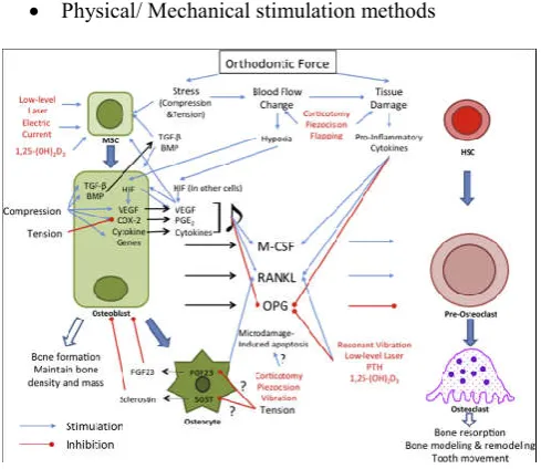
movement have been summarized in Figure 1 (Huang, 2014).
Methods of accelerating tooth movement
Methods to accelerate orthodontic tooth movement can be studied under following categories:
Drugs.
ISSN: 2230-9926
International Journal of Development Research
Vol. 08, Issue, 01, pp.18639-18643, January,2018
Article History:
Received 05th October, 2017
Received in revised form 26th November, 2017 Accepted 20th December, 2017
Published online 31st January, 2018
Key Words:
Accelerated orthodontic tooth movement, Corticotomy, Lasers,
Piezosurgery, Vibration.
Citation: Kundal, S. and Shokeen, T.2018. “Accelerated orthodontics- A review”, International Journal of Development Research, 08, (01), 18639-18643.
Surgical Methods.
[image:2.595.36.281.68.280.2] Physical/ Mechanical stimulation methods
Figure 1. Summary of cellular and molecular mechanisms underlying accelerated orthodontic tooth movement. Methods to
accelerate orthodontic tooth movement are shown in red. Blue arrow, Stimulation; red blunted arrow, inhibition; MSC, mesenchymal stem cell; HSC, hematopoietic stem cell; HIF,
hypoxia inducible factor; FGF, fibroblast growth factor.
DRUGS: Various experiments have been done using these molecules exogenously to enhance tooth movement both in animal experiments and humans (Amith Raja, 2016).
Cytokines: High concentration of cytokines such as interleukins IL-1, IL-2, IL-3, IL-6, IL-8 and tumour necrosis factor alpha (TNF) were found to play a major role in bone remodelling; moreover, interleukin-1 (IL-1) stimulates osteoclast function through its receptor on osteoclasts (Gurbax, 2017).
Prostaglandins: A series of experiments with rat tooth model demonstrated that injection of PGs increase the number of
osteoclasts (Yamasaki, 1980). The human studies depict
clearly that almost twice faster orthodontic tooth movement can be accomplished by local injection of prostaglandins (Yamasaki, 1984 and Anand K Patil, 2006).
Vitamin D: Vitamin D and especially its most active
metabolite which is 1,25-dihydroxyvitamin D3 (1,25(OH)2D3)
together with parathyroid hormone and calcitonin, regulates the amount of calcium and phosphorus in humans. Vitamin D is more effective in modulating bone turnover during orthodontic tooth movement because its effects on bone resorption and formation are balanced.
Parathormone: PTH affects osteoblasts cellular metabolic activity, gene transcriptional activity, and multiple protease secretions. PTH effects on osteoclasts occur through the production of RANKL, a protein that plays critical role in osteoclast formation and its activity. Uninterrupted raise of PTH leads to bone loss; intermittent shoert elevations of the hormone level can be anabolic for bone.
SURGICAL METHODS
The surgical technique to enhance the rate of tooth movement has been documented in many case reports previously. It is a clinically effective technique used for adult patients, where
[image:2.595.308.562.157.278.2]duration of orthodontic treatment may be critical in selective groups of patients. The PDL and alveolar bone remodelling are important parameters in tooth movement, and bone turnover is known to increase after bone grafting, fracture, and osteotomy. Various surgical approaches that have been tried to accelerate tooth movement include osteotomy, corticotomy and piezocision technique. The various surgical methods available are (Figure 2):
Figure 2. Different surgical techniques: A, Periodontally accelerated osteogenic orthodontics or Wilckodontics. B, Corticotomy modifications 1: monocortical piezosurgery 2: monocortical perforations 3: piezocision; C, PDL distraction D,
Dentoalveolar distraction
Corticotomy and Osteotomy
Corticotomy is one of the surgical procedures in which only the cortical bone is cut and perforated but not the medullary bone, suggesting that this will reduce the resistance of the cortical bone and accelerate tooth movements (Nimeri, 2013) Osteotomy is a procedure in which a segment of the bone is cut both into the cortical and medullary bone and is separated and then moved as a unit. It was first tried in orthodontics by Kole (Kole, 1959), where tooth movements were achieved between 6 and 12 months. The technique was further used by others, like Grenerson (Generson, 1978) et al. who used this for open bite treatments.
Advantages
It has been proven successfully by many authors, to accelerate tooth movement.
Bone can be augmented, thereby preventing periodontal
defects, which might arise, as a result of thin alveolar bone.
Disadvantages
High morbidity associated with the procedure.
Invasive procedure.
Chances of damage to adjacent vital structures.
Post-operative pain, swelling, chances of infection, avascular necrosis.
Low acceptance by the patient.
Wilckodontics
accelerated oestrogenic orthodontics (AOO) and periodontal accelerated oestrogenic orthodontics (PAOO). RAP was modified by adding bio-absorbable grafting material over the injured bone to enhance healing. This technique is reported to have postoperative stability and improved retention but more studies are still needed to be done. The drawbacks of these surgical techniques are their invasiveness and the acceleration was only in the first 3 to 4 months and declines with time to the same level of the controls (Nimeri, 2013).
Figure 3. Regional acceleratory phenomenon
Clinical Considerations of RAP
Clinical indications according to Wilcko brothers9 are: a) to accelerate or fasten corrective orthodontic tooth movement b) to facilitate the mechanically challenging orthodontic movements, c) to facilitate correction of moderate to severe skeletal malocclusions. PAOO is contraindicated in certain conditions like a) in patients with active periodontal disease, b) inadequately performed endodontic treatment, c) patients with history of prolonged corticosteroid usage, d) Patients on medication which interfere bone metabolism such as bisphosphonates or non-steroidal anti-inflammatory drugs (NSAIDs).
Advantages
It has been proven successfully by many authors to accelerate tooth movement.
Bone can be augmented, thereby preventing periodontal
defects, which might arise, as a result of thin alveolar bone.
Disadvantages
High morbidity associated with the procedure.
Invasive procedure.
Chances of damage to adjacent vital structures.
Post-operative pain, swelling, chances of infection, vascular necrosis.
Low acceptance by the patient.
Piezocision
Dibart (Dibart, 2011) et al in 2009, introduced a flapless method of corticotomy, using piezosurgery to reduce the
morbidity associated with conventional corticotomy
(Generson, 1978). Piezocision is a minimally-invasive surgical technique designed to accelerate orthodontic tooth movement
(OTM) in combination with orthodontic therapy. Piezocision is made without the need of a flap and perforations on the cortical bone are performed with a piezoelectric knife instead of a bur. Piezocision technique did not cause any periodontal damage as reported by Hassan13. Another benefit of this technique is that it can be used with Invisalign, which gives a better aesthetic appearance and less treatment time as reported by Keser14. Piezocision is a promising tooth acceleration technique because of its various advantages in periodontal, aesthetic, and orthodontic aspects. The vibrations of the piezotome are also claimed to contribute to a faster movement. Because of this, it represents a less aggressive surgical approach than corticotomy (Cadenas, 2015).
Advantages
Minimally invasive.
Better patient acceptance.
Disadvantages
Risk of root damage, as incisions and corticotomies are
“blindly” done.
Micro-osteoperforations
Micro-osteoperforation (MOP) is the only micro-invasive choice capable to speed up orthodontics. The procedure can be finished on chairside in very less time and does not need any advanced training procedures; hence, any trained clinician can perform it. In addition there is zero recovery time. MOP can be used in conjunction with other treatment modalities including but not restricted to, TADs, Invisalign (Align Technology), SureSmile (OraMetrix), and conventional braces (Amith Raja, 2016).
Dentoalveolar Distraction
Distraction osteogenesis is a procedure in which growth of the new bone by mechanical stretching of the pre-existing bone tissue. The technique of distraction osteogenesis involves mechanical stretching of the reparative bone tissue by a distraction device through an osteotomy or corticotomy site. With this technique, new bone is generated in the gap of osteotomy or corticotomy at the approximate rate of 1 mm per day. This has been used for lengthening mandibles than moving individual teeth. Liou16 et al. in 1998 found that when distractor was used in between premolars, able to achieve rates of tooth movement of up to 1.2mm/week (Dibart, 2011). Iseri (2005) et al. in a study said that they achieved tooth movement of 0.8mm per day by moving a canine and its associated block of bone into a premolar extraction space through a distractor appliance. There were no adverse effects such as periodontal problems, ankylosis and root resorption (Huang, 2014).
Physical/ Mechanical Stimulation
Surgical methods, regardless of technique, are still invasive to some degree, and hence have their associated complications. Hence, non-invasive methods have come to the fore. These modalities include lasers, vibration, direct electric current etc.
Lasers
One of the most promising approaches today is
has been shown in the midpalatal suture during rapid palatal expansion, and also stimulates bone regeneration after bone fractures and extraction site. It has been found that laser light stimulates the proliferation of osteoclast, osteoblast and fibroblasts, and thereby affects bone remodeling and accelerates tooth movement. The mechanism involved in the in the acceleration of tooth movement is by the production of ATP and activation of cytochrome C, that low-energy laser irradiation enhanced the velocity of tooth movement via RANK/RANKL and the macrophage colony-stimulating factor and its receptor expression.
Vibrations
Nishimura (Nishimura, 2008) in 2008, used a Ni-Ti expansion
spring on the 1st molar of Wistar rats, and applied a vibration of 60 Hz, 1 m/s2. They stated that the rats that received the vibration showed increased orthodontic tooth movement. Recently, a product by the name Acceledent has arrived at the market, which makes use of this technology. This device consists of an activator, which is the active part of the appliance that delivers the vibration impulses with a USB interface through which it can be connected to a computer to review the patient usage of the appliance, a mouthpiece that contacts the teeth. It is a portable device that can be charged similar to any other electronic device, and has to be worn for 20 minutes a day. Various case studies using this device have shown the treatment times to be reduced by up to 30-40% (Huang, 2014).
Acceledent System
It is a simple, removable and non-invasive appliance. Complements the orthodontic force applied by braces and works through the application of electromechanical vibrations. Just 20 minutes of daily use enhances the orthodontic force applied by braces or aligners to accelerate the rate of orthodontic tooth movement. It is designed to deliver this daily therapy - in the form of gentle micro vibrations - in a way that is comfortable and easy to use. The benefit is shortened orthodontic treatment time and all of the benefits associated with the fast track to correct malocclusion and a great smile. Patient Benefits:
Reduced treatment time without compromised
aesthetics
Less prone to caries or gum disease with shortened treatment
Clinical trial demonstrates excellent root resorption safety profile
Clinical Application for the Future
The administration of exogenous biological molecules to accelerate tooth movement during orthodontic treatments has been intensively tested on animal experiments. However, clinical trials on humans are limited since they are to be administered local injections which can be painful and discomforting to the patients. Also, their side effects were not tested for long periods of time. However, administrations of certain molecules have shown promising results; for example, cytokine, PTH, vitamin D, etc play an important role in bone remodelling and tooth movement. In the physical approach, the low level laser therapy is the most promising method; however, contradictory results are seen. This is due to the
different energies, duration and experimental design. Furthermore, most of these experiments were done in only few weeks, which is a very short time to notice any side effects. The surgical approach is the most clinically used and most tested with known predictions and stable results. However, it is invasive, aggressive and costly and patients are not open to the idea of surgery unless it is the only option needed to have a good occlusion. Piezocision technique is one of the newest techniques in accelerating tooth movement which has good clinical outcome and is considered the least invasive in the surgical approach.
Conclusion
Since long, orthodontic patients have been asking for shorter treatment times, and today, we do have methods that can accelerate orthodontic tooth movement safely. The current methods such as piezocision, micro-osteoperforations, lasers and vibration have reduced or eliminated the invasive nature of previous procedures used to achieve the Regional Acceleratory Phenomenon. Also, they come with additional advantages such as reduced rates of relapse, reduced orthodontic pain and reduced root resorption.
REFERENCES
Aalansari, S. et al. 2015. Biological principles behind accelerated tooth movement. Sem in Orthod., 21:151–161. Amith Raja, B. Muralidhar Reddy, Y. C.A.B. Sreekanth, B.
Vishnu Vardhan Reddy, G. Kranthi Praveen Raj, Reddy R.
2016. Speedy Orthodontics: A Comprehensive Review. Int
J Oral Health Med Res., 2(6):121-124.
Anand K patil et al. 2006. Advances in biology of orthodontic tooth movement- A review. J Ind Orthod Soc., 39: 155-164.
Cadenas de Llano-Perula M, Iglesias Linares A. 2015. Surgically-based methods to modify orthodontic tooth
movement: A literature review. Edorium J Dent.,
2015;2:35–42.
Dibart, S., Sebaoun, J. D., & Surmenian, J. 2011. Piezocision: a minimally invasive, periodontally accelerated orthodontic tooth movement procedure. Practical Osseous Surgery in
Periodontics and Implant Dentistry., 195.
Generson, R.M., Porter, J.M., Zell, A., Stratigos, G.T.. 1978. Combined surgical and orthodontic management of anterior open bite using corticotomy. J Oral Surg., 36(3):216–9.
Gurbax, S., Raahat, V. S., Roopsirat, K., Devinder, P. S. 2017. Accelerated Orthodontic Tooth Movement: A Review.
Mod Res Dent. 1(2).2017.
Hassan NHANE, Sa IT. The effect of using piezocision technique in orthodontic tooth movement on the periodontal condition. Egypt Dent J. 2011; 57:3047. 65. Huang H, Williams RC and Kyrkanides S. Accelerated
orthodontic tooth movement: molecular mechanisms. Am J Orthod Dentofacial Orthop 2014; 146: 620 – 632.
Iseri H, Kisnisci R, Bzizi N, Tuz H. Rapid canine retraction and orthodontic treatment with dentoalveolar distraction osteogenesis. Am J Orthod Dentofacial Orthop. 2005; 127(5):533–41.
Keser EI, Dibart S. Piezocision-assisted Invisalign treatment. Compend Contin Educ Dent. 2011; 32(2):46–8. 50–41 Kole H. Surgical operations on the alveolar ridge to correct
occlusal abnormalities. Oral Surg Oral Med Oral Pathol.
Liou EJ, Huang CS. Rapid canine retraction through distraction of the periodontal ligament. Am J Orthod
Dentofacial Orthop. 1998; 114(4):372–82.
Nimeri G, Kau CH, Abou-Kheir NS, Corona R: Acceleration of tooth movement during orthodontic treatment - a frontier in orthodontics. Prog Orthod 2013, 14: 42. 10.1186/2196-1042-14-42.
Nishimura M, Chiba M, Ohashi T, et al. Periodontal tissue activation by vibration: intermittent stimulation by
resonance vibration accelerates experimental tooth
movement in rats. Am J Orthod Dentofacial Orthop. 2008;
133:572–583.
Wilcko WM, Wilcko T, Bouquot JE, Ferguson DJ. Rapid orthodontics with alveolar reshaping: two case reports of decrowding. Int J Periodontics Restorative Dent. 2001; 21(1):9–19.
Yamasaki K et al. Clinical application of prostaglandin E,
(PGE,) upon orthodontic tooth movement. American
Journal of Orthodontics. 1984; 85: 508-518.
Yamasaki K, Miura F, Suda T. Prostaglandin as a mediator of bone resorption induced by experimental tooth movement in rats. Journal of Dental Research. 1980; 59: 1635-42.