Development and Evaluation of a Broad Bead-Based Multiplex
Immunoassay To Measure IgG Seroreactivity against Human
Polyomaviruses
Sergio Kamminga,
a,bEls van der Meijden,
aHerman F. Wunderink,
a*
Antoine Touzé,
cHans L. Zaaijer,
bMariet C. W. Feltkamp
aaDepartment of Medical Microbiology, Leiden University Medical Center, Leiden, The Netherlands
bDepartment of Blood-Borne Infections, Sanquin Research, Amsterdam, The Netherlands
cUMR 1282, Université François Rabelais/INRA, Tours, France
ABSTRACT
The family of polyomaviruses, which cause severe disease in
immunocom-promised hosts, has expanded substantially in recent years. To accommodate
measure-ment of IgG seroresponses against all currently known human polyomaviruses (HPyVs),
including the Lyon IARC polyomavirus (LIPyV), we extended our custom multiplex
bead-based HPyV immunoassay and evaluated the performance of this pan-HPyV
immunoas-say. The VP1 proteins of 15 HPyVs belonging to 13
Polyomavirus
species were expressed
as recombinant glutathione
S
-transferase (GST) fusion proteins and coupled to
fluores-cent Luminex beads. Sera from healthy blood donors and immunocompromised kidney
transplant recipients were used to analyze seroreactivity against the different HPyVs. For
BK polyomavirus (BKPyV), the GST-VP1 fusion protein-directed seroresponses were
com-pared to those obtained against BKPyV VP1 virus-like particles (VLP). Seroreactivity
against most HPyVs was common and generally high in both test populations. Low
se-roreactivity against HPyV9, HPyV12, New Jersey PyV, and LIPyV was observed. The assay
was reproducible (Pearson’s
r
2⬎
0.84,
P
⬍
0.001) and specific. Weak but consistent
cross-reactivity between the related viruses HPyV6 and HPyV7 was observed. The
serore-sponses measured by the GST-VP1-based immunoassay and a VP1 VLP-based
enzyme-linked immunosorbent assay were highly correlated (Spearman’s
⫽
0.823,
P
⬍
0.001).
The bead-based pan-HPyV multiplex immunoassay is a reliable tool to determine
HPyV-specific seroresponses with high reproducibility and HPyV-specificity and is suitable for use in
seroepidemiological studies.
KEYWORDS
immunoassay, immunology, polyomavirus, seroepidemiology
T
he
Polyomaviridae
family is a group of double-stranded DNA viruses that infect a
broad spectrum of hosts, including humans. The number of identified human
polyomaviruses (HPyVs) has substantially increased over recent years and currently
includes 13
Polyomavirus
species, which are listed in Table 1, including full virus names
and abbreviations (1). A novel polyomavirus recently identified in human skin samples,
named Lyon IARC polyomavirus (LIPyV), has not yet been assigned to a
Polyomavirus
species (2).
Several HPyVs are associated with severe disease, such as BK polyomavirus (BKPyV),
which is associated with nephropathy and hemorrhagic cystitis; JC polyomavirus
(JCPyV), which is associated with progressive multifocal leukoencephalopathy (PML); TS
polyomavirus (TSPyV), which is associated with a dysplastic hair follicle disorder called
trichodysplasia spinulosa; and MC polyomavirus (MCPyV), which is associated with
Merkel cell carcinoma (3–6). An association between HPyV6 and HPyV7 and pruritic and
dyskeratotic dermatosis has recently been proposed (7). In addition, HPyV7 might be
involved in thymomagenesis (8, 9). New Jersey polyomavirus (NJPyV) likely can cause
vasculitis, myositis, and retinal blindness (10).
Received5 October 2017 Returned for modification3 November 2017 Accepted2 January 2018
Accepted manuscript posted online5 January 2018
CitationKamminga S, van der Meijden E, Wunderink HF, Touzé A, Zaaijer HL, Feltkamp MCW. 2018. Development and evaluation of a broad bead-based multiplex immunoassay to measure IgG seroreactivity against human polyomaviruses. J Clin Microbiol 56:e01566-17.
https://doi.org/10.1128/JCM.01566-17.
EditorYi-Wei Tang, Memorial Sloan Kettering Cancer Center
Copyright© 2018 American Society for Microbiology.All Rights Reserved.
Address correspondence to Sergio Kamminga, [email protected], or Mariet C. W. Feltkamp, [email protected].
*Present address: Herman F. Wunderink, Department of Medical Microbiology, University Medical Center Utrecht, Utrecht, The Netherlands.
H.L.Z. and M.C.W.F. contributed equally to this article.
crossm
on May 16, 2020 by guest
http://jcm.asm.org/
The seroprevalence of well-studied polyomaviruses, for instance, BKPyV and JCPyV,
is generally high and comparable among geographically different populations (11–16).
Primary HPyV infections usually occur in childhood and are followed by asymptomatic
persistent infection throughout life, sometimes accompanied by little virus shedding
(12). Though HPyV infection is widespread and its pathology is diverse, symptomatic or
manifest HPyV infections are rare and usually limited to the immunocompromised and
the elderly (17). For most HPyVs, symptomatic infection occurs when the persistent
virus is no longer controlled by the immune system, a phenomenon often referred to
as virus reactivation. However, for some HPyVs primary infection coincident with severe
immunosuppression has been proposed to be the driver of symptomatic disease (18).
Although knowledge of the prevalence of HPyV infections is increasing, little is
known about the incidence and transmission of infection, in particular, of the recently
identified HPyVs, such as Saint Louis polyomavirus (STLPyV), HPyV12, NJPyV, and LIPyV.
One way of filling this knowledge gap is to develop HPyV species-specific serology.
In general, two viral protein 1 (VP1) antigen expression and presentation methods
are used to measure HPyV seroreactivity. One is based on insect cell-expressed VP1
assembled into VP1 virus-like particles (VLP). The other, used in this study, is based on
bacterially expressed glutathione
S
-transferase (GST)-VP1 fusion proteins. Here we
aimed to extend our present HPyV bead-based immunoassay measuring IgG
serore-sponses against the VP1 major capsid protein of HPyVs belonging to the species
Human
polyomavirus 1
(BKPyV),
5
(MCPyV),
6
(HPyV6),
7
(HPyV7),
8
(TSPyV), and
9
(HPyV9) (12)
to HPyVs belonging to the species
Human polyomavirus 2
(JCPyV),
3
(Karolinska
Institutet polyomavirus [KIPyV]),
4
(Washington University polyomavirus [WUPyV]),
10
(Malawi polyomavirus [MWPyV] and HPyV10),
11
(STLPyV),
12
(HPyV12), and
13
(NJPyV)
and LIPyV. MWPyV and HPyV10 belong to the same species and were both included
because they differ at eight amino acid positions in VP1, of which three might be
located in immunogenic loops important for antigen recognition (15, 19).
The performance of this new pan-HPyV multiplex immunoassay was evaluated in
this study by measuring seroreactivity in two pilot populations and by determining the
reproducibility and specificity of the assay. The GST-VP1 fusion protein bead-based
assay was also compared with a VP1 VLP-based serological assay for BKPyV.
MATERIALS AND METHODS
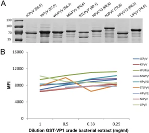
[image:2.585.41.547.84.253.2]Human polyomavirus serology assays.IgG seroreactivities against VP1 were measured using a customized Luminex xMAP assay, as previously described, albeit expanded to include all currently known HPyVs (12, 16, 20). In short, synthetic DNA sequences of VP1 (Table 1) (gBlocks; IDT, San Jose, CA, USA), either wild type (JCPyV, KIPyV, WUPyV, HPyV12, NJPyV, LIPyV) or codon optimized (MWPyV, HPyV10, STLPyV), were cloned into pGEX-5x-3 vectors (GE Healthcare Life Sciences, Chicago, IL, USA) and expressed as GST-VP1.tag fusion proteins inEscherichia coliBL21 Rosetta bacteria. Expression of each newly expressed GST-VP1 fusion protein was analyzed by glutathione-Sepharose 4B purification and SDS-PAGE (10%) separation, followed by Coomassie staining.
TABLE 1Nomenclature, origins, and GenBank accession numbers of the HPyVs used in the multiplex immunoassay
Species Virus (abbreviation) Original tissue (disease)
GenBank
accession no. Reference
HPyV1 BK polyomavirus (BKPyV) Urine JF894228 37
HPyV2 JC polyomavirus (JCPyV) Brain (PML) NC_001699 38
HPyV3 Karolinska Institutet polyomavirus (KIPyV) Nasopharynx NC_009238 39
HPyV4 Washington University polyomavirus (WUPyV) Nasopharynx NC_009539 40
HPyV5 Merkel cell polyomavirus (MCPyV) Skin (Merkel cell carcinoma) JF812999 5
HPyV6 Human polyomavirus 6 (HPyV6) Skin NC_014406 28
HPyV7 Human polyomavirus 7 (HPyV7) Skin NC_014407 28
HPyV8 Trichodysplasia spinulosa polyomavirus (TSPyV) Skin (TS spicule) NC_014361 6
HPyV9 Human polyomavirus 9 (HPyV9) Serum NC_015150 41
HPyV10 Malawi polyomavirus (MWPyV) Stool NC_018102 42
HPyV10 Human polyomavirus 10 (HPyV10) Skin (anal condyloma) JX262162 43
HPyV11 Saint Louis polyomavirus (STLPyV) Stool NC_020106 44
HPyV12 Human polyomavirus 12 (HPyV12) Liver NC_020890 34
HPyV13 New Jersey polyomavirus (NJPyV) Muscle NC_024118 10
Unassigned Lyon IARC polyomavirus (LIPyV) Skin NC_034253 2
on May 16, 2020 by guest
http://jcm.asm.org/
The GST-VP1.tag fusion protein was subsequently coupled to glutathione-casein-linked polystyrene beads (Bio-Rad Laboratories, Hercules, CA, USA.) Each bead is color coded by fluorescent dyes, which allows distinction between the different analytes in a single well. The coupling of the complete GST-VP1.tag fusion protein to the bead was verified on a Bio-Plex apparatus using mouse anti-tag antibodies (1:100; a kind gift from M. Pawlita), followed by anti-mouse immunoglobulin-phycoerythrin for detection (1:250; Jackson ImmunoResearch Laboratories Inc., West Grove, PA, USA) (which were incubated for 30 min each in the dark at room temperature).
In the HPyV multiplex immunoassay, serum samples (1:100) were incubated for 1 h in blocking buffer (1 mg/ml casein, 0.5% polyvinyl alcohol, 0.8% polyvinylpyrrolidone, 2.5% Super ChemiBlock [Chemicon International, Billerica, MA, USA], 2 mg/ml GST bacterial lysate in phosphate-buffered saline) to suppress potential nonspecific binding to the beads or to GST (20, 21). In the meantime, the GST-VP1 fusion proteins were coupled to glutathione-casein-linked polystyrene beads and the serum samples were subsequently incubated with the mixture of GST-VP1 beads (for 1 h in the dark at room temperature). For detection of a VP1-directed human IgG response, biotinylated goat anti-human IgG (H⫹L) was used (1:1,000; Jackson ImmunoResearch Laboratories Inc., West Grove, PA, USA), followed by streptavidin–R-phycoerythrin (SAPE; 1:1,000; Invitrogen, Waltham, MA, USA) (which were incubated for 30 min each in the dark at room temperature). As a positive control, a serially diluted mixture of four serum samples with known seroreactivity against various polyomaviruses was included in each test run (12). The seroreac-tivity was measured in a Bio-Plex 100 analyzer (Bio-Rad Laboratories, Hercules, CA, USA). Specific seroreactivity was defined by subtracting the median fluorescence intensity (MFI) values for both a blank sample and beads coupled to an irrelevant GST fusion protein (simian virus 40 small T antigen).
For a comparison between the in-house GST-VP1-based immunoassay and the VP1-VLP enzyme-linked immunosorbent assay (ELISA), 396 serum samples were analyzed in both assays for BKPyV IgG detection, as described previously (22). Our assay uses the VP1 protein from BKPyV genotype Ib1, while the VLP ELISA uses the VP1 protein from BKPyV genotype Ib2 (with 98.6% VP1 amino acid similarity existing between genotypes Ib2 and Ib1) (22).
HPyV12 and NJPyV VP1 seroreactivity confirmation. To demonstrate the antigenicity of the HPyV12 and NJPyV GST-VP1 antigens used, two synthetic peptides (HPyV12 VP1 [VPKSVTDVTAKIQC] and NJPyV VP1 [SIHPNDIAKLPEED]) were generated (GenScript, Nanjing, China) and used to immunize rabbits. These peptides were chosen on the basis of their expected antigenicity in VP1 (15, 19) and the low amino acid similarity with other HPyV VP1 proteins. The polyclonal rabbit antisera raised against these peptides were used in a 1:100 dilution for the recognition of GST-HPyV12- and NJPyV VP1-coupled beads (which were incubated for 30 min in the dark at room temperature). Detection was performed with anti-rabbit immunoglobulin-biotin (1:1,000; Dako, Santa Clara, CA, USA) and SAPE (which were incubated for 30 min each in the dark at room temperature).
Competition analysis.To gain further insight into cross-reactivity, VP1 antigen competition exper-iments were performed, as described previously (12). Serum samples with known seroreactivity were serially diluted from 1:100 to 1:409,600 and incubated with regular blocking buffer containing either GST or the GST-VP1 fusion proteins (⬃2 mg/ml). For this purpose, only serum samples with measured seroreactivity above 5,000 MFI at a 1:100 serum dilution were selected.
Study population.For evaluation of the HPyV multiplex serology assay, anonymized serum samples from a cohort of 87 healthy blood donors (HBD) (23) and a cohort of 65 immunocompromised kidney transplant recipients (KTR) (24) were tested. The participants gave written informed consent, and the study adhered to the Declaration of Helsinki principles.
Statistical analysis. Squared Pearson correlation coefficients (r2) were calculated to determine intertest reliability. The correlation between the assessed HPyVs was further examined by calculating Spearman rank correlation coefficients (). Statistical analysis was performed in IBM SPSS Statistics (version 23) software. When necessary, the significance level (␣⫽0.05) was adjusted according to the Bonferroni method for multiple comparisons.
RESULTS
Expression and coupling of HPyV VP1 to polystyrene beads.
To extend the
in-house multiplex immunoassay to all currently known HPyVs, the VP1 genes of JCPyV,
KIPyV, WUPyV, MWPyV, HPyV10, STLPyV, HPyV12, NJPyV, and LIPyV were individually
cloned and expressed as GST-VP1 fusion proteins. Expression of glutathione-purified
GST-VP1 fusion proteins was checked by Coomassie-stained SDS-PAGE and found to be
comparable for all HPyVs (Fig. 1A). GST-VP1-containing crude bacterial extracts were
purified and coupled to the glutathione-casein cross-linked beads. A tag sequence was
included at the C terminus of each GST-VP1 fusion protein to check for efficient antigen
binding and saturation of the beads. This was shown in a dilution series of
GST-VP1-containing crude bacterial extracts (Fig. 1B). For convenience, it was decided to use a
dilution of
⬃
1 mg/ml of each GST-VP1 crude extract in the HPyV VP1 multiplex
immunoassay.
Antigenicity of GST-VP1 in the HPyV multiplex immunoassay.
Serum samples from
HBD and immunocompromised KTR were tested to analyze the performance of the
HPyV multiplex immunoassay. A broad range of seroreactivities that spanned the entire
on May 16, 2020 by guest
http://jcm.asm.org/
dynamic range of the assay (0 to 25,000 MFI units) was observed. Overall, comparable
results were obtained for both sample sets (Fig. 2A and B). The measured
seroreactivi-ties against HPyV9, HPyV12, NJPyV, and LIPyV were generally lower than those against
most other HPyVs, with the exception of some outliers.
FIG 1Expression and coupling of HPyV VP1 to polystyrene beads. (A) Coomassie-stained SDS-PAGE gel showing glutathione-purified GST-VP1 bacterial lysates of JCPyV, KIPyV, WUPyV, MWPyV, STLPyV, HPyV10, NJPyV, HPyV12, and LIPyV. Numbers in parentheses display the molecular masses (in kilodaltons) of the GST-VP1 fusion proteins. The molecular masses (in kilodaltons) of the PageRuler prestained protein ladder (Thermo Fisher Scientific, Waltham, MA, USA) are indicated on the left. The lane for LIPyV was added at a later date. (B) Purification and coupling of GST-VP1.tag fusion proteins of JCPyV, KIPyV, WUPyV, MWPyV, HPyV10, STLPyV, HPyV12, NJPyV, and LIPyV to glutathione-casein cross-linked beads. GST-VP1-containing crude bacterial extracts were serially diluted (1 to 0.25 mg/ml). GST-VP1.tag cou-pling, detected by using anti-tag antibodies followed by anti-mouse immunoglobulin-phycoerythrin antibodies, is depicted as the median fluorescence intensity (MFI), measured in a Bio-Plex 100 analyzer.
FIG 2Seroresponses against each GST-HPyV VP1 antigen measured in the multiplex immunoassay. Seroreactivity was measured in a cohort of healthy blood donors (HBD;n⫽87) (A) and a cohort of kidney transplant recipients (KTR;n⫽65) (B). The results are depicted as the median fluorescence intensity (MFI), measured in a Bio-Plex 100 analyzer. Each circle represents one serum sample.
on May 16, 2020 by guest
http://jcm.asm.org/
[image:4.585.54.353.72.338.2] [image:4.585.46.542.517.706.2]To ensure the antigenicity of the HPyV12 and NJPyV VP1 preparations, polyclonal
rabbit antisera were raised against specific HPyV12- and NJPyV-derived immunogenic
peptides. These rabbit antisera recognized the relevant HPyV VP1 antigen (see Fig. S1
in the supplemental material), demonstrating the ability of our assay to detect HPyV12
and NJPyV antibody reactivity.
Reproducibility of the HPyV multiplex immunoassay.
The reproducibility of the
HPyV multiplex assay was determined by calculating the squared Pearson’s correlation
coefficients between repeated measurements while using beads independently
cou-pled to VP1 fusion proteins. These analyses were highly reproducible, with
r
2values
ranging from 0.84 to 0.98 (Fig. S2A to J). Furthermore, we compared the use of different
fluorescent beads for the same GST-VP1 fusion protein, which was tested for three
HPyVs (BKPyV, KIPyV, and HPyV10) and revealed reproducible results, with
r
2values
ranging from 0.77 to 0.95 (Fig. S3A to C). A historical comparison between
serore-sponses obtained in 2013 for six of the current HPyV targets with those for the HBD
population revealed highly reproducible results (
r
2range, 0.71 to 0.97) (Fig. S4A to F)
(12).
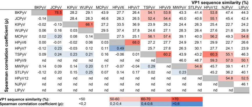
Specificity of the HPyV multiplex immunoassay.
Due to the VP1 amino acid
sequence similarity between different HPyV species, varying between 21.6% and 78.5%
(Fig. 3, red values at top right), one might expect epitope sharing and therefore a
certain degree of cross-reactivity among (related) HPyVs. To evaluate this, a correlation
matrix of the HPyV seroresponses was generated (Fig. S5), and Spearman rank
corre-lation coefficients were calculated for each HPyV combination for the HBD popucorre-lation
(Fig. 3, blue values at bottom left). The KTR population showed comparable data (not
shown). The lack of measured seroreactivity against HPyV9, HPyV12, NJPyV, and LIPyV
did not allow a meaningful correlation analysis, and these viruses were therefore
excluded from this analysis. Overall, we observed little correlation between the
sero-reactivities determined against the individual HPyVs. A moderate correlation between
HPyV6 and HPyV7 was observed in both the HBD population (Spearman’s
⫽
0.49,
P
⬍
0.001) and the KTR population (Spearman’s
⫽
0.44,
P
⬍
0.001). Despite 78.5% VP1
amino acid sequence similarity between BKPyV and JCPyV, no correlation between
these types was measured (Spearman’s
⫽ ⫺
0.14,
P
⫽
0.19). Between species (HPyV10)
members MWPyV and HPyV10, a high correlation was observed (Spearman’s
⫽
0.92,
P
⬍
0.001), which can be explained by their high VP1 amino acid sequence similarity
(98%) (Fig. 4).
FIG 3Summary of observed cross-reactivity between individual HPyV VP1 antigens. The data in the upper right show the percent VP1 sequence similarity based on a pairwise alignment obtained using Geneious software (version 10.0.9) with default ClustalW settings. The data in the lower left show Spearman correlation coefficients () calculated on the basis of the seroresponses measured against VP1 of the HPyV types tested in the HBD cohort. nd, Spearman correlation coefficients were not determined for these HPyVs.
on May 16, 2020 by guest
http://jcm.asm.org/
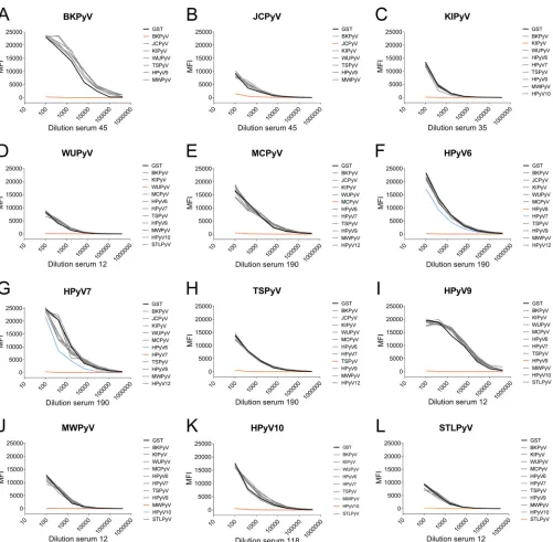
[image:5.585.44.478.74.258.2]To gain more insight into cross-reactivity, antigen competition experiments were
performed. In these experiments reactive serum samples were titrated and
preincu-bated with soluble GST-VP1 of various HPyVs before being exposed to antigenic beads
coated with the relevant HPyV VP1. Figure 5 shows some examples of the results of
these analyses. A complete overview of the selected serum samples tested in this way
can be found in Fig. S6. Overall, little competition between VP1 antigens from HPyVs
belonging to different species was observed. Preincubation with JCPyV VP1 did not
show a reduction in BKPyV seroreactivity in three out of four experiments (Fig. 5A and
S6A4 and A5); however, in one competition experiment, a substantial reduction was
seen (Fig. S6A3). Vice versa, preincubation with BKPyV VP1 reduced JCPyV seroreactivity
in two out of four competition experiments (Fig. S6B1 and B3). Between closely related
species HPyV6 and HPyV7, partial antigen competition indicative of limited
cross-reactivity was observed (Fig. 5F and G and S6F1, F2, and G2). As expected, HPyV10
species members MWPyV and HPyV10 showed high levels of cross-reactivity in this
analysis (Fig. 5J and K). Interestingly, in three out of six HPyV10 competition
experiments, preincubation with MWPyV VP1 did not block HPyV10 seroreactivity
(Fig. S6K1, K4, and K5). A summary of the results of the competition experiments is
shown in Table 2.
Comparison between the GST-VP1-based HPyV multiplex immunoassay and a
VP1 VLP-based ELISA.
To learn more about the antigenicity of the GST-VP1 fusion
proteins that we used, we compared the seroresponses measured for BKPyV in our
method to those obtained with a VLP-based ELISA. Although differences in especially
the presentation of conformational epitopes were anticipated, the BKPyV
seroreactivi-ties measured by both methods were quite similar (Fig. 6) (22). A high Spearman
correlation coefficient (
⫽
0.823,
P
⬍
0.001) was observed between the optical density
(OD) values obtained with the VP1 VLP ELISA and the MFI values obtained with the
GST-VP1 immunoassay.
DISCUSSION
Based on the performed evaluation, the broad HPyV multiplex immunoassay
de-scribed in this report provides highly reproducible and species-specific serological data.
Cross-recognizing antibody detection is sometimes seen between related HPyV species,
especially between HPyV6 and HPyV7, which was observed in other studies as well (25).
The mean correlation calculated between JCPyV and BKPyV seroreactivity was very low.
FIG 4Comparison of seroreactivity between MWPyV and HPyV10, both of which belong toPolyomavirusspecies 10. MWPyV and HPyV10 seroreactivities were measured in a cohort of healthy blood donors (HBD). Results are depicted as the median fluorescence intensity (MFI), measured in a Bio-Plex 100 analyzer, for MWPyV on thexaxis and for HPyV10 on theyaxis. The Spearman correlation coefficient is depicted. Each circle represents one serum sample, and the line represents the results of linear regression analyses.
on May 16, 2020 by guest
http://jcm.asm.org/
[image:6.585.121.289.72.253.2]Nevertheless, some serum samples clearly demonstrated cross-reactivity between these
two clinically relevant HPyVs. This observation deserves further study, since individual
seroresponses against JCPyV and perhaps against BKPyV as well (26) are used for
patient risk assessment regarding serious complications of HPyV-induced infection, for
example, PML (27). However, a limited role for cross-reactivity between HPyV6 and
HPyV7 serology (28) and between JCPyV and BKPyV serology (15, 19, 29, 30) has also
been described.
Apart from the cross-reactivity between two related HPyV species pairs that has
been described before for other serological platforms, the GST-VP1 bead-based
pan-FIG 5Analysis of cross-reactivity of polyomavirus seroresponses by VP1-specific competition. Titrated serum samples were preincubated with crude bacterial extract containing GST alone (black), with GST-VP1 of the autologous HPyV (orange), or with the nontarget heterologous HPyVs (gray). Blue lines indicate competition by VP1 other than the target analyte. Results are depicted as median fluorescence intensity (MFI), measured in a Bio-Plex 100 analyzer and shown for the seroresponses measured for BKPyV (A), JCPyV (B), KIPyV (C), WUPyV (D), MCPyV (E), HPyV6 (F), HPyV7 (G), TSPyV (H), HPyV9 (I); MWPyV (J), HPyV10 (K), and STLPyV (L).
on May 16, 2020 by guest
http://jcm.asm.org/
[image:7.585.44.545.67.558.2]HPyV assay seems to be a reliable tool for seroepidemiological HPyV studies. To what
extent this assay measures neutralizing antibodies was not investigated in this study,
but the high correlation between the BKPyV serological data obtained with this assay
and those obtained with the VP1 VLP-based ELISA suggests that the GST-VP1 fusion
proteins presented on glutathione-casein-coupled beads express conformational
epitopes. This was also previously suggested by highly comparable HPyV
seropreva-TABLE 2Summary of cross-reactivity observed among HPyV-VP1 antigens in individual serum samples
Source of VP1 antigen
Cross-reactivitya
BKPyV JCPyV KIPyV WUPyV MCPyV HPyV6 HPyV7 TSPyV HPyV9 MWPyV HPyV10 STLPyV
BKPyV ⫹⫹⫹⫹ ⫹⫹ ⫺ ⫺ ⫺ ⫺ ⫺ ⫺ ⫺ ⫺ ⫺ ⫺
JCPyV ⫹ ⫹⫹⫹⫹ ⫺ ⫺ ⫺ ⫺ ⫺ ⫺ ⫺ ⫺ ⫺ ⫺
KIPyV ⫺ ⫺ ⫹⫹⫹⫹ ⫺ ⫺ ⫺ ⫺ ⫺ ⫺ ⫺ ⫺ ⫺
WUPyV ⫺ ⫺ ⫺ ⫹⫹⫹⫹ ⫺ ⫺ ⫺ ⫺ ⫺ ⫺ ⫺ ⫺
MCPyV ⫺ ⫺ ⫺ ⫺ ⫹⫹⫹⫹ ⫺ ⫺ ⫺ ⫺ ⫺ ⫺ ⫺
HPyV6 ⫺ ⫺ ⫺ ⫺ ⫺ ⫹⫹⫹⫹ ⫹⫹ ⫺ ⫺ ⫺ ⫺ ⫺
HPyV7 ⫺ ⫺ ⫺ ⫺ ⫺ ⫹⫹ ⫹⫹⫹⫹ ⫺ ⫺ ⫺ ⫺ ⫺
TSPyV ⫺ ⫺ ⫺ ⫺ ⫺ ⫺ ⫺ ⫹⫹⫹⫹ ⫺ ⫺ ⫺ ⫺
HPyV9 ⫺ ⫺ ⫺ ⫺ ⫺ ⫺ ⫺ ⫺ ⫹⫹⫹⫹ ⫺ ⫺ ⫺
MWPyV ⫺ ⫺ ⫺ ⫺ ⫺ ⫺ ⫺ ⫺ ⫺ ⫹⫹⫹⫹ ⫹⫹⫹ ⫺
HPyV10 ⫺ ⫺ ⫺ ⫺ ⫺ ⫺ ⫺ ⫺ ⫺ ⫹⫹⫹⫹ ⫹⫹⫹⫹ ⫺
STLPyV ⫺ ⫺ ⫺ ⫺ ⫺ ⫺ ⫺ ⫺ ⫺ ⫺ ⫺ ⫹⫹⫹⫹
aArbitrary interpretation of the VP1 competition observed in the experiments whose results are shown in Fig. 5 and Fig. S6 in the supplemental material.⫺, no
reduction;⫹, slight reduction;⫹⫹, moderate reduction;⫹⫹⫹, high reduction;⫹⫹⫹⫹, complete reduction.
FIG 6Comparison between the GST-VP1 bead-based assay and the VLP-based ELISA for BKPyV. The seroreactivities of kidney transplantation donors (n⫽ 396) were measured by both the bead-based GST-VP1 immunoassay and the VP1 VLP ELISA for BKPyV. Each circle represents one serum sample, and the black line indicates the correlation between the bead-based measurement (MFI) and the ELISA (OD). (Adapted with permission from reference 22.)
on May 16, 2020 by guest
http://jcm.asm.org/
[image:8.585.41.543.84.228.2] [image:8.585.42.371.375.691.2]lence data obtained worldwide and independently with VP1 VLP and GST-VP1 fusion
proteins, for example, for TSPyV (11, 12, 24, 31, 32).
The high intraspecies cross-reactivity observed between MWPyV and HPyV10 did
not come as a surprise and probably resulted from their high VP1 amino acid sequence
similarity. Nevertheless, seroreactivity toward HPyV10 was not always abolished after
preincubation with the MWPyV VP1, indicating a subtle difference between some
epitopes of MWPyV and HPyV10, which could be explained by the fact that three of the
eight amino acid differences between MWPyV and HPyV10 might be located within the
antigenic loops (15, 19). The overall high degree of similarity between the seroreactivity
profiles of MWPyV and HPyV10, however, suggests no need for separate measurements
for both viruses when testing larger cohorts. The lack of samples seroreactive against
HPyV9, HPyV12, NJPyV, and LIPyV did not allow a thorough analysis of potential
cross-reactivity for these HPyVs. As a general remark, the possibility of cross-reactivity
by antibodies against yet unknown HPyVs cannot be excluded.
The aim of this study was to evaluate the abilities of the assay and not to determine
seroprevalence. As such, no seronegative cutoff determination was performed.
Sero-reactivity against most HPyVs was high in both the immunosuppressed KTR and the
HBD cohorts. The observed seroreactivity profile of HPyV9 was lower than that of other
polyomaviruses, in line with the findings presented in previous publications, including
ours (11, 12, 14, 33).
We observed limited seroreactivity against HPyV12, NJPyV, and LIPyV. For
compar-ison, to date no other serological data are available for NJPyV and LIPyV. For HPyV12,
one study reported a seroprevalence of 15 to 33% in healthy adults (34). Based on our
observed seroreactivity against HPyV12, presented in Fig. 2, we assume that in Dutch
populations the seroprevalence of HPyV12 is low. On the basis of the VP1 amino acid
sequence alignment, it was recently suggested that the translation initiation site of
HPyV12 VP1 is located 48 nucleotides (16 amino acids) downstream of the 5
=
end of the
VP1 open reading frame (35). We also analyzed the antigenicity of this shorter
GST-HPyV12 VP1 fusion protein and noticed no difference in GST-HPyV12 seroreactivity (not
shown). (After submission of the manuscript, the discoverers of HPyV12 published data
that convincingly show that HPyV12 is, in fact, a shrew-derived virus [36], suggesting
that HPyV12 does not circulate among humans and explaining the lack of HPyV12
seroreactivity found in our cohorts.)
To our knowledge, infection with NJPyV has been described only once, in an
immunocompromised kidney-pancreas transplant patient fleeing through sewage
wa-ter following Superstorm Sandy (10). Supported by the prompt recognition of NJPyV
VP1 by the rabbit polyclonal serum raised against NJPyV VP1 peptides, we are confident
that our assay is capable of measuring seroresponses against NJPyV. Therefore, we
interpret the lack of detectable seroresponses to be an indication that this
polyoma-virus does not represent a human polyomapolyoma-virus but, rather, represents a zoonotic
polyomavirus that was introduced into humans under exceptional conditions.
Alterna-tively, the lack of NJPyV seroresponses could suggest a difference in geographical
spread for NJPyV between North America and Europe, which is rather unusual for
(human) polyomaviruses. LIPyV also showed a low seroreactivity profile, suggesting the
possibility of environmental contamination of LIPyV in the original skin sample. A larger
seroprevalence study could help to elucidate this issue.
The comparison between a VLP-based ELISA and the bead-based assay showed a
clear monotonic relationship, despite the different methods in which conformational
epitopes are presented by both assays (Fig. 6). A close look at the kinetics of each assay
reveals a large dynamic range of the bead-based assay, which has a seemingly
increased sensitivity compared to that of the ELISA for the detection of seroresponses
in the lower reactivity range. For the purpose of seroepidemiology, we believe that
serological testing using HPyV VP1 expressed as a GST fusion protein or as a VP1 VLP
yields equally useful results. For individual use, for instance, to predict the risk of
developing polyomavirus-related disease such as PML, additional analyses and assay
validation are necessary.
on May 16, 2020 by guest
http://jcm.asm.org/
In conclusion, the custom-made pan-HPyV multiplex immunoassay is a reliable tool
for determination of HPyV-specific seroprevalences. It measures HPyV-specific IgG
seroreactivities with high reproducibility and specificity, can easily be extended in case
of new HPyV discoveries, and can potentially be combined with other (viral) antigens
of interest.
SUPPLEMENTAL MATERIAL
Supplemental material for this article may be found at
https://doi.org/10.1128/JCM
.01566-17
.
SUPPLEMENTAL FILE 1,
PDF file, 2.3 MB.
ACKNOWLEDGMENTS
This work was supported and funded by Sanquin Blood Supply Foundation, a
not-for-profit organization.
We declare no conflict of interest.
S.K., E.V.D.M., A.T., and M.C.W.F. conceived and designed the experiments. S.K.,
E.V.D.M., and A.T. performed the experiments. S.K. and E.V.D.M. analyzed the data. S.K.,
E.V.D.M., H.F.W., H.L.Z., and M.C.W.F. wrote the paper.
REFERENCES
1. Calvignac-Spencer S, Feltkamp MCW, Daugherty MD, Moens U, Ramqvist T, Johne R, Ehlers B. 2016. A taxonomy update for the family Polyoma-viridae. Arch Virol 161:1739 –1750.https://doi.org/10.1007/s00705-016 -2794-y.
2. Gheit T, Dutta S, Oliver J, Robitaille A, Hampras S, Combes J-D, McKay-Chopin S, Le Calvez-Kelm F, Fenske N, Cherpelis B, Giuliano AR, France-schi S, McKay J, Rollison DE, Tommasino M. 2017. Isolation and charac-terization of a novel putative human polyomavirus. Virology 506:45–54.
https://doi.org/10.1016/j.virol.2017.03.007.
3. Purighalla R, Shapiro R, McCauley J, Randhawa P. 1995. BK virus infection in a kidney allograft diagnosed by needle biopsy. Am J Kidney Dis 26:671– 673.https://doi.org/10.1016/0272-6386(95)90608-8.
4. Astrom KE, Mancall EL, Richardson EP. 1958. Progressive multifocal leuko-encephalopathy; a hitherto unrecognized complication of chronic lymphatic leukaemia and Hodgkin’s disease. Brain J Neurol 81:93–111.
https://doi.org/10.1093/brain/81.1.93.
5. Feng H, Shuda M, Chang Y, Moore PS. 2008. Clonal integration of a polyomavirus in human Merkel cell carcinoma. Science 319:1096 –1100.
https://doi.org/10.1126/science.1152586.
6. van der Meijden E, Janssens RWA, Lauber C, Bouwes Bavinck JN, Gor-balenya AE, Feltkamp MCW. 2010. Discovery of a new human polyoma-virus associated with trichodysplasia spinulosa in an immunocompro-mized patient. PLoS Pathog 6:e1001024.https://doi.org/10.1371/journal .ppat.1001024.
7. Nguyen KD, Lee EE, Yue Y, Stork J, Pock L, North JP, Vandergriff T, Cockerell C, Hosler GA, Pastrana DV, Buck CB, Wang RC. 2017. Human polyomavirus 6 and 7 are associated with pruritic and dyskeratotic dermatoses. J Am Acad Dermatol 76:932–940.e3. https://doi.org/10 .1016/j.jaad.2016.11.035.
8. Rennspiess D, Pujari S, Keijzers M, Abdul-Hamid MA, Hochstenbag M, Dingemans A-M, Kurz AK, Speel E-J, Haugg A, Pastrana DV, Buck CB, De Baets MH, zur Hausen A. 2015. Detection of human polyomavirus 7 in human thymic epithelial tumors. J Thorac Oncol 10:360 –366.https://doi .org/10.1097/JTO.0000000000000390.
9. Keijzers M, Rensspiess D, Pujari S, Abdul-Hamid MA, Hochstenbag M, Dingemans A-M, Kurz AK, Haugg A, Maessen JG, De Baets MH, zur Hausen A. 2015. Expression of pRb and p16INK4 in human thymic epithelial tumors in relation to the presence of human polyomavirus 7. Diagn Pathol 10:201.https://doi.org/10.1186/s13000-015-0418-6. 10. Mishra N, Pereira M, Rhodes RH, An P, Pipas JM, Jain K, Kapoor A, Briese
T, Faust PL, Lipkin WI. 2014. Identification of a novel polyomavirus in a pancreatic transplant recipient with retinal blindness and vasculitic my-opathy. J Infect Dis 210:1595–1599.https://doi.org/10.1093/infdis/jiu250. 11. Gossai A, Waterboer T, Nelson HH, Michel A, Willhauck-Fleckenstein M, Farzan SF, Hoen AG, Christensen BC, Kelsey KT, Marsit CJ, Pawlita M, Karagas MR. 2016. Seroepidemiology of human polyomaviruses in
a US population. Am J Epidemiol 183:61– 69.https://doi.org/10.1093/ aje/kwv155.
12. van der Meijden E, Bialasiewicz S, Rockett RJ, Tozer SJ, Sloots TP, Feltkamp MCW. 2013. Different serologic behavior of MCPyV, TSPyV, HPyV6, HPyV7 and HPyV9 polyomaviruses found on the skin. PLoS One 8:e81078.https://doi.org/10.1371/journal.pone.0081078.
13. Šroller V, Hamšíková E, Ludvíková V, Vochozková P, Kojzarová M, Fraiberk M, Saláková M, Morávková A, Forstová J, Neˇmecˇková Š. 2014. Seropreva-lence rates of BKV, JCV, and MCPyV polyomaviruses in the general Czech Republic population. J Med Virol 86:1560 –1568.https://doi.org/10.1002/ jmv.23841.
14. Šroller V, Hamšíková E, Ludvíková V, Musil J, Neˇmecˇková Š, Saláková M. 2016. Seroprevalence rates of HPyV6, HPyV7, TSPyV, HPyV9, MWPyV and KIPyV polyomaviruses among the healthy blood donors. J Med Virol 88:1254 –1261.https://doi.org/10.1002/jmv.24440.
15. Kean JM, Rao S, Wang M, Garcea RL. 2009. Seroepidemiology of human polyomaviruses. PLoS Pathog 5:e1000363.https://doi.org/10 .1371/journal.ppat.1000363.
16. Carter JJ, Paulson KG, Wipf GC, Miranda D, Madeleine MM, Johnson LG, Lemos BD, Lee S, Warcola AH, Iyer JG, Nghiem P, Galloway DA. 2009. Association of Merkel cell polyomavirus-specific antibodies with Merkel cell carcinoma. J Natl Cancer Inst 101:1510 –1522. https://doi.org/10 .1093/jnci/djp332.
17. Feltkamp MCW, Kazem S, van der Meijden E, Lauber C, Gorbalenya AE. 2013. From Stockholm to Malawi: recent developments in studying human polyomaviruses. J Gen Virol 94:482– 496.https://doi.org/10.1099/ vir.0.048462-0.
18. van der Meijden E, Horváth B, Nijland M, de Vries K, Rácz EK, Diercks GF, de Weerd AE, Groningen MCC, van der Blij-de Brouwer CS, van der Zon AJ, Kroes ACM, Hedman K, van Kampen JJA, Riezebos-Brilman A, Felt-kamp MCW. 2016. Primary polyomavirus infection, not reactivation, as the cause of trichodysplasia spinulosa in immunocompromised patients. J Infect Dis 215:1080 –1084.https://doi.org/10.1093/infdis/jiw403. 19. Moens U, Van Ghelue M, Song X, Ehlers B. 2013. Serological
cross-reactivity between human polyomaviruses. Rev Med Virol 23:250 –264.
https://doi.org/10.1002/rmv.1747.
20. Waterboer T, Sehr P, Michael KM, Franceschi S, Nieland JD, Joos TO, Templin MF, Pawlita M. 2005. Multiplex human papillomavirus serology based on in situ-purified glutathione S-transferase fusion proteins. Clin Chem 51:1845–1853.https://doi.org/10.1373/clinchem.2005.052381. 21. Waterboer T, Sehr P, Pawlita M. 2006. Suppression of non-specific
bind-ing in serological Luminex assays. J Immunol Methods 309:200 –204.
https://doi.org/10.1016/j.jim.2005.11.008.
22. Wunderink HF, van der Meijden E, van der Blij-de Brouwer CS, Mallat MJK, Haasnoot GW, van Zwet EW, Claas ECJ, de Fijter JW, Kroes ACM, Arnold F, Touzé A, Claas FHJ, Rotmans JI, Feltkamp MCW. 2016.
on May 16, 2020 by guest
http://jcm.asm.org/
plantation donor-recipient pair seroreactivity against BK polyomavirus predicts viremia and nephropathy after kidney transplantation. Am J Transplant 17:161–172.https://doi.org/10.1111/ajt.13880.
23. van der Meijden E, Wunderink HF, van der Blij-de Brouwer CS, Zaaijer HL, Rotmans JI, Bavinck JNB, Feltkamp MCW. 2014. Human polyomavirus 9 infection in kidney transplant patients. Emerg Infect Dis 20:991–999.
https://doi.org/10.3201/eid2006.140055.
24. van der Meijden E, Kazem S, Burgers MM, Janssens R, Bavinck JNB, de Melker H, Feltkamp MCW. 2011. Seroprevalence of trichodysplasia spinulosa-associated polyomavirus. Emerg Infect Dis 17:1355–1363.
https://doi.org/10.3201/eid1708.110114.
25. Nicol JTJ, Robinot R, Carpentier A, Carandina G, Mazzoni E, Tognon M, Touzé A, Coursaget P. 2013. Age-specific seroprevalences of Merkel cell polyomavirus, human polyomaviruses 6, 7, and 9, and trichodysplasia spinulosa-associated polyomavirus. Clin Vaccine Immunol 20:363–368.
https://doi.org/10.1128/CVI.00438-12.
26. Wunderink HF, van der Meijden E, van der Blij-de Brouwer CS, Zaaijer HL, Kroes ACM, van Zwet EW, Rotmans JI, Feltkamp MCW. 2017. Stability of BK polyomavirus IgG seroreactivity and its correlation with preceding viremia. J Clin Virol 90:46 –51.https://doi.org/10.1016/j.jcv.2017.03.015. 27. Bloomgren G, Richman S, Hotermans C, Subramanyam M, Goelz S, Natarajan A, Lee S, Plavina T, Scanlon JV, Sandrock A, Bozic C. 2012. Risk of natalizumab-associated progressive multifocal leukoencephalopathy. N Engl J Med 366:1870 –1880.https://doi.org/10.1056/NEJMoa1107829. 28. Schowalter RM, Pastrana DV, Pumphrey KA, Moyer AL, Buck CB. 2010. Merkel cell polyomavirus and two previously unknown polyomaviruses are chronically shed from human skin. Cell Host Microbe 7:509 –515.
https://doi.org/10.1016/j.chom.2010.05.006.
29. Viscidi RP, Clayman B. 2006. InSerological cross reactivity between polyomavirus capsids, p 73– 84.InAshan N (ed), Polyomaviruses and human diseases. Springer, New York, NY.
30. Egli A, Infanti L, Dumoulin A, Buser A, Samaridis J, Stebler C, Gosert R, Hirsch HH. 2009. Prevalence of polyomavirus BK and JC infection and replication in 400 healthy blood donors. J Infect Dis 199:837– 846.
https://doi.org/10.1086/597126.
31. Fukumoto H, Li T-C, Kataoka M, Hasegawa H, Wakita T, Saeki H, Suzuki T, Katano H. 2015. Seroprevalence of trichodysplasia spinulosa-associated polyomavirus in Japan. J Clin Virol 65:76 – 82.https://doi.org/ 10.1016/j.jcv.2015.02.014.
32. Chen T, Mattila PS, Jartti T, Ruuskanen O, Söderlund-Venermo M, Hed-man K. 2011. Seroepidemiology of the newly found trichodysplasia spinulosa-associated polyomavirus. J Infect Dis 204:1523–1526.https:// doi.org/10.1093/infdis/jir614.
33. Antonsson A, Pawlita M, Feltkamp MCW, Bouwes Bavinck JN, Euvrard S, Harwood CA, Naldi L, Nindl I, Proby CM, Neale RE, Waterboer T. 2013. Longitudinal study of seroprevalence and serostability of the human
polyomaviruses JCV and BKV in organ transplant recipients. J Med Virol 85:327–335.https://doi.org/10.1002/jmv.23472.
34. Korup S, Rietscher J, Calvignac-Spencer S, Trusch F, Hofmann J, Moens U, Sauer I, Voigt S, Schmuck R, Ehlers B. 2013. Identification of a novel human polyomavirus in organs of the gastrointestinal tract. PLoS One 8:e58021.https://doi.org/10.1371/journal.pone.0058021.
35. Norkiene M, Stonyte J, Ziogiene D, Mazeike E, Sasnauskas K, Gedvilaite A. 2015. Production of recombinant VP1-derived virus-like particles from novel human polyomaviruses in yeast. BMC Biotechnol 15:68.https:// doi.org/10.1186/s12896-015-0187-z.
36. Gedvilaite A, Tryland M, Ulrich RG, Schneider J, Kurmauskaite V, Moens U, Preugschas H, Calvignac-Spencer S, Ehlers B. 2 November 2017. Novel polyomaviruses in shrews (Soricidae) with close similarity to human polyomavirus 12. J Gen Virol.https://doi.org/10.1099/jgv.0.000948. 37. Gardner S, Field A, Coleman D, Hulme B. 1971. New human papovavirus
(B.K.) isolated from urine after renal transplantation. Lancet 297: 1253–1257.https://doi.org/10.1016/S0140-6736(71)91776-4.
38. Padgett B, Zurhein G, Walker D, Eckroade R, Dessel B. 1971. Cultiva-tion of papova-like virus from human brain with progressive multi-focal leucoencephalopathy. Lancet 297:1257–1260.https://doi.org/10 .1016/S0140-6736(71)91777-6.
39. Allander T, Andreasson K, Gupta S, Bjerkner A, Bogdanovic G, Persson MAA, Dalianis T, Ramqvist T, Andersson B. 2007. Identification of a third human polyomavirus. J Virol 81:4130 – 4136.https://doi.org/10.1128/JVI .00028-07.
40. Gaynor AM, Nissen MD, Whiley DM, Mackay IM, Lambert SB, Wu G, Brennan DC, Storch GA, Sloots TP, Wang D. 2007. Identification of a novel polyomavirus from patients with acute respiratory tract infections. PLoS Pathog 3:e64.https://doi.org/10.1371/journal.ppat.0030064. 41. Scuda N, Hofmann J, Calvignac-Spencer S, Ruprecht K, Liman P, Kühn J,
Hengel H, Ehlers B. 2011. A novel human polyomavirus closely related to the African green monkey-derived lymphotropic polyomavirus. J Virol 85:4586 – 4590.https://doi.org/10.1128/JVI.02602-10.
42. Siebrasse EA, Reyes A, Lim ES, Zhao G, Mkakosya RS, Manary MJ, Gordon JI, Wang D. 2012. Identification of MW polyomavirus, a novel polyoma-virus in human stool. J Virol 86:10321–10326.https://doi.org/10.1128/ JVI.01210-12.
43. Buck CB, Phan GQ, Raiji MT, Murphy PM, McDermott DH, McBride AA. 2012. Complete genome sequence of a tenth human polyomavirus. J Virol 86:10887.https://doi.org/10.1128/JVI.01690-12.
44. Lim ES, Reyes A, Antonio M, Saha D, Ikumapayi UN, Adeyemi M, Stine OC, Skelton R, Brennan DC, Mkakosya RS, Manary MJ, Gordon JI, Wang D. 2013. Discovery of STL polyomavirus, a polyomavirus of ancestral re-combinant origin that encodes a unique T antigen by alternative splic-ing. Virology 436:295–303.https://doi.org/10.1016/j.virol.2012.12.005.