0095-1137/05/$08.00⫹0 doi:10.1128/JCM.43.8.3760–3768.2005
Copyright © 2005, American Society for Microbiology. All Rights Reserved.
Identification of Medically Important Molds by an
Oligonucleotide Array†
Chen Ren Hsiao,
1Liyin Huang,
1Jean-Philippe Bouchara,
2Richard Barton,
3Hsin Chieh Li,
1and Tsung Chain Chang
1*
Department of Medical Laboratory Science and Biotechnology, School of Medicine, National Cheng Kung University, Tainan, Taiwan, Republic of China1; Host-Parasite-Interaction Study Group (UPRES-EA 3142), Laboratory of
Parasitology and Mycology, University Hospital, Angers, France2; and School of Biochemistry
and Microbiology, University of Leeds, United Kingdom3
Received 22 February 2005/Returned for modification 1 April 2005/Accepted 2 May 2005
Infections caused by fungi have increased in recent years. Accurate and rapid identification of fungal pathogens is important for appropriate treatment with antifungal agents. On the basis of the internal transcribed spacer 1 (ITS 1) and ITS 2 sequences of the rRNA genes, an oligonucleotide array was developed to identify 64 species (32 genera) of clinically important filamentous (or dimorphic) fungi. These 64 species included fungi causing superficial, cutaneous, subcutaneous, and invasive infections. The method consisted of PCR amplification of the ITS regions using a pair of universal primers, followed by hybridization of the digoxigenin-labeled PCR products to a panel of species- or group-specific oligonucleotides immobilized on a nylon membrane. Of 397 fungal strains (290 target and 107 nontarget strains) tested, the sensitivity and specificity of the array was 98.3% (285/290) and 98.1% (105/107), respectively. Misidentified strains were usually those belonging to the same genus of the target species or having partial homology with oligonucleotide probes on the membrane. The whole procedure can be finished within 24 h starting from isolated colonies; reproductive structures, which are essential for the conventional identification methods, are not needed. In conclusion, the present array is a powerful tool for identification of clinically important filamentous fungi and may have the potential to be continually extended by adding further oligonucleotides to the array without significantly increasing the cost or complexity.
The identification of molds (filamentous fungi) can be chal-lenging, and accuracy will depend on the organism and the experience of the clinical microbiologist (20). Conventional methods for fungal identification in the clinical laboratory are based on morphological and physiological tests. These meth-ods often require several days or even weeks and may be inaccurate (30). In the last few decades, invasive mycoses have become a major cause of infectious morbidity and mortality in patients receiving immunosuppressive chemotherapy for can-cer or organ transplantation and in immunodeficient patients, such as individuals with AIDS (2, 3, 9, 14, 33). Since invasive mycoses are often associated with a poor prognosis, the early, rapid, and accurate identification of the pathogenic fungi is important for timely and appropriate management.
In recent years, numerous DNA-based methods have been developed to diagnose mycotic infections and to identify pathogenic fungi (6). PCR methods are particularly promising because of their simplicity, specificity, and sensitivity. Genes of the 18S rRNA (27, 41, 47) and 28S rRNA (15, 28, 38) have been extensively used for molecular identification. Recently, Hall et al. (20) evaluated the commercial MicroSeq D2 large-subunit rRNA gene fungal sequencing kit (Applied Biosys-tems, Foster City, Calif.) and drew the conclusion that the kit
offers the promise of being an accurate identification system. However, they noted that 33% of clinical isolates could not be identified due to a limited sequence database in this commer-cial system. In addition, the MicroSeq kit requires an auto-matic sequencer, which is not yet standard equipment for most clinical microbiology laboratories. A number of studies have described probes, restriction fragment length polymorphism, or other methods to identify unique ribosomal DNA sequences (26, 35, 44). Although these published methods are useful for the identification of fungal species, they can identify only one or a limited number of species. The ribosomal internal tran-scribed spacer (ITS) regions also have been widely used as targets to detect and identify human fungal pathogens (5, 7, 10, 21, 23, 24, 31–33, 43).
DNA array (or DNA chip) technology has been found to be a useful tool to identify a variety of bacteria, especially for those bacteria that are difficult to differentiate by conventional methods or when the identification procedures may take a long time (1, 17, 45). Wu et al. (47) recently developed an array of oligonucleotide probes to detect airborne fungi. The probes were designed on the basis of the 18S rRNA gene and were used to detect 31 species belonging to 15 genera. The aim of this study was to investigate the feasibility of using a panel of oligonucleotide probes designed on the basis of the ITS re-gions to identify 64 mold species (32 genera) of clinical impor-tance.
MATERIALS AND METHODS
Fungal strains.A total of 397 strains including 290 target (64 species) and 107 nontarget strains were used (see Table S1 in the supplemental material) (sup-* Corresponding author. Mailing address: Department of Medical
Laboratory Science and Biotechnology, School of Medicine, National Cheng Kung University, 1 University Road, Tainan 701, Taiwan, Re-public of China. Phone: 886-6-2353535, ext. 5790. Fax: 886-6-2363956. E-mail: [email protected].
† Supplemental material for this article may be found at http://jcm .asm.org.
3760
on May 15, 2020 by guest
http://jcm.asm.org/
plemental Table S1 and supplemental Fig. S2 from this study are supplied as supplemental files). Of the 290 target strains, 233 were reference strains obtained from culture collection centers and 57 were clinical isolates. Of the 107 nontarget strains, 93 were reference strains and 14 were clinical isolates. Reference strains were obtained from the American Type Culture Collection (Manassas, Va.), Bioresources Collection and Research Center (BCRC, Hsichu, Taiwan, Repub-lic of China), and CBS (Centraalbureau voor Schimmelcultures, The Nether-lands). Clinical isolates were obtained from the Mycology Reference Centre, Division of Microbiology University of Leeds, Leeds, UK (strain designated with a prefix of LM), and from the Laboratory of Parasitology and Mycology of Angers University Hospital (Angers, France) (strain designated with a prefix of LMA). Morphological identification of clinical isolates to the species level was accomplished using established procedures including microscopic and macro-scopic characteristics (11, 30). All fungal strains were subcultured on Sabouraud dextrose agar (Difco, Detroit, Mich.), incubated at 30°C until there was evidence of apparent hyphal growth, and then used for DNA extraction.
DNA extraction.Mycelium (approximately 0.5⫻0.5 cm) was scraped into a 2-ml screw cap tube (Azygen Sientific, Union City, Calif.) containing 300 mg of 0.5-mm-diameter zirconium/silica beads (Biospec Products, Bartlesville, Okla.) in 1 ml of sterilized water using an inoculation loop. The mycelial suspension was shaken for 5 min at a speed of 4,200 rpm in a mechanical cell disrupter (Mini-Beadbeater, Biospec Products). An aliquot (0.1 ml) of the disrupted cell suspen-sion was transferred to a 1.5-ml centrifuge tube and centrifuged at 8,000⫻gfor 10 min in a microcentrifuge. Fungal DNA in the supernatant was extracted by a genomic DNA extraction kit (Viogene, Taipei, Taiwan).
ITS amplification and sequencing.The ITS sequences of some species that were not available in the database of GenBank were determined in this study (see Table S1 in the supplemental material). The fungus-specific, universal prim-ers ITS 1 (5⬘-TCCGTAGGTGAACCTGCGG-3⬘) and ITS 2 (5⬘-GCTGCGTT CTTCATCGATGC-3⬘) (46) were used to amplify a small conserved portion of the 18S ribosomal DNA, the adjacent ITS 1, and a small portion of the 5.8S ribosomal DNA. An additional pair of primers, ITS 3 (5⬘-GCA TCGATGAAG AACGCAGC-3⬘) and ITS 4 (5⬘-TCCTCCGCTTATTGATATGC-3⬘) (46), were used to amplify a conserved portion of the 5.8S ribosomal DNA, the intervening ITS 2, and a small portion of the 28S ribosomal DNA. PCR was performed with 5l (1 to 5 ng) of template DNA in a total reaction volume of 50l consisting of 10 mM Tris-HCl (pH 8.3), 50 mM KCl, 1.5 mM MgCl2, 0.8 mM
deoxyribo-nucleoside triphosphates (0.2 mM each), 0.7M primer (each), TaqDNA polymerase (1.25 U), and 50l of a mineral oil overlay. PCR was carried out with an OmniGen thermal cycler (Hybaid Limited, Middlesex, UK) under the fol-lowing conditions: initial denaturation, 94°C, 3 min; 35 cycles of denaturation (94°C, 1 min), annealing (60°C, 1 min), and extension (72°C, 1 min); and final extension, 72°C, 5 min. A negative control was performed with each test run by replacing the template DNA with sterilized water in the PCR mixture. PCR products were purified with a PCR-M CleanUp kit (Viogene, Taipei, Taiwan) and were sequenced on a model 377 sequencing system (Applied Biosystems, Taipei, Taiwan).
To amplify the ITS regions for array hybridization, the primer pair ITS 1 (5⬘-TCCGTAGGTGAACCTGCGG-3⬘) and ITS 4 (5⬘-TCCTCCGCTTATTGA TATG-3⬘) (46) was used to amplify a fragment encompassing ITS 1, the 5.8S rRNA gene, ITS 2, and partial regions of the 18S and 28S ribosomal DNA. The reverse primer ITS 4 was labeled with a digoxigenin molecule at its 5⬘end and was synthesized by MDBio Inc. (Taipei, Taiwan). PCR conditions were the same as described in the previous section. DNA extracted fromSaccharomycodes ludwigiiBCRC 21378 was used as a positive control for each run of PCR and array hybridization.
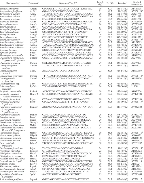
Design of oligonucleotide probes.Species- or group-specific oligonucleotide probes (21- to 30-mers) used for identification of 64 fungal species are listed in Table 1. Probe design was based on sequence data from the ITS 1 or 2 regions; these sequences were either available in the GenBank database or determined in this study. Probe selection was facilitated by using visual sequence alignment employing the PrettyBox command of the Wisconsin Genetics Computer Group package (version 10.3; Accelrys Inc., San Diego, Calif.). Areas displaying se-quence divergence among different species were analyzed for probe selection. A total of 58 probes, including one positive control (a probe designed on the basis of the ITS 2 region ofSaccharomycodes ludwigiiBCRC 21378), were used for fabrication of the oligonucleotide array (Table 1). The designed probes were checked for internal repeat, self-biding, secondary structure, and GC content by using Vector NTI software (Invitrogen Corporation, Carlsbad, Calf.). Seven additional bases of thymine were added to the 3⬘end of each probe (4). The specificity of the prospective sequence was first analyzed with a fungal ITS database developed in our laboratory and with sequences available in the Gen-Bank using BLAST.
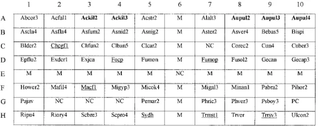
Fabrication of oligonucleotide arrays.The arrays (1.1 by 0.9 cm) (Fig. 1) were made in batches of 20. The oligonucleotide probes were diluted 1:1 (final con-centration 10M) with a tracking dye solution (30% [vol/vol] glycerol, 40% [vol/vol] dimethyl sulfoxide, 1 mM disodium EDTA, 0.15% [wt/vol] bromophe-nol blue, and 10 mM Tris-HCl, pH 7.5). The probe solutions were drawn into different wells of a round-bottom 96-well microtiter plate and spotted onto a positively charged nylon membrane (Roche, Mannheim, Germany) with an au-tomatic arrayer (Wittech, Taipei, Taiwan) using a solid pin (500m in diameter). The array contained 80 dots, including 58 dots for identification of 64 medically relevant molds (32 genera), 1 for positive control (probe code PC), and 5 for negative controls (probe code NC; tracking dye only) (Fig. 1). In addition, 16 dots (probe code M) contained 5⬘-digoxigenin-labeled ITS 4 (final concentration 0.16M); these dots formed a cross on the array after hybridization and were used as markers to visually locate hybridized probes (see Fig. S2 in the supple-mental material). Once all probes had been applied, the membrane was air-dried and exposed to shortwave UV (Stratalinker 1800; Stratagen, La Jolla, Calf.) for 30 s. Unbound oligonucleotides were removed by two washes (2 min each) at room temperature in 0.5⫻SSC (1⫻SSC is 0.15 M NaCl plus 0.015 M sodium citrate)–0.1% sodium dodecyl sulfate (SDS). The arrays were stored at room temperature for further use.
Hybridization procedures.Except where otherwise indicated, the hybridiza-tion procedures were carried out at room temperature (approximately 27°C) with a shaking speed of 60 rpm. Most reagents, except buffers, were included in the DIG Nucleic Acid Detection kit (catalog no. 1175041; Roche). Each array was prehybridized at 50°C for 2 h with 1 ml of hybridization solution (5⫻SSC, 1% [wt/vol] blocking reagent, 0.1%N-laurylsarcosine, 0.02% SDS) in an individual well of a 24-well cell culture plate. The digoxigenin-labeled PCR product am-plified from an isolate was heated in a boiling water bath for 5 min and imme-diately cooled on an ice bath. Ten microliters of denatured PCR products of the test organism and of the positive control (Saccharomycodes ludwigii) were diluted with 0.3 ml of hybridization solution and added to each well. Hybridization was conducted at 55°C for 90 min. After removing the nonhybridized PCR products, the array was washed four times (5 min each) in 1 ml of 0.25⫻SSC–0.1% SDS, followed by incubation for 1 h with 1 ml of blocking solution (1% [wt/vol] blocking reagent dissolved in maleic acid buffer [0.1 M maleic acid, 0.15 M NaCl, pH 7.5]). After removal of the blocking solution, 0.3 ml of alkaline phosphatase-conjugated sheep antidigoxigenin antibodies (diluted 1:2,500 in blocking solu-tion) was added to each well and incubated for 1 h. The array was washed three times (each 15 min) in 1 ml of washing solution (0.3% [vol/vol] Tween 20 in maleic acid buffer), followed by washing in 1 ml of detection buffer (0.1 M Tris-HCl, 0.15 M NaCl, pH 9.5) for 5 min. Finally, 0.3 ml of alkaline phosphatase substrate (a stock solution of nitroblue tetrazolium chloride/5-bromo-4-chloro-3-indolylphosphate diluted 1:50 in detection buffer) was added to each well and incubated at 37°C without shaking. Color development was visible between 30 min and 1 h after the start of the reaction.
Definition of sensitivity and specificity.A fungal strain was identified as one of the 64 target species when the probe (or one of several probes) designed for the species and the positive control probe (see Fig. S2 in the supplemental material) were hybridized. Sensitivity was defined as the number of target strains correctly identified (true positives) divided by total number of target strains tested (36). When a strain hybridized to its corresponding group-specific probe, the strain was considered to be correctly identified. Specificity was defined as the number of nontarget strains producing negative hybridization reactions (true negatives) divided by total number of nontarget strains tested (36). Occasionally, the PCR product of one strain hybridized to more than one species-specific probe on the array (i.e., cross-hybridization). Under these conditions, the strain was identified as the species corresponding to the probe that produced the most intense hy-bridization signal as judged visually. When a strain produced discrepant identi-fication results between the conventional methods and the array, the ITS 1 and 2 regions of the isolate were amplified by PCR and sequenced for species clarification.
RESULTS
Probe development.In the beginning of this study, between one and seven probes (data not shown) were designed for identification of each species (or a group of species) and a total of 213 probes were synthesized to identify 64 fungal species listed in Table S1 in the supplemental material. Through ex-tensive hybridization screening, many probes cross-reacted with heterologous species or produced no hybridization signals
on May 15, 2020 by guest
http://jcm.asm.org/
TABLE 1. Oligonucleotide probes used in this study
Microorganism Probe codea Sequence (5⬘to 3⬘)b Length
(bp) Tm(°C) Locationc
GenBank accession no.
Absidia corymbifera Abcor3 CTGGGCTTCTAGTTGATGGCATTTAGTTGC 30 57.9 146–175 (2) AF117938 Acremonium falciforme Acfal1 GGAAGCCCCCTGCGGGCACAA 21 49.7 39–59 (2) AY830120d
Acremonium kiliense Ackil2 TCTGATTTTATTGTGAATCTCTGAGGGGCG 30 55.2 111–140 (1) AJ621775 Acremonium kiliense Ackil3 TGATTTTATTGTGAATCTCTGAGGGGCGAA 30 53.8 113–142 (1) AJ621775 Acremonium strictum Acstr2 CAGCCTCCCCTGCGTAGTAGCA 22 55.3 422–443 (2) AJ621771 Alternaria alternata Alalt3 CGCACTCTCTATCAGCAAAGGTCTAGCATC 30 57.9 461–490 (2) AY625056 Aspergillus clavatus Ascla3 CGACACCAACCCAATYTTTCTAAGGT 26 52.1 518–543 (2) AY373847 Aspergillus flavus Asfla4 CGAACGCAAATCAATCTTTTTCCAGGT 27 51.6 512–538 (2) AY373848 Aspergillus fumigatus Asfum2 GCCAGCCGACACCCAACTTTATTTTTCTAA 30 55.2 213–242 (2) AY230140 Aspergillus nidulans Asnid2 GCGTCTCCAACCTTATTTTTCTCAGGT 27 53.1 483–509 (2) AY373888 Aspergillus niger Asnig2 ACGTTTTCCAACCATTCTTTCCAGGT 26 51.3 517–542 (2) AY373852 Aspergillus terreus Aster2 CCGACGCATTTWTTGTGCAACTTGTTT 27 51.6 493–520 (2) AJ001333 Aspergillus versicolor Asver4 ACGTCTCCAACCATTTTCTTCAGGT 25 50.9 486–510 (2) AY830119d
Aureobasidium pullulans Aupul2 ATTTCTAACAACGCTCTTTGGGTCGGTACG 30 56.5 454–483 (2) AF121283 Aureobasidium pullulans Aupul3 TCAAAGGAGAGGACTTCTGCCGACTGAAAC 30 57.9 456–485 (2) AY139395 Aureobasidium pullulans Aupul4 GGCGTAGTAGAATTTATTCGAACGTCTGTC 30 55.2 428–457 (2) AY139395 Beauvera bassiana Bebas5 GGGACCTCAAACTCTTGTATTCCAGCATC 29 56.4 166–194 (1) AJ560691 Bipolaris spicifera Bispi TCTTTGGCCCGCCAAAGACTCGCCTTAAA 29 64.1 395–423 (2) AY253918 Blastomyces dermatitidis Blder2 CCGCTAGAACTTCTGGTGAACGATTGACAT 30 56.5 163–192 (1) AF038358 Chaetomium cochlioides/
C. globosum/C. funicola
Chcgf1e GGCCTCTCTGAGTCTTCTGTACTGAATAAG 30 56.5 157–186 (1) AJ279450
Chaetomium funicola Chfun2 CGTAGTAGCATATCTTTGTCTCGCTCAGG 29 56.4 436–464 (2) AJ279450 Cladophialophora
bantiana
Clban5 TCTTCTCCCTCATGTGGGAAACATTGCA 28 54.8 544–571 (2) AB091211
Cladophialophora carrionii
Clcar2 AGGCCACGGTCCTCTCCTCTAA 22 53.4 524–545 (2) AB109180
Cokeromyces recurvatus Corec2 TTTAGACTTTGGGGCCGCCCAAATAATACT 30 55.2 157–186 (2) AY830118d
Cunninghamella bertholletiae
Cuber3 CACTCTCGGCCTAAATATAAGGCTCGAC 28 56.3 590–617 (2) AF254930
Cunninghamellaspp. Cun4 GAGATAAATTATTACTGGTCCTGGTGATTC 30 52.4 527–556 (2) AF254931 Epidermophyton
floccosum
Epflo2 TCCATAGGTGGTTCAGTCTGAGCGTT 26 54.4 279–304 (1) 213646
Exophiala dermatitidis Exder1 ACTCTTGAATCAAATCGTGTCCAATGTCTG 30 53.8 157–186 (1) AF050270 Exophiala werneckii
(Hortaea werneckii)
Hower2 GTCCGTCTCTAAGCGTTGTGAATAGCGATC 30 57.9 387–416 (2) AY128704
Exophiala jeanselmei Exjea CCAAACGTGTCTTGTCTGAGTAAACGTC 28 54.8 160–187 (1) AY163556 Fonsecaea compacta/
F. pedrosoi
Focpe CTCACGGGAACACTTTTTTTTTTAAGGT 28 50.4 167–194 (2) AY830117d
Fusarium moniliforme/ F. oxysporum/ F. pallidoroseum
Fumope AGTAGTAAAACCCTCGTTACTGGTAATCGT 30 53.8 448–477 (2) AY462580
Fusarium moniliform Fumon CGAGTCAAATCGCGTTCCCCAAATTG 26 54.4 395–420 (2) AY533376 Fusarium solani Fusol2 AGTAGCTAACACCTCGCGACTGGAGA 26 56.0 446–471 (2) AF129105 Geotrichum candidum Gecan CTCTCTTGGAATTGCWTTGCTYTTCTAAA 29 51.5 291–319 (2) AJ279451 Geotrichum capitatum Gecap3 GTGCAACAAGCTGTGTTGAATCTTTC 26 49.7 369–388 (2) AF455443 Malbranchea filamentosa Mafil4 CGGCGCTGGTCAGAACCAAATCTTTTA 27 54.6 463–489 (2) AY177301 Microsporum audouinii/
M. canis/M. ferrugineum
Macf1e TGGCCTAACGCACCATGTATTATTCAGGT 29 55.0 706–734 (2) AJ252333
Microsporum cookei Micok4 GCCTTGACTGGACTCCTTTGTCCGTTAAAT 30 56.5 152–181 (2) AY830116d
Microsporum gallinae Migal3 GGCCTCGTTTCAATAATTGTCGTTAGAGAAT 31 54.0 536–566 (2) AJ000620 Microsporum gypseum Migyp3 CCGGTTTTCTGGCCTAGTTTTAGTTAGGGAT 31 56.6 582–612 (2) AG168128 Microsporum nanum Minan1 AGGAACGATCAAAACACGCGAACAC 25 52.6 115–139 (1) AB049927 Paracoccidioides
brasiliensis
Pabra2 TTCGGAGCTTTGACGTCTGAGACCTATCAT 30 56.5 158–187 (1) AY631237
Paecilomyces javanicuss Pajav TAGTACTCCAACGCGCACCGGGAA 24 55.7 99–122 (2) AY830115d
Penicillium marneffei Pemar2 GTCACCACCATATTTACCACGG 22 50.6 468–487 (2) AB049132 Phialophora richardsiae Phric3 CCACTAAAACTCTTCTGTATCTCGCGTACC 30 56.5 111–140 (1) AY179948 Phialophora verrucosa Phver3 CCAGGACCCGGTCCTTCTCCTTTAAC 26 57.6 521–546 (2) AF397135 Piedriaia hortaivar.hortai Pihor2 AAACGATCCGCCCAGCGAGAAT 22 51.6 106–127 (2) AY839755d
Pseudallescheria boydii Psboy3 GGTTGCCTTCTGCGTAGTAAGTCTCTTTTG 30 56.5 579–608 (2) AY228118 Rhizopus oryzae Riory4 GCAGGAATATTACGCTGGTCTCAGGATCTT 30 56.5 486–515 (2) AB097271 Rhizomucor pusillus Ripu4 ATCCGTTCAAGCTACCCGAACAATTTGTAT 30 53.8 169–198 (2) AF117934 Saccharomycodes ludwigii Saludf CTTGAAATTGTTGCCTAGCAAAGAAGAA 28 55.4 522–549 (2) AB056135
Scopulariopsis brevicaulis Scbre3 TGCGTAGTAGATCCTACATCTCGCATCG 28 56.3 500–527 (2) AY625065 Scytalidium dimidiatum/
S. hyalinum
Sydhe AACTCCGGTCAGTGAACGTTGCC 23 53.7 161–183 (2) AY213688
Scedosporium prolificans Scpro4 GGCCAGCCGTCAAACCCTCTATTCTTAT 28 56.3 467–494 (2) AY228117
Continued on facing page
on May 15, 2020 by guest
http://jcm.asm.org/
with homologous species. Finally, 58 probes were selected for fabrication of the array (Fig. 1). For most fungi, a single probe was designed for each species. However, some group-specific probes (Fig. 1, probe codes underlined; Table 1) were de-signed, i.e., two or more closely related species shared a probe that could not differentiate species within the group. This is due to the fact that some genetically related species have high similarities of both ITS 1 and 2 sequences. For example, the
probe Chcgf1 was used to identify three species (Chaeotomium
cochlioides,C. globosum, andC. fumicola). Other
group-spe-cific probes were Focp (Fonsecaea compacta/F. pedrosoi),
Fu-mop (Fusarium moniliforme/F. oxysporum/F. pallidoroseum),
Macf1 (Microsporum audouinii/M. canis/M. ferrugineum), Sydh
(Scytalidium dimidiatum/S. hyalinum), Trmst1 (Trichophyton mentagrophytes/T. schoenleinii/T. tonsurans), and Trrsv3 (Trichophyton rubrum/T. soudanense/T. violaceum). The probe
Cun4 was a genus-specific (Cuminghamella) probe.
Furthermore, strains of some species were found to have high intraspecies divergence of ITS sequences and multiple probes (Fig. 1, probe codes in bold face; Table 1) were used to identify a single species. For example, probes Ackil2 and
Ackil3 were used to identifyAcremonium kiliense, and probes
Aupul2, Aupul3, and Aupul4 were designed for identification
ofAureobasidium pullulans. The melting temperatures (Tm) of
probes ranged from 49.7 to 57.9°C (Table 1).
[image:4.585.42.551.81.193.2]Hybridization of reference strains to the oligonucleotide array.A total of 326 reference strains, including 233 target and 93 nontarget strains, were tested by hybridization to the array. Fig. S2 in the supplemental material shows the hybridization results of reference strains of different species. Of the 233 target strains, 228 (97.9%) were correctly identified to the
species or group level, 2 (Acremonium falciformeCBS 101427
and Acremonium strictum BCRC 32290) were misidentified,
and 3 (Acremonium strictum CBS 102295, Bipolaris spicifera
CBS 418.67, and Hortaea werneckii ATCC 58301) were not
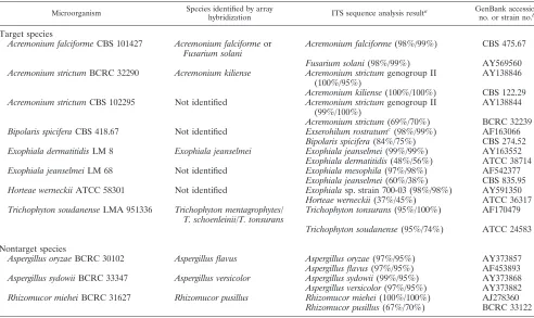
identified (no hybridization signal) (see Table S1 in the sup-plemental material and Table 2). Strains that produced dis-crepant identification by the conventional methods and the array method were further analyzed in Table 2.
A. falciformeCBS 101427 hybridized to probes Acfal1
(de-signed for A. falciforme) and Fusol2 (designed for Fusarium
solani) with almost equal hybridization signals, and no unam-biguous species designation for the strain was obtained by the
array method.A. strictumBCRC 32290 was misidentified asA.
kiliense(Table 2). The ITS 1 and 2 regions ofA. strictumBCRC 32290 were sequenced and compared with sequences in the
[image:4.585.137.449.561.686.2]FIG. 1. Layout of oligonucleotide probes on the array (1.1 by 0.9 cm). The probe “PC” (G10) was designed on the basis of the ITS 2 region ofSaccharomycodes ludwigiiBCRC 21378 and used as a positive control. Probes coded “NC” (E6 and G2 to G4) were negative controls (tracking dye only). Probes coded “M” were ITS 4 labeled with digoxigenin at its 5⬘end and were used as position markers. Group-specific probes are underlined, and multiple probes used to identify a single species are in bold face. The corresponding sequences of probes are listed in Table 1.
TABLE 1—Continued
Microorganism Probe codea Sequence (5⬘to 3⬘)b Length
(bp) Tm(°C) Locationc
GenBank accession no.
Trichophyton rubrum/ T. soudanense/ T. violaceum
Trrsv3e CCGCCCTGGCCCCAATCTTTATA 23 53.7 614–636 (2) Z97993
Trichophyton verrucossum Trver GCCTTCCCCCAAATCTCTCTGAGATW 26 54.4 607–632 (1) Z98003 Trichophyton
mentagrophytes/ T. tonsurans/ T. schoenleinii
Trmmiste GCCTCAAAATCTGTTTTATACTTATCAGGT 30 51.1 619–648 (2) Z97999
Ulocladium consortiale Ulcon2 CCAAGGTCAGCATCCACAAAGCCTT 25 54.2 477–501 (2) AY278837
a
Oligonucleotide probes are arranged on the array as indicated in Fig. 1. b
Seven additional bases of thymine were added to the 3⬘end of each probe. c
The location of probe is shown by the nucleotide number of either ITS 1 or ITS 2; the number (1 or 2) in parenthesis indicates the ITS region from which the probe was designed.
d
ITS sequences determined in this study and submitted to GenBank. e
A group-specific probe used to detect several closely related species. f
The probe on the array was used as a positive control of assay.
on May 15, 2020 by guest
http://jcm.asm.org/
GenBank and in our database. The results revealed that both
ITS sequences ofA. strictumBCRC 32290 had 100% similarity
with those of the type strain (A. kilienseCBS 122.29) in our
database. However, A. strictum BCRC 32290 had sequence
similarities of 100% (ITS 1) and 95% (ITS 2), respectively, with A. strictum genogroup II (GenBank accession no. AY138844) (Table 2).
Of the three strains not identified (Acremonium strictum
CBS 102295,Bipolaris spiciferaCBS 418.67, andHortaea
wer-neckii ATCC 58301),A. strictum CBS 102295 had sequence similarities of 99% (ITS 1) and 100% (ITS 2), respectively, withA. strictumgenogroup II (AY138844), but the similarities were only 69% (ITS 1) and 70% (ITS 2), respectively, with type
strain A. strictum BCRC 32239 (Table 2). B. spicifera CBS
418.67 had sequence similarities of 98% (ITS 1) and 99% (ITS
2), respectively, withExserohilum rostratum(a new name ofB.
rostratum) (AF163066). However,B. spiciferaCBS 418.67 dis-played sequence similarities of only 84% (ITS 1) and 75% (ITS
2), respectively, withB. spiciferaCBS 274.52 in our database.
Therefore, the designation of CBS 418.67 asB. spiciferamay be
questionable.H. werneckiiATCC 58301 had high ITS
similar-ities (98 to 99%) withExophialasp. strain NYSDOH 700-03
(AY591350) by a BLAST search in GenBank (Table 2).
How-ever,H. werneckii ATCC 58301 had sequence similarities of
only 37% (ITS 1) and 45% (ITS 2), respectively, withH.
wer-neckii ATCC 36317 (type strain) in our database. For this
reason, the designation of the strain ATCC 58301 asH.
wer-neckiimay be incorrect. Despite the above-described
inconsis-tent results, a third strain ofA. strictum(BCRC 32239) and a
second strain ofB. spicifera(CBS 274.52) were correctly
iden-tified (see Table S1 and Fig. S2, panels 4 and 19, in the sup-plemental material).
Of the 93 nontarget reference strains, two were misidentified
by the array.Aspergillus sydowiiBCRC 33347 andRhizomucor
mieheiBCRC 31627 were misidentified asA. versicolorandR. pusillus, respectively (see Table S1 in the supplemental mate-rial and Table 2). A BLAST search revealed that the species designation of the two reference strains is correct. Misidenti-fications of the two strains were apparently caused by their partial ITS sequence homology with the above-indicated target
species (Table 2). AlthoughR. mieheiBCRC 31627 had low
ITS similarities (67 to 70%) withR. pusillus, sequence
align-ment indicated that the probe (Ripu4) designed forR. pusillus
had an 85% similarity with a partial region of the ITS 2 ofR.
mieheiBCRC 31627.
In addition to hybridization to their species-specific probes,
each of the strainsAcremonium falciformeCBS 475.67 (see Fig.
2, panel 2, in the supplemental material),Alternaria alternata
BCRC 30501 (panel 5), Aspergillus nidulans ATCC 11267
(panel 12), Aspergillus versicolor BCRC 30225 (panel 15),
[image:5.585.44.536.80.373.2]Scopulariopsis brevicaulisATCC 7903 (panel 55), and Ulocla-dium consortialeCBS 105.21 (panel 65) cross-hybridized to an extra heterologous probe on the array. However, the hybrid-ization signals caused by the heterologous probes were much
TABLE 2. List of fungal strains that produced discrepant identification by the conventional methods and array hybridization
Microorganism Species identified by array
hybridization ITS sequence analysis result
a GenBank accession no. or strain no.b
Target species
Acremonium falciformeCBS 101427 Acremonium falciformeor Fusarium solani
Acremonium falciforme(98%/99%) CBS 475.67
Fusarium solani(98%/99%) AY569560 Acremonium strictumBCRC 32290 Acremonium kiliense Acremonium strictumgenogroup II
(100%/95%)
AY138846
Acremonium kiliense(100%/100%) CBS 122.29 Acremonium strictumCBS 102295 Not identified Acremonium strictumgenogroup II
(99%/100%)
AY138844
Acremonium strictum(69%/70%) BCRC 32239 Bipolaris spiciferaCBS 418.67 Not identified Exserohilum rostratumc(98%/99%) AF163066
Bipolaris spicifera(84%/75%) CBS 274.52 Exophiala dermatitidisLM 8 Exophiala jeanselmei Exophiala jeanselmei(99%/99%) AY163552
Exophiala dermatitidis(48%/56%) ATCC 38714 Exophiala jeanselmeiLM 68 Not identified Exophiala mesophila(97%/98%) AF542377
Exophiala jeanselmei(60%/38%) CBS 835.95 Horteae werneckiiATCC 58301 Not identified Exophialasp. strain 700-03 (98%/98%) AY591350
Horteae werneckii(37%/45%) ATCC 36317 Trichophyton soudanenseLMA 951336 Trichophyton mentagrophytes/
T. schoenleinii/T. tonsurans
Trichophyton tonsurans(95%/100%) AF170479
Trichophyton soudanense(95%/74%) ATCC 24583
Nontarget species
Aspergillus oryzaeBCRC 30102 Aspergillus flavus Aspergillus oryzae(97%/95%) AY373857 Aspergillus flavus(97%/95%) AF453893 Aspergillus sydowiiBCRC 33347 Aspergillus versicolor Aspergillus sydowii(99%/95%) AY373868 Aspergillus versicolor(97%/95%) AY373882 Rhizomucor mieheiBCRC 31627 Rhizomucor pusillus Rhizomucor miehei(100%/100%) AJ278360
Rhizomucor pusillus(67%/70%) BCRC 33122
a
Values in parenthesis are the ITS 1/ITS 2 sequence similarities of the discrepant strain with the indicated fungal species. b
The ITS sequences of the discrepant strain were compared with the indicated GenBank accession number or the closest reference strain in our database. c
Exserohilum rostratumis a new name ofBipolaris rostratum.
on May 15, 2020 by guest
http://jcm.asm.org/
weaker than those produced by their homologous probes, and according to the protocol, these species were correctly identi-fied by the array. The hybridization results for three selected
nontarget strains (Absidia coeruleaBCRC 30897, Penicillium
lividumBCRC 31673, andRhizopus azygosporusBCRC 31158) are also shown (see Fig. S2, panels 68 to 70, in the supplemen-tal material). For the three nontarget species, no probes were hybridized except the probe for positive control.
Hybridization of clinical isolates to the oligonucleotide
ar-ray.A total of 71 clinical isolates, including 57 target and 14
nontarget strains, were tested. Of the 57 target strains, 54 were correctly identified (see Table S1 in the supplemental
mate-rial).Exophiala dermatitidisLM 8 andTrichophyton soudanense
LMA 951336 were identified asE. jeanselmeiandTrichophyton
mentagrophytes/T. schoenleinii/T. tonsurans, respectively, by the array method (Table 2). ITS sequence comparison revealed
that the identifications ofE. dermatitidisLM 8 andT.
soudane-nse LMA 951336 might have been misidentifications of E.
jeanselmeiandT. tonsurans, respectively, because of their high ITS similarities with the later two species and low similarities
with their respective reference strains (Table 2).E. jeanselmei
LM 68 was not identified, and ITS sequence analysis demon-strated that its ITS had similarities of 97% (ITS 1) and 98%
(ITS 2), respectively, withE. mesophila (GenBank accession
no. AF542377), which was not a target strain of this study
(Table 2). Therefore, the clinical isolatesE. dermatitidisLM 8,
E. jeanselmeiLM 68, andT. soudanenseLMA 951336 should
represent misidentifications ofE. jeanselmei,E. mesophila, and
T. tonsurans, respectively, or their close relatives. On the basis of these results, 100% (57/57) of clinical isolates were correctly identified by the array method. All 14 nontarget clinical iso-lates produced no hybridization signal with all probes on the array (see Table S1 in the supplemental material).
Performance of the array for fungal identification. Of the 233 target reference strains, 228 (97.9%) were correctly iden-tified, and of the 57 target clinical isolates, 57 (100%) were correctly identified (see Table S1 in the supplemental mate-rial). Irrespective of strain sources, an overall sensitivity of 98.3% (285/290) was obtained by the array method. Of the 93
nontarget reference strains, two (A. sydowiiBCRC 33347 and
Rhizomucor miehei BCRC 31627) were misidentified and a specificity of 97.9% (91/93) was obtained. For the 14 nontarget clinical isolates, a specificity of 100% (14/14) was obtained. In summary, a specificity of 98.1% (105/107) was obtained irre-spective of the sources of nontarget strains.
Hybridization of multiple microorganisms to an array.An array could be used to simultaneously identify several different microorganisms if these microorganisms belonged to distant
taxa. For example, the PCR products ofAbsidia corymbifera
BCRC 33078 and Aspergillus fumigatus BCRC 32120 could
concurrently hybridize to their respective probes on an array (see Fig. S2, panel 66, in the supplemental material). Another
example was thatAspergillus flavus BCRC 30007 and
Penicil-lium marneffeiCBS 334.59 could be identified on a chip (see Fig. S2, panel 67, in the supplemental material).
Detection limit of the array. Serial 10-fold dilutions of
DNAs extracted from two strains (Aspergillus fumigatusBCRC
32120 andRhizopus oryzaeBCRC 31108) were used to
deter-mine the detection limit. The present method was able to
detect fungal genomic DNA at a level as low as 10 pg (data not shown).
DISCUSSION
In this study, an oligonucleotide array was developed to identify 64 species (32 genera) of clinically relevant molds. The 64 species covered pathogens causing superficial, cutaneous, subcutaneous, and invasive infections. Several species of
di-morphic fungi (Blastomyces dermatitidis,Paracoccidioides
bra-siliensis,Penicillium marneffei) (see Table S1 in the supplemen-tal material) were also included. The prominent feature of the present method is that it combines the various methods into a single standardized protocol encompassing DNA extraction, PCR amplification of the ITS regions, and hybridization of the PCR products to the array. Another important characteristic is that visualization of fungal reproductive structures, which is essential for classical identification, is not needed for the present method. Since the primer pairs (ITS 1 and 4) used in this study can also amplify the ITS regions of yeasts (5, 31, 46), it is expected that clinically relevant yeasts also could be iden-tified by the same approach used in this study.
Traditionally, the identification of molds causing infections involves isolation of organisms and relies on the expertise of the clinical microbiologist. Identification of filamentous fungi is therefore technical demanding and time consuming, and inaccurate identification is not rare. Taylor et al. (42) have pointed out that there are distinctions between the “evolution-ary species” (theoretical species) concept and the “operational species” concept that may include the “morphological species” concept, “biological species” concept, and “phylogenetic spe-cies” concept. It has been noted that in general a single mor-phological species can contain multiple biological or phyloge-netic species (42). Besides, strains belong to the same species may display different morphological characteristics at different growth stages. In this study, it was found that even some
ref-erence strains (Bipolaris spiciferaCBS 418.67 andHortaea
wer-neckiiATCC 58301) obtained from collection centers may be misidentified.
The good performance (sensitivity and specificity) of the present array might be due to the fact that the fungal ITS sequences have low intraspecies variation and high interspecies divergence (10, 19, 21, 24, 32). Until now, there has been no report defining the criteria for maximum intraspecies variation in the ITS regions. However, the criteria have been established for the D1/D2 region of large-subunit rRNA genes. Fell et al. (16) established a database of 230 species of basidiomycetous yeasts and suggested that strains differing by two or more nucleotides in the D1/D2 region represented different taxa. Kurtzman and Robnett (29) studied approximately 500 yeast species and proposed that strains showing greater than 1% substitutions in the approximately 600-nucleotide D1/D2 re-gion were likely to be different species. However, the fungal ITS region is more variable than the D1/D2 region. We have sequenced both ITS 1 and 2 regions of many fungal species to establish the intraspecies variation of medically important molds (data not shown) and found that, in general, 2% diver-gence of both regions (ITS 1 and 2) seems to be a reasonable threshold to differentiate species. Some intraspecies variations
on May 15, 2020 by guest
http://jcm.asm.org/
of ITS 1 (or ITS 2) may be⬎2%; however, it was rarely found
that the divergence of both ITS regions is⬎2%.
Although a single probe was designed to identify each indi-vidual species under most conditions, some exceptions existed.
For example, multiple probes were used to identify
Acremo-nium kiliense(Ackil2 and Ackil3) andAureobasidium pullulans
(Aupul2, Aupul3, and Aupul4) (Table 1 and Fig. 1) because strains within the two species have high intraspecies divergence of ITS sequences. Fig. S2 (panel 16) in the supplemental
ma-terial shows thatA. pullulansBCRC 32064 hybridized to the
probe Aupul2, whileA. pullulansBCRC 31981 hybridized to
other two probes (Aupul3 and Aupul4) (panel 17). In contrast, some species within a genus displayed high ITS homology and the design of species-specific probes was difficult. Therefore, several probes were designed to identify a “group” of closely related species. For example, the probe Chcgf1 (Table 1) was
used to identify species ofChaeotomium cochlioides,C.
funi-cola, andC. globosum. However,C. funicolahad its own
spe-cies-specific probe (Chfun2) (Table 1). Therefore, when both probe Chcgf1 and probe Chfun2 were hybridized, the
micro-organism wasC. funicola(see Fig. S2, panel 22, in the
supple-mental material). If only the probe Chcgf1 was hybridized, the
strain might belong toC. cochlioidesorC. globosum(panels 21
and 23). Similarly, the probe Fumop was designed to identify
Fusarium moniliforme,F. oxysporum, andF. pallidoroseum, but
an additional probe (Fumon) was designed forF. moniliforme
(Table 1). Therefore, F. moniliforme could be differentiated
fromF. oxysporum(panel 34) andF. pallidoroseum(panel 35) by hybridization to probes of both Fumop and Fumon (panel 33).
In this study, four strains (Acremonium strictumCBS 102295,
Bipolaris spicifera CBS 418.67, Exophiala jeanselmei LM 68, andHortaea werneckiiATCC 58301) were not identified (Table
2). The anamorphic characteristics ofA. strictumCBS 102295
are as follows: conidiophores simple, occasionally branched, phialides slender (arising from submerged or slightly fasiculate aerial hyphae), conidia grouped in slimy heads (cylindrical or ellipsoidal, hyaline). These characteristics are typical of the
speciesA. strictum(11).A. strictumCBS 102295 was not
iden-tified, and this might be due to genetic diversity among isolates of the species. Novicki et al. (39) found a
greater-than-ex-pected variation among strains previously identified asA.
stric-tum. In sequence analyses of the ITS regions and the D1/D2
variable domain of the 28S ribosomal DNA, five clinical
iso-lates phenotypically identified as A. strictum were found to
have ⬍99% similarities to the A. strictum type strain (CBS
346.70) at the ITS and 28S loci. But a sixth isolate
phenotyp-ically identified as anAcremoniumsp. had⬎99% similarities to
the type strain at both loci. They concluded that five out of the
six clinical isolates belong to species other thanA. strictumor
that theA. strictumtaxon is genetically diverse.
Bipolaris spicifera CBS 418.67 had sequence similarities of
98% (ITS 1) and 99% (ITS 2), respectively, withExserohilum
rostratum(a new name ofB. rostratum) (GenBank accession
no. AF163066). However, B. spicifera CBS 418.67 displayed
sequence similarities of only 84% (ITS 1) and 75% (ITS 2),
respectively, with B. spicifera CBS 274.52 in our database.
Therefore, the designation of CBS 418.67 asB. spiciferamay be
questionable. The conidiophores of CBS 418.67 are erect, un-branched, septate, and regularly zig-zagged in the apical part.
The conidia of CBS 418.67 are brown, cylindrical with rounded ends, and have three distosepta (conidia subdivided by inner wall layer only). The above-described characteristics of CBS
418.67 are typical of the species ofB. spicifera(11) and may be
why CBS 418.67 was given the nameB. spicifera.
For Exophiala jeanselmei LM 68 (a not-identified clinical isolate), its ITS sequences had similarities of 97% (ITS 1) and
98% (ITS 2), respectively, withE. mesophila(GenBank
acces-sion no. AF542377), which was not a target strain of this study
(Table 2). Members of genusExophialaare often difficult to
identify to the species level because of their variable
morpho-logical appearances (40). The microscopic morphologies ofE.
jeanselmeiLM 68 andE. mesophilaare similar; both produce annellides that are slender, tubular, sometimes branched, and characteristically tapered to a narrow, elongated tip. Their conidia are oval and gather in clusters at the end and sides of the conidiophore.
Hotaea werneckiiATCC 58301 had high ITS similarities (98
to 99%) withExophialasp. NYSDOH 700-03 (AY591350) by
a BLAST search in GenBank (Table 2). At the early stage, ATCC 58301 produce pale or dark brown yeast-like cells.
These cells (2 to 5 by 5 to 10m) are actually annellides and
are round at one end while tapered and elongated with stria-tions at the other end where conidia are formed. The one- or two-celled annelloconidia form and accumulate at annellidic points along the hyphae. The above-described microscopic
characteristics of ATCC 58301 are typical of the species H.
werneckii(11), and this might explain why ATCC 58301 was
given the nameH. werneckii.
Invasive aspergillosis is a leading cause of infection among patients undergoing treatment for hematological malignancies, solid organ transplantation, and hematopoietic stem cell
trans-plantation (8, 10, 37).Aspergillus fumigatusremains the most
frequent cause of invasive aspergillosis, followed byA. flavus,
A. terreus,A. niger,A. nidulans, andA. versicolor(12, 22). These
six importantAspergillusspecies were all included in this study,
and probes designed for them showed satisfactory hybridiza-tion results (see Fig. S2, panels 10 to 15, in the supplemental material). The only exception was one strain (BCRC 302225) of A. versicolorthat cross-hybridized with the probe Asnid2
designed forA. nidulans(see Fig. S2, panel 15, in the
supple-mental material). However, the probe Asver4, designed to
identifyA. versicolor, produced much a stronger hybridization
signal than the probe Asnid2.
Recently, Husain et al. (22) prospectively studied 53 liver and heart transplant recipients and found that invasive
infec-tions due to filamentous fungi other than Aspergillusspecies
were significantly more likely to be associated with dissemi-nated and central nervous system infections than were those
due toAspergillus species. The associated mortality rate was
100% for zygomycosis, 80% for non-Aspergillus
hyalohyphomy-cosis, and 54% for aspergillosis. Non-Aspergillusmolds causing
sever infections in organ transplant recipients include
zygomy-cetes,Fusariumspp.,Pseudallescheria boydii(Scedosporium
ap-iospermum), Scedosporium prolificans, and dematiaceous
molds (6). Probes for most of these important non-Aspergillus
species were included in the current study (Table 1).
Some species of dermatophytes are phylogenetically and taxonomically very closely related (19, 25, 34). A high level of ITS homology was observed for several common dermatophyte
on May 15, 2020 by guest
http://jcm.asm.org/
species. Our in-house ITS database demonstrates that the
se-quence similarities betweenTrichophyton rubrumATCC 28188
andT. violaceumATCC 28944 are 0.98 (ITS 1) and 0.95 (ITS 2), respectively, whereas the similarities are 1.0 (ITS 1) and
0.97 (ITS 2), respectively, betweenT. rubrumATCC 28188 and
T. soudanenseATCC 24583. Sequence similarities of 0.96 (ITS
1) and 0.99 (ITS 2) were observed betweenT. mentagrophytes
CBS 361.62 andT. tonsuransATCC 56186, and were 0.95 (ITS
1) and 0.94 (ITS 2) betweenT. schoenleiniiATCC 22775 andT.
tonsurans ATCC 56186. For this reason, the group-specific
probe Trrsv3 was designed to identify species ofT. rubrum,T.
soudanense, and T. violaceum, whereas Trmst1 was used for
identification ofT. mentagrophytes,T. schoenleinii, andT.
ton-surans. Additional probes designed from other loci such as the D1/D2 or ribosomal intergenic regions (13) may help to gen-erate species-specific probes of dermatophytes. However, sep-arate PCR or multiplex PCR using different sets of primers will be needed to amplify these different regions (13).
Although the Tm values of some probes listed in Table 1
were lower than the array hybridization temperature (55°C), clear hybridization signals were still obtained for these probes (see Fig. S2 in the supplemental material). For example,
probes Acfal1 (Tm ⫽ 49.7°C) and Gecap3 (Tm ⫽ 49.7°C),
designed to identify Acremonium falciforme and Geotrichum
capitatum, respectively, produced discernible signals (see Fig. S2, panels 2 and 38, in the supplemental material). This might be partially due to the use of relatively low stringency buffer
(high ionic strength [5⫻SSC] and low detergent concentration
[0.02% SDS]) for hybridization reaction. Volokhov et al. (45)
used several probes havingTmvalues (40 to 44°C) lower than
the hybridization temperature (45°C) for identification of
Lis-teriaspp. and still obtained good hybridization results. In
con-trast, probes Acstr2 (Tm⫽55.3°C) and Blder2 (Tm⫽56.5°C),
used to identifyAcremonium strictumandBlastomyces
derma-titidis, respectively, produced relatively weak hybridization sig-nals (see Fig. S2, panels 4 and 20, in the supplemental mate-rial). The secondary structure of probes may influence the hybridization efficiency (13).
In conclusion, species identification of clinically important molds by the present oligonucleotide array is reliable and can be used as an effective alternative to the conventional identi-fication methods. The whole procedure can be finished within 24 h starting from isolated colonies, and visualization of fungal reproductive structures is not required. The array used in this study has the potential to be expanded by including more probes in order to identify more species.
ACKNOWLEDGMENT
This project was supported by grants (NSC 93-2323-B006-007 and NSC 93-2314-B006-117) from the National Science Council, Taiwan, Republic of China.
REFERENCES
1.Adamczyk, J., M. Hesselsoe, N. Iversen, M. Horn, A. Lehner, P. H. Nielsen, M. Schloter, P. Roslev, and M. Wagner.2003. The isotope array, a new tool that employs substrate-mediated labeling of rRNA for determination of microbial community structure and function. Appl. Environ. Microbiol.69:
6875–6887.
2.Baddley, J. W., T. P. Stroud, D. Salzman, and P. G. Pappas.2001. Invasive mold infections in allogeneic bone marrow transplant recipients. Clin. Infect. Dis.32:1319–1324.
3.Barnes, R. A., D. W. Denning, E. G. V. Evans, R. J. Hay, C. C. Kibbler, A. G. Prentice, M. D. Richardson, M. M. Roberts, T. R. Rogers, D. C. Speller,
D. W. Warnock, and R. E. Warren.1996. Fungal infections: a survey of laboratory services for diagnosis and treatment. Commun. Dis. Rep. Rev.
6:R69–R75.
4.Brown, T. J., and R. M. Anthony.2000. The addition of low numbers of 3⬘ thymine bases can be used to improve the hybridization signal of oligonu-cleotides for use within arrays on nylon supports. J. Microbiol. Methods
42:203–207.
5.Chang, H. C., S. N. Leaw, A. H. Huang, T. L. Wu, and T. C. Chang.2001. Rapid identification of yeasts in positive blood cultures by a multiplex PCR method. J. Clin. Microbiol.39:3466–3471.
6.Chen, S. C. A., C. L. Halliday, and W. Meyer.2002. A review of nucleic acid-based diagnostic tests for systemic mycoses with an emphasis on poly-merase chain reaction-based assays. Med. Mycol.40:333–357.
7.Chen, Y., J. D. Eisner, M. M. Kattar, S. L. Rassoulian-Barrett, K. Lafe, U. Bui, A. P. Limaye, and B. T. Cookson.2001. Polymorphic internal tran-scribed spacer region 1 DNA sequences identify medically important yeasts. J. Clin. Microbiol.39:4042–4051.
8.Cornet, M., L. Fleury, C. Maslo, J. F. Bernard, G. Brucker, and the Invasive Aspergillosis Surveillance Network of the Assistance Publique-Hopitaux de Paris.2002. Epidemiology of invasive aspergillosis in France: a six-year multicentric survey in the Greater Paris area. J. Hosp. Infect.51:288–296. 9.Dasbach, E. J., G. M. Davies, and S. M. Teutsch.2000. Burden of
aspergil-losis-related hospitalizations in the United States. Clin. Infect. Dis.31:1524– 1528.
10.De Aguirre, L., S. F. Hurst, J. S. Choi, J. H. Shin, H. P. Hinrikson, and C. J. Morrison.2004. Rapid differentiation ofAspergillusspecies from other med-ically important opportunistic molds and yeasts by PCR-enzyme immunoas-say. J. Clin. Microbiol.42:3495–3504.
11.De Hoog, G. S., J. Guarro, J. Gene, and M. J. Figueras.2000. Atlas of clinical fungi, 2nd ed. Centraalbureau voor Schimmelcultures, Utrecht, The Neth-erlands, and University Rovirai Virgili, Reus, Spain.
12.Denning, D. W.1998. Invasive aspergillosis. Clin. Infect. Dis.26:781–803. 13.Diaz, M. R., and J. W. Fell.2004. High-throughput detection of pathogenic
yeasts of the genusTrichosporon. J. Clin. Microbiol.42:3696–3706. 14.Ellis, M. E., H. Al-Abdely, A. Sandridge, W. Greer, and W. Ventura.2001.
Fungal endocarditis: evidence in the world literature, 1965–1995. Clin. In-fect. Dis.32:50–62.
15.Evertsson, U., H. J. Monstein, and A. G. Johansson.2000. Detection and identification of fungi in blood using broad-range 28S rDNA PCR amplifi-cation and species-specific hybridisation. Acta Pathol. Microbiol. Immunol. Scand.108:385–392.
16.Fell, J. W., T. Boekhout, A. Fonseca, G. Scorzetti, and A. Statzell-Tallman.
2000. Biodiversity and systematics of basidiomycetous yeasts as determined by large-subunit rDNA D1/D2 domain sequence analysis. J. Syst. Evol. Microbiol.50:1351–1371.
17.Fukushima, M., K. Kakinuma, H. Hayashi, H. Nagai, K. Ito, and R. Kawaguchi.2003. Detection and identification ofMycobacteriumspecies isolates by DNA microarray. J. Clin. Microbiol.41:2605–2615.
18.Geiser, D. M., J. I. Pitt, and J. W. Taylor.1998. Cryptic speciation and recombination in the aflatoxin-producing fungusAspergillus flavus. Proc. Natl. Acad. Sci. USA95:388–393.
19.Gra¨ser, Y., M. El Fari, R. Vilgalys, A. F. A. Kuijpers, G. S. De Hoog, W. Presber, and H. Tietz.1999. Phylogeny and taxonomy of the family Arthro-dermataceae(dermatophytes) using sequence analysis of the ribosomal ITS region. Med. Mycol.37:105–114.
20.Hall, L., S. Wohlfiel, and G. D. Roberts.2004. Experience with the MicroSeq D2 large-subunit ribosomal DNA sequencing kit for identification of fila-mentous fungi encountered in the clinical laboratory. J. Clin. Microbiol.
42:622–626.
21.Henry, T., P. C. Iwen, and S. H. Hinrichs.2000. Identification ofAspergillus
species using internal transcribed spacer regions 1 and 2. J. Clin. Microbiol.
38:1510–1515.
22.Husain, S., B. D. Alexander, P. Munoz, R. K. Avery, S. Houston, T. Pruett, R. Jacobs, E. A. Dominguez, J. G. Tollemar, K. Baumgarten, C. M. Yu, M. M. Wagener, P. Linden, S. Kusne, and N. Singh.2003. Opportunistic mycelial fungal infections in organ transplant recipients: emerging impor-tance of non-Aspergillusmycelial fungi. Clin. Infect. Dis.37:221–229. 23.Iwen, P. C., A. G. Freifeld, T. A. Bruening, and S. H. Hinrichs.2004. Use of
a panfungal PCR assay for detection of fungal pathogens in a commercial blood culture system. J. Clin. Microbiol.42:2292–2293.
24.Iwen, P. C., S. H. Hinrichs, and M. E. Rupp.2002. Utilization of the internal transcribed spacer regions as molecular targets to detect and identify human fungal pathogens. Med. Mycol.40:87–109.
25.Jackson, C. J., R. C. Barton, and E. G. V. Evans.1999. Species identification and strain differentiation of dermatophyte fungi by analysis of ribosomal-DNA intergenic spacer regions. J. Clin. Microbiol.37:931–936.
26.Kappe, R., C. N. Okeke, C. Fauser, M. Maiwald, and H.-G. Sonntag.1998. Molecular probes for the detection of pathogenic fungi in the presence of human tissue. J. Med. Microbiol.47:811–820.
27.Kobayashi, M., H. Sonobe, T. Ikezoe, E. Hakoda, Y. Ohtsuki, and H. Tagu-chi.1999. In situ detection ofAspergillus18S ribosomal RNA in invasive pulmonary aspergillosis. Intern. Med.38:563–569.
on May 15, 2020 by guest
http://jcm.asm.org/
28.Kurtzman, C. P., and C. J. Robnett.1997. Identification of clinically impor-tant ascomycetous yeasts based on nucleotide divergence in the 5⬘end of the large-subunit (26S) ribosomal DNA gene. J. Clin. Microbiol.35:1216–1223. 29.Kurtzman, C. P., and C. J. Robnett.1998. Identification and phylogeny of ascomycetous yeasts from analysis of nuclear large subunit (26S) ribosomal DNA partial sequences. Antonie Leeuwenhoek73:331–371.
30.Larone, D. H.2002. Medically important fungi: a guide to identification, 4th ed. Washington, D.C.
31.Li, Y. L., S. N. Leaw, J. H. Chen, H. C. Chang, and T. C. Chang.2003. Rapid identification of yeasts commonly found in positive blood cultures by ampli-fication of the internal transcribed spacer regions 1 and 2. Eur. J. Clin. Microbiol. Infect. Dis.22:693–696.
32.Lindsley, M. D., S. F. Hurst, N. J. Iqbal, and C. J. Morrison.2001. Rapid identification of dimorphic and yeast-like fungal pathogens using specific DNA probes. J. Clin. Microbiol.39:3505–3511.
33.Luo, G., and T. G. Mitchell.2002. Rapid identification of pathogenic fungi directly from cultures by using multiplex PCR. J. Clin. Microbiol.40:2860– 2865.
34.Makimura, K., T. Mochizuki, A. Hasegawa, K. Uchida, H. Saito, and H. Yamaguchi.1998. Phylogenetic classification ofTrichophyton mentagrophytes
complex strains based on DNA sequences of nuclear ribosomal internal transcribed spacer 1 regions. J. Clin. Microbiol.36:2629–2633.
35.Martin, C., D. Roberts, M. van Der Weide, R. Rossau, G. Jannes, T. Smith, and M. Maher.2000. Development of a PCR-based line probe assay for identification of fungal pathogens. J. Clin. Microbiol.38:3735–3742. 36.McClure, F. D.1990. Design and analysis of quantitative collaborative
stud-ies: minimum collaborative program. J. Assoc. Off. Anal. Chem.73:953–960. 37.McNeil, M. M., S. L. Nash, R. A. Hajjeh, M. A. Phelan, L. A. Conn, B. D. Plikaytis, and D. W. Warnock.2001. Trends in mortality due to invasive mycotic diseases in the United States, 1980–1997. Clin. Infect. Dis.33:641– 647.
38.Ninet, B., J. Isabell, O. Bontems, B. Lechenne, O. Jousson, R. Panizzon, D. Lew, and M. Monod.2003. Identification of dermatophyte species by 28S ribosomal DNA sequencing with a commercial kit. J. Clin. Microbiol.41:
826–830.
39.Novicki, T. J., K. LaFe, L. Bui, U. Bui, R. Geise, K. Marr, and B. T. Cookson.
2003. Genetic diversity among clinical isolates ofAcremonium strictum de-termined during an investigation of a fatal mycosis. J. Clin. Microbiol.41:
2623–2628.
40.Porteous, N. B., A. M. Grooters, S. W. Redding, E. H. Thompson, M. G. Rinaldi, G. S. De Hoog, and D. A. Sutton.2003. Identification ofExophiala mesophilaisolated from treated dental unit waterlines. J. Clin. Microbiol.
41:3885–3889.
41.Skladny, H., D. Buchheidt, C. Baust, F. Krieg-Schneider, W. Seifarth, C. Leib-Mosch, and R. Hehlmann.1999. Specific detection ofAspergillus spe-cies in blood and bronchoalveolar lavage samples of immunocompromised patients by two-step PCR. J. Clin. Microbiol.37:3865–3871.
42.Taylor, J. W., D. J. Jacobson, S. Kroken, T. Kasuga, D. M. Geiser, D. S. Hibbett, and M. C. Fisher.2000. Phylogenetic species recognition and spe-cies concepts in fungi. Fungal Genet. Biol.31:21–32.
43.Turenne, C. Y., S. E. Sanche, D. J. Hoban, J. A. Karlowsky, and A. M. Kabani.1999. Rapid identification of fungi by using the ITS 2 genetic region and an automated fluorescent capillary electrophoresis system. J. Clin. Mi-crobiol.37:1846–1851.
44.Velegraki, A., M. E. Kambouris, G. Skiniotis, M. Savala, A. Mitroussia-Ziouva, and N. J. Legakis.1999. Identification of medically significant fungal genera by polymerase chain reaction followed by restriction enzyme analysis. FEMS Immunol. Med. Microbiol.23:303–312.
45.Volokhov, D., A. Rasooly, K. Chumakov, and V. Chizhikov.2002. Identifi-cation ofListeriaspecies by microarray-based assay. J. Clin. Microbiol.40:
4720–4728.
46.White, T., T. Burns, S. Lee, and J. Taylor.1990. Amplification and direct sequencing of fungal ribosomal RNA genes for phylogenetics, p. 315–322.In
M. A. Innis, D. H. Gelfand, J. J. Sninsky, and T. J. White (ed.), PCR protocols. A guide to methods and applications. Academic Press, Inc., San Diego, Calif.
47.Wu, Z., Y. Tsumura, G. Blomquist, and X.-R. Wang.2003. 18S rRNA gene variation among common airborne fungi, and development of specific oli-gonucleotide probes for the detection of fungal isolates. Appl. Environ. Microbiol.69:5389–5397.