Copyright © 2002, American Society for Microbiology. All Rights Reserved.
Six-Year Molecular Analysis of
Burkholderia cepacia
Complex Isolates
among Cystic Fibrosis Patients at a Referral Center for
Lung Transplantation
David G. Heath,
1† Kathy Hohneker,
2Charlene Carriker,
3Kelly Smith,
1Jonathan Routh,
1John J. LiPuma,
4Robert M. Aris,
2David Weber,
2,3,5and Peter H. Gilligan
1,6*
Clinical Microbiology-Immunology Laboratories1and Department of Hospital Epidemiology,3University of North Carolina
Hospitals, and Microbiology-Immunology,6Medicine,2and Pediatrics,5The University of North Carolina at Chapel Hill,
Chapel Hill, North Carolina, and Department of Pediatrics and Communicable Diseases,
The University of Michigan, Ann Arbor, Michigan4
Received 28 June 2001/Returned for modification 26 October 2001/Accepted 24 December 2001
Over a 6-year period,Burkholderia cepaciacomplex species were isolated from cystic fibrosis (CF) patients receiving care at The University of North Carolina Hospitals (clinic CF patients) and from those referred from other treatment centers. Fifty-six isolates collected from 30 referred patients and 26 clinic CF patients were characterized by pulsed-field gel electrophoresis (PFGE) and were assayed by PCR to detect the cable pilin gene,cblA. PFGE results indicated that six separate clusters (clusters A to F) were present among the 56 isolates and that three clusters (clusters A, B, and E) consisted only of isolates from referred patients infected withB. cepaciacomplex isolates prior to referral. However, one cluster (cluster C) consisted of isolates from four CF patients, and hospital records indicate that this cluster began with an isolate that came from a referred patient and that spread to three clinic CF patients. Cluster D consisted of two isolates from clinic CF patients, and hospitalization records are consistent with nosocomial, patient-to-patient spread.cblAwas present in only 4 of the 56 isolates and included isolates in cluster E from the referred patients. Our results indicate a lack of spread of a previously characterized, transmissible clone from referred patients to our clinic CF population. Only two instances of nosocomial, patient-to-patient spread could be documented over the 6-year period. An additional spread of an isolate (cluster F) from a referred patient to a clinic patient could not be documented as nosocomial and may have been the result of spread in a nonhospitalized setting. The majority (36 of 56) of ourB. cepaciacomplex-infected CF patients harbor isolates with unique genotypes, indicating that a diversity of sources account for infection. These data suggest that CF patients infected withB. cepaciacomplex and referred for lung transplantation evaluation were not a major source ofB. cepaciacomplex strains that infected our resident CF clinic population.
The Burkholderia cepacia complex is composed of at least
seven closely related species or genomovars consisting of B.
cepacia(genomovar I),B. multivorans(genomovar II),B.
sta-bilis(genomovar IV),B. vietnamiensis(genomovar V), andB.
ambifaria(genomovar VII), and genomovars III and VI, with
species designations for genomovars III and VI still pending (4, 8).
Nearly 4% of cystic fibrosis (CF) patients in the United
States are infected with a member of theB. cepaciacomplex
(13). Of these patients, some will develop the cepacia syn-drome, a rapidly fatal necrotizing pneumonia with bacteremia (9, 10, 26). Previous studies have shown that transmissible
clones ofB. cepaciaexist, and subsequent infection with these
clones of both healthy uninfected CF patients and CF patients seeking lung transplantation can be devastating (9, 11, 12, 22,
25). Therefore, screening of CF patients for the detection ofB.
cepaciacomplex and management of patients once infection is
documented are extremely important. Furthermore, CF
pa-tients infected withB. cepaciacomplex isolates are stigmatized,
being segregated from the general CF patient population in terms of clinical care and social interaction. In many North
American transplant centers, infection withB. cepaciacomplex
is a strict contraindication for lung transplantation in CF pa-tients (14).
There are more than 120 lung transplantation centers in North America. The University of North Carolina (UNC) Hos-pitals is one of the few CF centers in North America that will
perform lung transplantation for CF patients infected withB.
cepaciacomplex. For this reason, about 14% of adult CF
pa-tients (i.e., roughly three times the national percentage) re-ferred to the UNC Hospitals for double lung transplantation
are infected withB. cepaciacomplex.
Some transmissible clones ofB. cepaciacomplex exist, such
as the cable pilin-positive (cblA⫹) electropherotype (ET) ET
12 clone responsible for epidemic transmission in both Canada and the United Kingdom (7, 9, 11, 21, 22, 25). Therefore, we believed that it was important to study the CF patient popu-lation at the UNC Hospitals by asking three questions. First, have referred patients brought a previously characterized, transmissible clone into our (the UNC Hospitals) center, and has it been transmitted to our clinic CF patient population? Second, are any new, previously uncharacterized, transmissible
* Corresponding author. Mailing address: Clinical Microbiology-Im-munology Laboratories, University of North Carolina Hospitals CB 7600, Chapel Hill, NC 27514. Phone: (919) 6313. Fax: (919) 966-0486. E-mail: pgilliga@unch.unc.edu.
† Present address: Department of Pathology and Area Laboratory Services, Landstuhl Regional Medical Center, Landstuhl, Germany.
1188
on May 15, 2020 by guest
http://jcm.asm.org/
clones evident among our referred patients, and has transmis-sion from referred patients to our local clinic CF population
occurred? Third, how much intracenter spread ofB. cepacia
complex has occurred?
MATERIALS AND METHODS
Characterization of UNC Hospitals CF center and infection control.AllB. cepaciacomplex isolates were recovered from CF patients between 1 January 1995 and 1 March 2001. We studied two distinct patient populations from which B. cepaciacomplex organisms were isolated. One consisted of 30 CF patients who were referred to the UNC Hospitals, often for lung transplantation evaluation, already infected withB. cepaciacomplex. The second population was of 26 CF patients who received their routine care at the UNC Hospitals and who became newly infected during a study period from 1 January 1997 to March 2001. For these patients to be considered “newly infected,” prior cultures of samples from these patients at our institution had to be negative forB. cepaciaand the organism had to be isolated during the study period.For the year 2000, 451 CF patients received care at the UNC Hospitals, with 251 of these patients being pediatric patients, while 200 were adult patients. Nineteen (9.5%) of the 200 adult patients and 8 (3.2%) of the 251 pediatric patients were infected withB. cepaciacomplex. A total of 107 CF patients are awaiting lung transplantation at the UNC Hospitals, with 15 (14%) of these patients infected withB. cepacia complex. As part of the evaluation of possible nosocomial transmission among CF patients whose isolates were in clusters C, D, and F, all patient charts were reviewed by a nurse trained in infection control. The review included production of a time line of all hospital admissions including hospital location, clinic visits, physical and occupational therapy visits, pulmonary rehabilitation visits, and evaluations in specialty clinics (e.g., pulmonary function laboratory, radiology, and transplantation clinics). Patients with knownB. cepaciacomplex colonization requiring inpatient hospitalization were admitted to private rooms and placed on contact precautions. When pediatric patients withB. cepaciacomplex were seen in the outpatient clinic, they either were scheduled to be seen on a different day than other CF patients or were scheduled to be seen at the end of the day, after all patients not infected or colonized withB.cepaciahad left the facility. Until 1998, adult CF patients were seen in the general pulmonary clinics in order to minimize contact with each other. In 1998, the formation of a dedicated adult CF clinic prompted the institution of several infection control measures, including patient education, masking of all patients upon arrival at the clinic and in all
common areas, strict adherence to hand-washing procedures, and disinfection of rooms between patients. The UNC Hospitals Lung Transplant Clinic adopted similar infection control guidelines in 2000.
B.cepaciaisolation, PFGE, andcblAanalysis.All strains were isolated on eitherPseudomonas cepaciaagar orB. cepaciaselective agar prepared in-house and were further characterized biochemically and by PCR genomovar analysis as described previously (6, 16, 19). Once a member of theB. cepaciacomplex was isolated from a CF patient, a subculture was prepared from a single colony and stocks were prepared in skim milk and frozen at⫺70°C.
Pulsed-field gel electrophoresis (PFGE) was conducted as described previ-ously, with the following revisions (7). All cultures were grown overnight in tryptic soy broth and adjusted to an optical density at 600 nm of 1. One milliliter was removed and centrifuged for 5 min to pellet the cells. The cells were then resuspended in formalin (3.8% [vol/vol]) (5) and allowed to sit on ice for 1 h. The cells were pelleted and rinsed three times in TE (10 mM Tris-HCl, 10 mM EDTA [pH 8.0]) before being placed in agarose blocks. Genomic DNA was restricted withSpeI (New England Biolabs); and electrophoresis was conducted for 24 h in 0.5⫻TBE (Tris-borate, EDTA), with initial and final pulse times of 1.2 and 54 s, respectively, by using a CHEF Mapper system (Bio-Rad). An ET 12 strain ofB. cepaciacomplex was kindly provided by R. Goldstein, Boston University School of Medicine (25). Gels were analyzed visually according to the criteria of Tenover et al. (27).
All isolates ofB. cepaciacomplex were tested by PCR for the presence of the cable pilin gene,cblA, as described previously (22).
RESULTS
Fifty-six B. cepacia complex isolates cultured from 56 CF
patients were characterized for their PFGE patterns
(geno-types) after digestion with SpeI (Table 1). Genotypes were
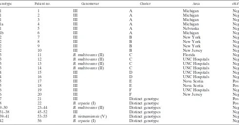
[image:2.587.49.539.84.340.2]considered related if they differed by three or fewer bands (27). In total, 42 distinct genotypes were present, which likely indi-cates a wide diversity in the sources of infection. There were 26 genomovar II (B. multivorans) isolates, 25 genomovar III iso-lates, two genomovar I (B. cepacia) isoiso-lates, and three geno-movar V (B. vietnamiensis) isolates. Our PFGE analysis re-vealed that six clusters of PFGE patterns were present within
TABLE 1. Summary of genotypes and clusters ofB. cepaciacomplex isolates recovered from January 1995 to March 2001
Genotype Patient no. Genomovar Cluster Area cblAa
1 1 III A Michigan Neg
1 2 III A Michigan Neg
1 3 III A Michigan Neg
1a 4 III A Michigan Neg
1 5 III A Nebraska Neg
1b 6 III A Michigan Neg
2 7 III B New York Neg
2 8 III B New York Neg
2 9 III B New York Neg
2 10 III B New Jersey Neg
3 11 B. multivorans(II) C Florida Neg
3 12 B. multivorans(II) C UNC Hospitals Neg
3 13 B. multivorans(II) C UNC Hospitals Neg
3 14 B. multivorans(II) C UNC Hospitals Neg
4 15 III D UNC Hospitals Neg
4 16 III D UNC Hospitals Neg
5 17 III E Nova Scotia Pos
5 18 III E Nova Scotia Pos
6 19 III F UNC Hospitals Neg
6 20 III F New Jersey Neg
7 21 III Distinct genotype Pos
8 22 B. cepacia(I) Distinct genotype Pos
9–30 23–44 B. multivorans(II) Distinct genotypes Neg
31–38 45–52 III Distinct genotypes Neg
39–41 53–55 B. vietnamiensis(V) Distinct genotypes Neg
42 56 B. cepacia(I) Distinct genotype Neg
aNeg, negative; Pos, positive.
on May 15, 2020 by guest
http://jcm.asm.org/
the CF population at the UNC Hospitals during this study period (Fig. 1). Three clusters consisted of strains from mul-tiple patients (clusters A to C), while clusters D to F consisted of isolates from only two patients each. The cluster A genotype occurred in six patients, five of whom had previously been seen at CF centers in Michigan and who were infected prior to care at the UNC Hospitals. The fifth patient whose isolate was in
this cluster was also infected withB. cepaciaprior to referral,
but the patient was originally from Nebraska and it is not known whether there is a link between this patient and the other patients whose isolates were in this cluster. Isolates from patients 1 through 3 and 5 shared identical genotypes, while isolates from patient 4 differed from those from patients 1 through 3 and 5 by two bands and isolates from patient 6 differed from those from patients 1 through 3 and 5 by three bands. All were genomovar III isolates.
Likewise, cluster B isolates (Fig. 1) occurred in those re-ferred patients seen previously at CF centers within the met-ropolitan New York City-New Jersey area and infected prior to care at the UNC Hospitals. Again, all were genomovar III isolates, and there was no evidence of the spread of this cluster to any of our clinic CF patients.
Interestingly, cluster C (Fig. 1) began with patient 11 from Florida, who sought care at the UNC Hospitals for a possible double lung transplantation and who was already infected with B. multivorans. This patient had a lengthy hospitalization from 7 June 1998 until 13 August 1998 and again from 21 August 1998 until 13 March 1999 (Fig. 2). Patient 12 was also hospi-talized from 5 through 11 August 1998, during the hospitaliza-tion of patient 11, and subsequently became culture positive
forB. multivorans on 5 November 1998. On the same date,
patient 12 was also seen in the adult CF clinic. Patient 13 was a pediatric patient hospitalized from 29 October 1998 until 12 November 1998 and was later (11 February 1999) culture
pos-itive forB. multivorans. Patient 14 was hospitalized from 13
through 21 December 1999 and had an overlapping hospital-ization with patient 12. Patient 14 was then culture positive for
B. multivoranson 25 May 2000. Although these patients did not
share the same hospital floor, they did share common hospital services (physical therapy, pulmonary function laboratory, and
radiology). Since theB. multivoransisolates from all of these
patients share the same genotype and overlapping hospitaliza-tions were evident, we conclude that nosocomial, patient-to-patient spread occurred among these patient-to-patients either directly or indirectly.
Three small clusters (clusters D to F) (Fig. 1) consisting of isolates from two patients each were also detected by PFGE, and isolates in one of these clusters (cluster E) were from
siblings who were infected withB. cepaciaand who had been
referred to the UNC Hospitals (Fig. 1). As with cluster C isolates, the isolates in cluster D were from two patients who also had overlapping hospitalizations on the same hospital unit that were clearly documented (data not shown). These two patients represent part of the UNC Hospitals CF clinic popu-lation. The isolates in cluster F consisted of isolates from patient 19, who became infected while receiving care at the UNC Hospitals, and patient 20, who was from New Jersey and
who came to the UNC Hospitals already infected with B.
cepaciagenomovar III. We could find no evidence of
[image:3.587.74.504.73.330.2]overlap-ping hospitalizations or shared hospital services between these two patients. Therefore, we suspect that patient 19 may have
FIG. 1. PFGE banding patterns of six clusters (clusters A to F) ofB. cepaciacomplex isolates recovered from CF patients at the UNC Hospitals CF center. The cluster C gel contains aB. multivoransstrain designated 23 for purposes of comparison. The cluster E gel contains the Toronto strain, designated ET, and acblA⫹genomovar III strain, designated 21, for the purposes of comparison. Numbers on the left are molecular weights.
on May 15, 2020 by guest
http://jcm.asm.org/
become infected with an isolate of the same genotype as that from patient 20 in a nonhospitalized setting (23).
Epidemic clone ET 12, isolated from CF centers in both the United Kingdom and North America, expresses cable pilin on its surface (25). For this reason, we performed PCR on our
isolates to see if thecblAgene was present, especially in our
cluster isolates. Our results show that the cblA gene was
present in only four isolates, two of which were from cluster E
(isolates from a sibling pair). A comparison of theSpeI
restric-tion patterns between theB. cepacia cblA⫹isolates from the
siblings (patients 17 and 18) and patient 21 with that of the highly transmissible Toronto strain is shown in Fig. 1. The
results indicate that while the SpeI patterns for the isolates
from the siblings are an exact match, the genotypes produced by isolates from the siblings, patient 21, and the ET 12 clone differ from each other by more than seven fragments and are therefore unrelated (25). A fourth patient infected with a
cblA⫹strain also showed a uniqueSpeI genotype, as expected,
since this isolate was a genomovar I strain (data not shown),
while the othercblA⫹isolates were genomovar III strains
(Ta-ble 1).
DISCUSSION
In an earlier study, we analyzedB. cepaciacomplex isolates
from five transplant patients and 17 clinic patients and found
no evidence of transmission of B. cepacia complex isolates
among these patients (24). However, we did find a strain ofB.
cepaciacomplex in a patient referred for transplantation
eval-uation that resulted in the recognition of a large nosocomial outbreak at the institution from which he was referred (9). This caused us to be concerned that patients referred for
transplan-tation might transmit B. cepacia complex strains to patients
receiving their routine care for CF at our institution. Of the six clones found in clusters in this study, isolates of four previously uncharacterized clones (clusters B, C, E, and F) were present in our referred patients, while cluster A has been reported previously (12) and cluster D isolates were not present in patients referred for transplantation. Cluster B iso-lates were present in four referred patients from the
metro-politan New York City-New Jersey area. To our knowledge,
there is no published study of an epidemic clone of the B.
cepaciacomplex from CF patients from that geographic area.
Therefore, we cannot comment on the transmissibility of this clone except to say that it was present in these four referred CF patients and did not spread to our clinic CF patients.
Cluster C comprised a clone ofB. multivorans.Among the
isolates in theB. cepaciacomplex, transmissibility is typically
thought to be associated withB. cepaciagenomovar III
organ-isms (2, 7, 9, 11, 21, 22, 25). Person-to-person spread of B.
multivoransis not as frequently described, and most patients
infected withB. multivoransare infected with unique strains (1,
14). Overlapping hospitalizations could be traced for all four patients infected with cluster C isolates. Cluster E consisted of only two isolates from referred patients who were siblings, but
the isolates were cblA⫹. Again, there was no evidence of
spread from these referred patients to our clinic CF patient population. Cluster F consisted of isolates from two patients
and represented the spread ofB. cepaciagenomovar III from
a referred patient to a clinic CF patient. However, we were unable to show any link between these two patients through overlapping hospitalizations, clinic visits, or hospital services and therefore consider this cluster not to have occurred noso-comially.
A closer analysis of the isolates in cluster A indicated that four of five referred patients from Michigan were infected with isolates with identical genotypes (cluster A) when they arrived at the UNC Hospitals and that the isolate from the fifth patient (Fig. 1, patient 4) possessed a genotype that differed from that of the isolates from the first four patients by only two bands.
Digestion withXbaI and PFGE of the isolate from patient 4
[image:4.587.79.503.77.220.2]allowed us to confirm by direct comparison with the PFGE results of Kumar et al. (12) that these isolates were part of the cluster of genetically related isolates from five CF centers in Michigan (data not shown). We were initially alarmed at find-ing this cluster among our referred patients. However, we found no evidence of the spread of isolates in this cluster to any of the other CF patients screened in this study. Cluster A also included an isolate from a single referred patient from Ne-braska. We have not been able to verify whether this patient
FIG. 2. Time line graph showing the hospitalization course of patients (Pt.) 11 to 14. Horizontal bars represent inpatient status, with the admission date given at the beginning of the bars and the discharge date given at the ends. Vertical bars represent dates of concurrent admission resulting in possible cross infection. #,⫹, andⴱ, the dates that patients 12, 13, and 14, respectively, first became positive forB. multivoransinfection (see text).
on May 15, 2020 by guest
http://jcm.asm.org/
attended some of the same CF camps or CF centers as the rest of the patients whose isolates were in cluster A.
One of the shortcomings of our study design was that we studied only a single isolate from each patient. We had two instances in which isolates from sibling pairs had different genotypes. Patient 14 is a sibling of patient 23, and both pa-tients were infected with the same genomovar (B. multivorans) but the genotypes of the isolates were not related. In another instance, we noted that isolates from siblings (patient 10, whose isolate was in cluster B, and patient 20, whose isolate was in cluster F) differed in their genotypic patterns. Since we studied only one isolate from each patient, it is possible that the siblings may have been infected with isolates with common genotypes that were not detected. We previously showed that
over time patients harborB. cepaciacomplex isolates with the
same genotype (24).
Overall, the results of our genotype analysis indicate that the
spread of a previously characterized, transmissible clone ofB.
cepaciafrom referred patients to our clinic CF patient
popu-lation has not occurred. Other studies have indicated that a high percentage of patients at CF centers often harbor en-demic, transmissible clones (2, 12, 15, 17, 23, 25, 29). In con-trast, our PFGE results did not indicate the presence of a common, transmissible clone among our clinic CF patient pop-ulation. Our results more closely parallel those of a recent study (20) indicating that hospitals with a segregation policy tend to have patients infected with unique strains. Docu-mented nosocomial spread involved 4 of 26 (15%) of our clinic
B. cepaciacomplex-infected patients (patients 12 to 14, whose
isolates were in cluster C, and patient 16, whose isolate was in cluster D).
A recent publication indicated that in the United Kingdom
thecblAgene may be used as a marker to identify strains with
an enhanced capacity for spread (3). We found thecblAgene
in only 4 of the 56B. cepaciaisolates that we examined. Two of
the four isolates were from referred siblings from Canada and were in cluster E. However, the genotype for cluster E isolates
differed from that of the epidemic, cblA⫹ET 12 strain, also
isolated from Canadian patients, by more than seven frag-ments, and they are therefore considered genetically
unre-lated. There was no evidence of spread ofcblA⫹clones in our
patient population. Our data are consistent with those of LiPuma and colleagues (18), who found that only 1 of 606
isolates carried thecblAgene. These data suggest that other
factors are important in the transmissibility of the genomovar III organisms.
The most frequently recoveredB. cepacia complex species
from CF patients are genomovar III (18), and recent data indicate that CF transplant patients infected with genomovar III suffer higher rates of mortality than those infected with another genomovar (1). However, it is apparent from our study
and those of others thatB. multivoranscan also be frequently
recovered (8, 18, 28). In fact, our results indicate thatB.
mul-tivoranswas slightly more prevalent than genomovar III among
our CF patient population (26 and 25 patients, respectively). Our results also revealed that 36 of the 56 CF patients seeking care at the UNC Hospitals harbor strains with unique geno-types, so their sources of infection are likely to be diverse.
In conclusion, our study ofB. cepaciacomplex-infected CF
patients indicated that transmission of an isolate from a
re-ferred patient to our clinic CF patient population occurred in only two instances (with isolates in clusters C and F), but nosocomial transmission could clearly be documented for only one of these isolates (a cluster C isolate). Intracenter trans-mission of isolates in one, two-patient cluster (cluster D) oc-curred within our clinic patient population. Our clinic patients
were infected with a variety of different genotypes of the B.
cepaciacomplex. These data suggest that CF patients who were
infected with B. cepacia complex isolates and who were
re-ferred for lung transplantation evaluation were not a major
source of theB. cepaciacomplex organisms that infected our
resident CF clinic population.
ACKNOWLEDGMENT
This work was supported in part by a grant from the Cystic Fibrosis Foundation (to J.J.L.).
REFERENCES
1.Aris, R. M., J. Routh, J. LiPuma, D. G. Heath, and P. H. Gilligan.2001. Increased mortality due toBurkholderia cepaciacomplex infection in cystic fibrosis patients after lung transplantation. Am. J. Respir. Crit. Care Med. 164:2102–2106.
2.Chen, J. S., K. A. Witzmann, T. Spilker, R. J. Fink, and J. J. LiPuma.2001. Endemicity and inter-city spread ofBurkholdria cepaciagenomovar III in cystic fibrosis patients. J. Pediatr.139:643–649.
3.Clode, F. E., M. E. Kaufmann, H. Malnick, and T. L. Pitt.2000. Distribution of genes encoding putative transmissibility factors among epidemic and non-epidemic strains ofBurkholderia cepaciafrom cystic fibrosis patients in the United Kingdom. J. Clin. Microbiol.38:1763–1766.
4.Coenye, T., E. Mahenthiralingam, D. Henry, J. J. LiPuma, S. Laevens, M. Gillis, D. P. Speert, and P. Vandamme.2001.Burkholderia ambifariasp. nov., a novel member of theBurkholderia cepaciacomplex including biocontrol and cystic fibrosis-related isolates. Int. J. Syst. Evol. Microbiol.51:1481–1490. 5.Gibson, J. R., K. Sutherland, and R. J. Owen.1994. Inhibition of DNase activity in PFGE analysis of DNA fromCampylobacter jejuni. Lett. Appl. Microbiol.19:357–358.
6.Gilligan, P. H., and S. Whittier. 1999.Burkholderia,Stentotrophomonas, Ralstonia, Brevundomonas,Comamonas, and Acidovorax, p. 526–538.In P. R. Murray, E. J. Baron, M. A. Pfaller, F.C. Tenover, and R.H. Yolken (ed.), Manual of clinical microbiology, 7th ed. American Society for Micro-biology, Washington, D.C.
7.Goldstein, R., L. Sun, R.-Z. Jiang, U. Sajjan, J. F. Forstner, and C. Cam-panelli.1995. Structurally variant classes of pilus appendage fibers coex-pressed fromBurkholderia(Pseudomonas)cepacia. J. Bacteriol.177:1039– 1052.
8.Henry, D. A., E. Mahenthiralingham, P. Vandamme, T. Coenye, and D. Speert.2001. Phenotypic methods for determining genomovar status of the Burkholderia cepaciacomplex. J. Clin. Microbiol.39:1073–1078.
9.Holmes, A., R. Nolan, R. Taylor, R. Finley, M. Riley, R. Z. Jiang, S. Stein-bach, and R. Goldstein.1999. An epidemic ofBurkholderia cepacia trans-mitted between patients with and without cystic fibrosis. J. Infect. Dis.179: 1197–1205.
10.Isles, A., I. Maclusky, M. Corey, R. Gold, C. Prober, P. Fleming, and H. Levison.1984.Pseudomonas cepaciainfection in cystic fibrosis: an emerging problem. J. Pediatr.104:206–210.
11.Johnson, W. M., S. D. Tyler, and K. R. Rozee.1994. Linkage analysis of geographic and clinical clusters inPseudomonas cepaciainfections by mul-tilocus enzyme electrophoresis and ribotyping. J. Clin. Microbiol.31:924– 930.
12.Kumar, A., S. Dietrich, W. Schneider, R. Jacobson, F. P. Downes, B. E. Robinson-Dunn, R. Honicky, J. Smith, and R. Martin.1997. Genetic relat-edness ofBurkholderia(Pseudomonas)cepaciaisolates from five cystic fibro-sis centers in Michigan. Respir. Med.91:485–492.
13.LiPuma, J. J.1998.Burkholderia cepacia: management issues and new in-sights. Clin. Chest Med.19:473–486.
14.LiPuma, J. J.2001.Burkholderia cepacia: a contraindication to lung trans-plantation in CF? Transplant. Infect. Dis.3:150–161.
15.LiPuma, J. J., S. E. Dasen, D. W. Nielson, R. C. Stern, and T. L. Stull.1990. Person-to-transmission ofPseudomonas cepaciabetween patients with cystic fibrosis. Lancet336:1094–1096.
16.LiPuma, J. J., B. J. Dulaney, J. D. McMenamin, P. W. Whitby, T. L. Stull, T. Coenye, and P. Vandamme.1999. Development of rRNA-based PCR assays for identification ofBurkholderia cepaciacomplex isolates recovered from cystic fibrosis patients. J. Clin. Microbiol.37:3167–3170.
17.LiPuma, J. J., J. E. Mortensen, S. E. Dasen, T. D. Edlind, D. V. Schidlow,
on May 15, 2020 by guest
http://jcm.asm.org/
J. Burns, and T. L. Stull.1988. Ribotype analysis ofPseudomonas cepacia from cystic fibrosis treatment centers. J. Pediatr.113:859–862.
18.LiPuma, J. J., T. Spiker, L. Gill, P. W. Campbell, L. Liu, and E. Ma-henthiralingham.2001. Disproportionate distribution ofBurkholderia cepa-ciacomplex species and transmissibility factors in cystic fibrosis. Am. J. Respir. Crit. Care Med.164:92–96.
19.Mahenthiralingham, E., J. Bischof, S. K. Byrne, C. Radomski, J. E. Davies, Y. Av-Gay, and P. Vandamme.2000. DNA-based diagnostic approaches for the identification ofBurkholderia cepaciacomplex,Burkholderia vietnamien-sis,Burkholderia multivorans,Burkholderia stabilis, andBurkholderia cepacia genomovars I and III. J. Clin. Microbiol.38:3165–3173.
20.Paul, M., M. Pegler, and R. Benn.1998. Molecular epidemiology of Burk-holderia cepaciain two Australian cystic fibrosis centres. J. Hosp. Infect. 38:19–26.
21.Pitt, T. L., M. E. Kaufmann, P. S. Patel, L. C. Gaskin, and D. M. Livermore. 1996. Type characterisation and antibiotic susceptibility of Burkholderia (Pseudomonas)cepacia isolates from patients with cystic fibrosis in the United Kingdom and the Republic of Ireland. J. Med. Microbiol.44:203– 210.
22.Sajjan, U. S., L. Sun, R. Goldstein, and J. F. Forstner.1995. Cable (Cbl) type II pili of cystic fibrosis-associatedBurkholderia(Pseudomonas)cepacia: nu-cleotide sequence of thecblAmajor subunit pilin gene and novel morphology of the assembled appendage fibers. J. Bacteriol.177:1030–1038.
23.Smith, D. L., L. B. Gumery, E. G. Smith, D. E. Stableforth, M. E. Kaufmann, and T. L. Pitt.1993. Epidemic ofPseudomonas cepaciain an adult cystic
fibrosis unit: evidence of person-to-person transmission. J. Clin. Microbiol. 31:3017–3022.
24.Steinbach, S., L. Sun, R.-U. Jiang, P. Flume, P. Gilligan, T. M. Egan, and R. Goldstein.1994. Transmissibility ofPseudomonas cepaciainfection in clinic patients and lung transplant recipients with cystic fibrosis. N. Engl. J. Med. 331:981–987.
25.Sun, L., R. Z. Jiang, S. Steinbach, A. Holmes, C. Campanelli, J. Forstner, U. Sajjan, Y. Tan, M. Riley, and R. Goldstein.1995. The emergence of a highly transmissible lineage ofcbl⫹Pseudomonas(Burkholderia)cepaciacausing
CF centre epidemics in North America and Britain. Nat. Med.1:661–666. 26.Tablan, O. C., T. L. Chorba, D. V. Scidlow, J. W. White, K. A. Hardy, P. H.
Gilligan, W. M., Morgan, L. A. Carson, W. J. Martone, J. M. Jason, et al. 1985.Pseudomonas cepaciacolonization in patients with cystic fibrosis: risk factors and clinical outcome. J. Pediatr.107:382–387.
27.Tenover, F. C., R. D. Arbeit, R. V. Goering, P. A. Mickelsen, B. E. Murray, D. A. Persing, and B. Swaminathan.1995. Interpreting chromosomal DNA restriction patterns produced by pulsed-field gel electrophoresis: criteria for bacterial strain typing. J. Clin. Microbiol.33:2233–2239.
28.Vandamme, P., B. Holmes, M. Vancanneyt, T. Coenye, B. Hoste, T. Coop-man, H. Revets, S. Lauwers, M. Gillis, K. Kersters, and J. R. W. Govan. 1997. Occurrence of multiple genomovars ofBurkholderia cepaciain cystic fibrosis patients and proposal ofBurkholderia multivoranssp. nov. Int. J. Syst. Bacteriol.47:1188–1200.
29.Yamagishi, Y., J. Fujita, K. Takigawa, K. Negayama, T. Nakazawa, and J. Takahara.1993. Clinical features ofPseudomonas cepaciapneumonia in an epidemic among immunocompromised patients. Chest103:1706–1709.