Helicobacter hepaticusinfection in mice is being used as an animal model for elucidating the pathogenesis of gastrointestinal and biliary diseases in humans.H. hepaticus, which forms a spreading film on selective agar, is not amenable to routine quantitative counts of organisms in tissues using a CFU method. In this study, a fluorogenic PCR-based assay was developed to quantitatively detectH. hepaticusin mouse ceca and feces using the ABI Prism 7700 sequence detection system. A pair of primers and a probe for this assay were generated from theH. hepaticus cdtBgene (encoding subunit B of theH. hepaticuscytolethal distending toxin). Using this assay, the sensitivity for detection of H. hepaticuschromosomal DNA prepared from pure culture was 20 fg, which is equivalent to approximately 14 copies of theH. hepaticusgenome based on an estimated genome size of⬇1.3 Mb determined by pulsed-field gel electrophoresis.H. hepaticuspresent in feces and cecal samples from H. hepaticus-infected mice was readily quantified. The selected PCR primers and probe did not generate fluorescent signals from eight other helicobacters (H. canis,H. cineadi,H. felis, H. mustelae, H. nemestrinae, H. pullorum, H. pylori, andH. rodentium).A fluorescent signal was detected from 20 ng ofH. bilisDNA but with much lower sensitivity (106-fold) than from H. hepaticusDNA. Therefore, this assay can be used with high
sensitivity and specificity to quantify H. hepaticus in experimentally infected mouse models as well as in naturally infected mice.
Helicobacter hepaticus, an enterohepatic member of the ge-nusHelicobacter, colonizes the lower gastrointestinal tract, in-cluding the cecum, colon, and hepatobiliary system of mice (4, 5).H. hepaticusinfection can cause chronic active hepatitis and typhlocolitis in immunocompetent mice (6, 7, 22, 23) and can also lead to liver carcinoma in male mice of susceptible strains (4, 6, 9, 22, 23). Natural and experimental infection withH. he-paticus in certain immunodeficient mice can induce in-flammatory bowel disease (1, 3, 10, 21). These studies have prompted the increased use of murine models withH. hepati-cusinfection to begin elucidating the possible roles of helico-bacters in the development of gastrointestinal diseases in hu-mans, particularly liver carcinogenesis and inflammatory bowel disease.
Progress in studying the pathogenic role ofH. hepaticusin gastrointestinal diseases has been hampered by the difficulty in quantifying this pathogen in naturally and experimentally in-fected mice. The most commonly used technique is quantita-tive culture on selecquantita-tive agar plates, yielding CFU per unit (often gram) of sample weight. This technique is insensitive and inaccurate in quantifying CFU ofH. hepaticusbecause of the organism’s fastidious growth requirements, and more im-portantly,H. hepaticusforms a spreading film on agar plates, making it impossible to count individual colonies (4). Recently, a fluorogenic TaqMan assay has been developed (12). It has been reported that this technique can detect approximately two copies of theYersinia enterocoliticagenome in blood sam-ples (17). In this report, a sensitive and specific fluorogenic
PCR assay for quantifyingH. hepaticusin murine cecum and feces is described.
MATERIALS AND METHODS
Bacterial strains.Ten members of the genusHelicobacter, i.e.,H. hepaticus
type strain 3B1 (ATCC 51448),H. bilisATCC 51630,H. canisATCC 43772,H. cineadiATCC 35683,H. felisATCC 49179, H. mustelaeATCC 43772,H. nem-estrinaeATCC 49396,H. pyloriNCTC 11639,H. pullorumATCC 12825, andH. rodentiumATCC 700285, were used in this study. These bacteria were grown under microaerobic conditions (5% O2, 10% H2, and 85% N2) on blood agar
plates (Remel, Lenexa, Kans.) at 37°C for 3 to 5 days.
Experimental infection of mice withH. hepaticus.Viral antibody-free and helicobacter-free female A/JCr mice were purchased from the National Cancer Institute (Frederick, Md.). Mice were housed under environmental conditions of 22°C, 40 to 70% humidity, 15 air changes/h, and a 12 h-12 h light-dark cycle. Eight-week-old female A/JCr mice (n⫽2) received 0.2 ml of fresh inoculum of
H. hepaticustype strain ATCC 51448 by oral gavage every other day for a total of three doses. At 6 months postinfection, feces of the helicobacter-free (n⫽2) and experimentally infected mice were collected, and these mice were then necropsied. Ceca and feces from these mice were stored at⫺20°C until use.
Preparation of DNA.Chromosomal DNA from bacterial cultures and total DNA from mouse cecum were prepared using a High Pure PCR kit according to the instructions of the supplier (Roche Molecular Biochemicals, Indianapolis, Ind.). For the isolation of DNA from mouse feces, five fecal pellets were sus-pended in 1 ml of phosphate-buffered saline, followed by brief centrifugation in a microcentrifuge. DNA was isolated from 200l of the supernatant using a QIAampDNA minikit according to the protocol of the supplier (Qiagen, Valen-cia, Calif.). The concentration of DNA was determined using Genequant (Am-ersham Pharmacia Biotech, Piscataway, N.J.).
Design of primers and probe.Two primers and a probe for fluorogenic PCR assays in the ABI Prism TaqMan 7700 sequence detection system (PE Biosys-tems, Foster City, Calif.) were derived from theH. hepaticus cdtBgene (encoding subunit B of bacterial cytolethal distending toxin) with the aid of the software Primer Express (PE Biosystems) (2, 25). The nucleotide sequences of the for-ward primer (cdtBF), reverse primer (cdtBR), and probe (cdtBP) are given in Fig. 1. This pair of PCR primers produces an 81-bp PCR DNA fragment. The probe was labeled with FAM (6-carboxyfluorescein, a fluorescent reporter) and with TAMARA (6-carboxytetramethylrhodamine, a fluorescent quencher) at its 3⬘end. These sequences were compared with the corresponding regions of the
* Corresponding author. Mailing address: Massachusetts Institute of Technology, 16–873, 77 Massachusetts Ave., Cambridge, MA 02139. Phone: (617) 253-5518. Fax: (617) 258-5708. E-mail: [email protected].
2598
on May 15, 2020 by guest
http://jcm.asm.org/
cdtBgenes inCampylobacter jejuni, H. bilis,andH. caniswith the Lasergene software package (DNAStar Inc., Madison, Wis.) (2, 15).
Real-time quantitation.A 50-l mixture contained 10l of template DNA (duplicate); 1⫻commercial buffer A; 3.5 mM MgCl2; 200M (each) dATP,
dCTP, dGTP, and 400M dUTP; 0.5l of uracil-N-glycosylase, a 400 nM concentration of each primer, 100 nM probe, and 0.25l of AmpliTaq Gold polymerase. Thermocycling was performed using the default setting recom-mended by the manufacturer: 50°C for 2 min, 95°C for 10 min, and then 40 to 45 repeats of 95°C for 15 s and 60°C for 60 s. Serial dilutions ofH. hepaticusDNA, including 2⫻107, 2⫻106, 2⫻104, 2⫻102, 2⫻101, and 2 fg, were used to
generate a standard curve.
PCR data interpretation.The methodology of the fluorogenic TaqMan assay has been recently described (12). For real-time quantification using the ABI Prism 7700 sequence detection system (PE Biosystems), the fluorescence inten-sity at any given PCR cycle, which is represented by a⌬Rvalue, was calculated by SDS software (PE Biosystems). The⌬Rvalue was obtained as Rn⫹minus
Rn⫺, in which Rn⫹and Rn⫺represent the emission intensity of the reporter
divided by the emission intensity of a passive reference at any given time during PCR amplification with and without a target, respectively. The Ct value is the
threshold PCR cycle at which the fluorescence intensity in the reaction is signif-icantly higher than background, due to release of free fluorescent reporter by cleavage of the probe during PCR amplification. Therefore, the Ct value is inversely related to the number of template copies.
RESULTS
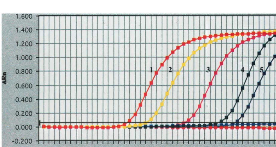
Sensitivity of quantitative PCR using TaqMan. To deter-mine the detection limit of this assay, chromosomal DNA from pureH. hepaticuscultures was used as the PCR template. The concentrations of the primers and probe for optimal amplifi-cation were 400 and 100 nM, respectively. As shown in Fig. 2, 20 fg of DNA could be detected, whereas there was no signif-icant difference in fluorescent signal intensity between 2 fg of this DNA and the no-template controls. The genome size of
[image:2.612.65.550.74.194.2]H. hepaticuswas previously estimated as 1.3 Mb using pulsed-FIG. 1. Sequence comparison between theH. hepaticusprimers and probe and the corresponding regions of thecdtBgenes in selected bacteria. Hh,H. hepaticus; Cj,C. jejuni; Hb,H. bilis; Hc,H. canis. Numbers indicate the positions of the respective primers and probe in the nucleotide sequence determined by Young et al. (25). The nucleotides different from those present in theH. hepaticus cdtBgene are indicated in boldface. The sequence of primer cdtBR is reverse and complementary to the sequence shown here. ForH. bilisandH. canis, only the partial sequence of thecdtBgene is available (2).
FIG. 2. Detection limitation of the TaqMan PCR assay forH. hepaticusgenomic DNA. Amplification plots 1 to 6 were generated from 2⫻ 107, 2⫻106, 2⫻104, 200, 20, and 2 fg, respectively. Ther2value from the linear regression in this assay is⬎0.99.
VOL. 39, 2001 QUANTITATIVE PCR DETECTION OF H. HEPATICUS 2599
on May 15, 2020 by guest
http://jcm.asm.org/
[image:2.612.74.534.459.705.2]field gel electrophoresis (16). Since the molecular weight for 1 bp is 650, the 1.3-MbH. hepaticusgenome is equal to approx-imately 8.45⫻108g/mol, which contains 6.0⫻1023molecules/
mol (Avogadro’s number). Thus, 1 g of the H. hepaticus
genomic DNA contains 7.1 ⫻ 1014 molecules and 20 fg is
equivalent to⬇14 copies. We demonstrate that 14 or more copies of theH. hepaticusgenome in a sample can be detected in this assay.
Specificity of primer and probe.To investigate the specificity of the primers and the probe used in the TaqMan PCR assay, genomic DNA samples from nine species of helicobacters, i.e.,
H. bilis, H. canis, H. cineadi, H. felis, H. mustelae, H. nemestri-nae, H. pylori, H. pullorum, and H. rodentium,were assayed using these primers and probe. The enterohepatic helico-bactersH. bilis, H. canis,andH. pullorumhave been shown to have cytolethal distending toxin activity that causes progressive cell enlargement and eventual death (2, 24, 25). There were two reasons for selecting theH. hepaticus cdtBgene as a PCR target in this assay. First, the designed primers and probe will be used to investigate the role of cytolethal distending toxin in the pathogenesis ofH. hepaticus.Second, the partial sequences of the cdtB gene in H. bilis and H. canis, which are close relatives ofH. hepaticus,have been determined, so they were used for sequence comparison with that ofH. hepaticus(2). These limited nucleotide sequences of thecdtBgene inH. bilis
andH. canis encompass only the forward primer cdtBF and part of the cdtBP sequence. Sequence comparison revealed that cdtBF and cdtBP display little sequence similarity to the
H. canis cdtBgene but have significant sequence identity to the
H. bilis cdtBgene (Fig. 1). In addition, these primers and probe did not display significant sequence similarity to thecdtBgene
inC. jejuni(Fig. 1). In the TaqMan PCR assay, Ct values for 20 and 2 ng of theH. hepaticus genomic DNA are 16 and 20, respectively (Fig. 3). In contrast, there was no fluorescent sig-nal detected from 20 ng of eight selected non-H. hepaticus
helicobacterial DNA templates. A weak signal (Ct⫽ ⬇36.4), which is approximately equivalent to 20 fg of H. hepaticus
DNA, was detected from 20 ng ofH. bilisDNA. The results demonstrate that these primers and probe can be used to detectH. hepaticuswith high specificity.
Detection of theH. hepaticusgenome present in mouse ce-cum and feces in TaqMan PCR assay.To test the applicability of this assay in samples from experimentally infected mice, chromosomal DNA was prepared from ceca and feces of two A/JCr mice experimentally inoculated with H. hepaticusand two control (helicobacter-free) mice. Subsequently, theH. he-paticusgenomic DNA (20 ng and 0.2 pg) was spiked into 20 ng of either cecal or fecal DNA prepared from the helicobacter-free mouse and then analyzed using the TaqMan PCR assay for investigating if the cecal and fecal DNA preparations in-terfere with the PCR amplification ofH. hepaticusDNA. The spikedH. hepaticusDNA produced Ct values (16.4 at 20 ng and 36.5 at 2 ng) similar to those for the pure standard DNA (Fig. 2, plots 1 and 2), demonstrating that there is no inhibitory effect on the PCR amplification in the presence of murine chromosomal DNA. Twenty nanograms of each DNA tem-plate in parallel with the H. hepaticus DNA standards was evaluated using the TaqMan PCR assay with the primer pair cdtBF-cdtBR and probe cdtBP. The quantity ofH. hepaticus
DNA in each sample was calculated using the standard curve (Fig. 4) and then converted into the number of copies of the
[image:3.612.72.536.68.322.2]H. hepaticusgenome (Table 1). Using the same quantity of the FIG. 3. Evaluation of the specificity of the primers and probe forH. hepaticus. Amplification plots 1 and 2 were generated using 2⫻107and
2⫻106fg ofH. hepaticuschromosomal DNA, respectively, whereas plots 3 to 11 were produced using 2⫻107fg of chromosomal DNA from H. bilis, H. canis, H. cineadi,H. felis, H. mustelae, H. nemestrinae, H. pullorum, H. pylori,andH. rodentium, respectively.
on May 15, 2020 by guest
http://jcm.asm.org/
initial DNA templates from the ceca and feces, different copy numbers of theH. hepaticusgenome were detected: 13.6⫻104
and 9.1 ⫻ 103 in the cecum and feces, respectively, of one
mouse (mouse I) and 6.2⫻104and 7.8⫻103in the cecum and
feces, respectively, of a second mouse (mouse II). In the two uninfected control mice, noH. hepaticusDNA was detected in any of the samples. These data demonstrate thatH. hepaticus
present in murine ceca and feces was specifically and sensi-tively quantified using this assay.
DISCUSSION
Quantitative analysis of a microbial pathogen in its host provides information useful in elucidating the mechanisms uti-lized by the pathogen to elicit disease and to evade immune defenses. This analysis is also useful in evaluating the efficacy of vaccines and new drugs for eradicating specific pathogens. Quantitative culture has been widely used to quantify bacterial pathogens which form single colonies on agar plates. In the case ofH. hepaticus,which does not form single colonies (4),
an alternative technique is needed for its quantification. In this study, a rapid, sensitive, and reproducible fluorogenic PCR assay in the ABI Prism 7700 sequence detection system was developed to quantifyH. hepaticusin samples from experimen-tally infected mice. PCR primers producing an 81-bp amplicon and a probe were generated from theH. hepaticus cdtBgene. This technique is readily applicable to DNA templates pre-pared from ceca and feces of mice and will be a powerful tool in determining the pathogenic role ofH. hepaticusin the in-duction of murine gastrointestinal and biliary diseases as an animal model for understanding the development of similar human diseases. More recently, the copy numbers of theH. hepaticusgenome in ceca from experimentally infected A/JCr (n⫽23) and C57BL/6 (n⫽20) mice were determined by this technique; the numbers ofH. hepaticusin A/JCr and C57BL/6 mice were significantly different (P⬍ 0.003; 7.14⫻ 105 and
2.27⫻ 107 bacteria/ng of mouse DNA, respectively) (M. T.
Whary, Z. Ge, and J. G. Fox, unpublished data).
The designed primers and probe did not generate PCR amplification signals from eight helicobacters, includingH. py-lori, H. felis, H. mustelae, H. pullorum, H. cineadi, H. rodentium, H. nemestrinae, andH. canis,or from mouse DNA. PCR am-plification fromH. bilisDNA was detected (Fig. 3); however, this amplification was much lower in sensitivity (Ct⫽ ⬇36.4 forH. bilisat 20 ng) than that forH. hepaticus(Ct⫽ ⬇16.3 at 20 ng). This cross-amplification betweenH. hepaticusand H. bilis could be due to the fact that primer cdtBF and probe cdtBP share significant sequence similarity to the correspond-ing regions of theH. bilis cdtBgene (Fig. 1). In addition, the reverse primer cdtBR should be expected to have a similar degree of sequence identity to the corresponding region of the
H. bilis cdtBgene, since there is 69.6% nucleotide sequence identity in the 702-bp region of thecdtBgene betweenH. bilis
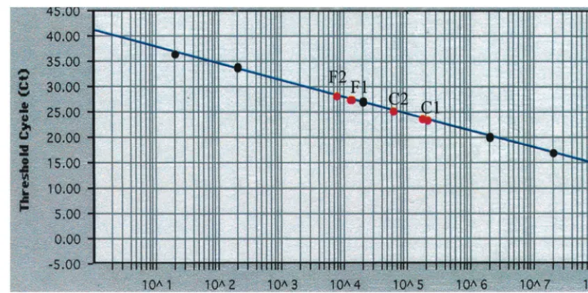
[image:4.612.93.515.71.281.2]andH. hepaticus(2). It has been reported that the mismatched FIG. 4. Quantitative detection ofH. hepaticuspresent in mouse ceca and feces. The quantities ofH. hepaticusgenomic DNA used for the standard curve are indicated on thexaxis, and the corresponding Ct values are given on theyaxis. Twenty nanograms of total DNA from each sample was used in this assay. C1 (cecum) and F1 (feces), DNA from mouse I; C2 (cecum) and F2 (feces), DNA from mouse II.
TABLE 1. Detection ofH. hepaticuspresent in mouse tissues and feces
Sample
(20 ng) Cta H. hepaticusQuantity (fg) ofDNA Copy no.b
Mouse I
Cecum (C1) 23.81 1.95⫻105 136,500
Feces (F1) 27.68 1.3⫻104 9,100
Mouse II
Cecum (C2) 25.46 6.2⫻104 43,400
Feces (F2) 28.45 7.8⫻103 5,460
aThe Ct values for the samples from the control mice were greater than 39. bThe copy numbers were calculated by converting 1 fg into 0.7 copy of theH.
hepaticusgenome.
VOL. 39, 2001 QUANTITATIVE PCR DETECTION OF H. HEPATICUS 2601
on May 15, 2020 by guest
http://jcm.asm.org/
[image:4.612.53.292.608.701.2]DNA. It is worth noting that the detection limit forH. biliswith the primer pair we used is 106-fold more insensitive than that
forH. hepaticus, since the Ct value for 20 ng ofH. bilisDNA is approximately equivalent to that for 20 fg of H. hepaticus
DNA. In experimental infections, this nonspecific amplifica-tion should have no or little impact on the final copy number ofH. hepaticus. In addition, the presence of theH. bilisDNA in a sample can be easily discriminated using the H. bilis -specific primers targeting the 16S rRNA gene (18). Further-more, additional primers and probes with higher specificity for
H. hepaticuscan be designed when more information on theH. hepaticusgenome is available.
In theory, a single copy of a target nucleotide sequence can be detected in a sample under optimal conditions using the ABI Prism 7700 sequence detection system. However, many factors, such as the purity and complexity of a PCR template and concentrations of Mg2⫹, primers, and the probe, could
significantly influence this sensitivity. In our assay, the detec-tion limit is 20 fg, which is approximately equivalent to 14 copies of the H. hepaticusgenome. Although the theoretical detection limit could not be achieved, this sensitivity is much higher than those of other available techniques and should be sufficient for quantifying H. hepaticusin experimental animal models and probably in natural infections as well. Further investigations on the dynamic relationship between the colo-nization site or number of H. hepaticus organisms and the inflammatory state in infected mice during chronic infection will increase our understanding of the mechanism of helico-bacter-induced gastrointestinal and liver diseases.
ACKNOWLEDGMENTS
This research was supported by a grant from the ACLAM Founda-tion and NIH grants R01 CA 67529 and R01 DK 52413.
REFERENCES
1.Cahill, R. J., C. J. Foltz, J. G. Fox, C. A. Dangler, F. Powrie, and D. B. Schauer.1997. Inflammatory bowel disease: an immunity-mediated condi-tion triggered by bacterial infeccondi-tion withHelicobacter hepaticus. Infect. Im-mun.65:3126–3131.
2.Chien, C. C., N. S. Taylor, Z. Ge, D. B. Schauer, V. B. Young, and J. G. Fox.
2000. Identification ofcdtBhomologues and cytolethal distending toxin ac-tivity in enterohepaticHelicobacterspp. J. Med. Microbiol.49:525–534. 3.Chin, E. Y., C. A. Dangler, J. G. Fox, and D. B. Schauer.2000.Helicobacter
hepaticusinfection triggers IBD in TCR alpha beta mutant mice. Comp. Med.50:586–592.
4.Fox, J. G., F. E. Dewhirst, J. G. Tully, B. J. Paster, L. Yan, N. S. Taylor, M. J.
9.Hailey, J. R., J. K. Haseman, J. R. Bucher, A. E. Radovsky, D. E. Malarkey, R. T. Miller, A. Nyska, and R. R. Maronpot.1998. Impact ofHelicobacter hepaticusinfection in B6C3F1 mice from twelve National Toxicology Pro-gram two-year carcinogenesis studies. Toxicol. Pathol.26:602–611. 10. Kullberg, M. C., J. M. Ward, P. L. Gorelick, P. Caspar, S. Hieny, A. Cheever,
D. Jankovic, and A. Sher.1998.Helicobacter hepaticus triggers colitis in specific-pathogen-free interleukin-10 (10)-deficient mice through an IL-12- and gamma interferon-dependent mechanism. Infect. Immun.66:5157– 5166.
11. Kwok, S. D. E. Kellogg, N. McKinney, D. Spasic, L. Goda, C. Levenson, and J. J. Sninsky.1990. Effects of primer-template mismatches on the polymer-ase chain reaction: human immunodeficiency virus type 1 model studies. Nucleic Acids Res.18:999–1005.
12. Livak, K. J., S. J. Flood, J. Marmaro, W. Giusti, and K. Deetz.1995. Oligonucleotides with fluorescent dyes at opposite ends provide a quenched probe system useful for detecting PCR product and nucleic acid hybridiza-tion. PCR Methods Appl.4:357–362.
13. Major, J. G., Jr.1992. A rapid PCR method of screening for small mutations. BioTechniques12:40, 42, 44.
14. Nassal, M., and A. Rieger.1990. PCR-based site-directed mutagenesis using primers with mismatched 3⬘-ends. Nucleic Acids Res.18:3077–3078. 15. Pickett, C. L., E. C. Pesci, D. L. Cottle, G. Russell, A. N. Erdem, and H.
Zeytin.1996. Prevalence of cytolethal distending toxin production in Campy-lobacter jejuniand relatedness ofCampylobactersp.cdtBgene. Infect. Im-mun.64:2070–2078.
16. Saunders, K. E., K. J. McGovern, and J. G. Fox.1997. Use of pulsed-field gel electrophoresis to determine genomic diversity in strains ofHelicobacter hepaticusfrom geographically distant locations. J. Clin. Microbiol.35:2859– 2863.
17. Sen, K.2000. Rapid identification ofYersinia enterocoliticain blood by the 5⬘
nuclease PCR assay. J. Clin. Microbiol.38:1953–1958.
18. Shen, Z., Y. Feng, and J. G. Fox.2000. Identification of enterohepatic
Helicobacterspecies by restriction fragment-length polymorphism analysis of the 16S rRNA gene. Helicobacter5:121–128.
19. Taylor, D. E., Z. Ge, D. Purych, T. Lo, and K. Hiratsuka.1997. Cloning and sequence analysis of two copies of a 23S rRNA gene fromHelicobacter pylori
and association of clarithromycin resistance with 23S rRNA mutations. An-timicrob. Agents Chemother.41:2621–2628.
20. Theroux, S. J., and R. J. Davis.1992. Rapid screening of cloned DNA fragments for specific mutations. Nucleic Acids Res.20:915.
21. Ward, J. M., M. R. Anver, D. C. Haines, J. M. Melhorn, P. Gorelick, L. Yan, and J. G. Fox.1996. Inflammatory large bowel disease in immunodeficient mice naturally infected withHelicobacter hepaticus. Lab. Anim. Sci.46:15–20. 22. Ward, J. M., J. G. Fox, M. R. Anver, D. C. Haines, C. V. George, M. J. Collins, Jr., P. L. Gorelick, K. Nagashima, M. A. Gonda, R. V. Gilden, J. G. Tully, R. E. Russell, B. J. Paster, F. E. Dewhirst, J. C. Conovan, L. M. Anderson, and J. M. Rice.1994. Chronic active hepatitis and associated liver tumors in mice caused by a persistent bacterial infection with a novel Heli-cobacterspecies. J. Natl. Cancer. Inst.86:1222–1227.
23. Whary, M. T., T. J. Morgan, C. A. Dangler, K. J. Gaudes, N. S. Taylor, and J. G. Fox.1998. Chronic active hepatitis induced byHelicobacter hepaticusin the A/JCr mouse is associated with a Th1 cell-mediated immune response. Infect. Immun.66:3142–3148.
24. Young, V. B., C. C. Chien, K. A. Knox, N. S. Taylor, D. B. Schauer, and J. G. Fox.2000. Cytolethal distending toxin in avian and human isolates of Heli-cobacter pullorum. J. Infect. Dis.182:620–623.
25. Young, V. B., K. A. Knox, and D. B. Schauer.2000. Cytolethal distending toxin sequence and activity in the enterohepatic pathogenHelicobacter he-paticus. Infect. Immun.68:184–191.

![Crystal structure of N [(E) (1,3 benzodioxol 5 yl)methylidene] 4 chloroaniline](data:image/gif;base64,R0lGODlhAQABAIAAAP///wAAACH5BAEAAAAALAAAAAABAAEAAAICRAEAOw==)