Copyright © 2000, American Society for Microbiology. All Rights Reserved.
Antipneumococcal Activity of Telithromycin by Agar Dilution,
Microdilution, E Test, and Disk Diffusion Methodologies
TODD A. DAVIES,1LINDA M. KELLY,1MICHAEL R. JACOBS,2ANDPETER C. APPELBAUM1*Department of Pathology, Hershey Medical Center, Hershey, Pennsylvania 17033,1and Department of Pathology, Case Western Reserve University,
Cleveland, Ohio 441062
Received 13 October 1999/Returned for modification 26 November 1999/Accepted 3 January 2000
Agar dilution and microdilution (both in air) and E test and disk diffusion (both in air and CO2) were used
to test the activity of telithromycin against 110 erythromycin-susceptible and 106 erythromycin-resistant pneumococci. The MICs at which 50 and 90% of strains are inhibited (MIC50s and MIC90s, respectively) for
erythromycin-susceptible strains varied between 0.008 and 0.016g/ml and 0.016 and 0.03g/ml when the samples were incubated in air. By comparison, telithromycin MIC50s and MIC90s for erythromycin-resistant
strains were in air 0.03 to 0.125 and 0.125 to 0.5 g/ml, respectively. When agar dilution was used as the reference method, essential agreement was found for 112 of 216 strains (51.9%) for microdilution, 168 of 216 (77.8%) for E test in air, and 132 of 216 (61.1%) for E test in CO2. With the exception of four strains tested by
E test in CO2, all organisms were susceptible to a proposed telithromycin susceptibility breakpoint of <1
g/ml. By disk diffusion with 15-g telithromycin disks, all strains but one had zones of inhibition>19 mm
in diameter when incubated in CO2, while all strains had zone diameters of>22 mm when incubated in air.
Zone diameters in air were generally 4 to 5 mm larger than in CO2. By all methods, MICs and zones of all
erythromycin-resistant strains occurred in clusters separated from those seen with erythromycin-susceptible strains. The results for macrolide-resistant strains withermandmefresistance determinants were similar. The results show that (i) telithromycin is very active against erythromycin-susceptible and -resistant strains irrespective of macrolide resistance mechanism; (ii) susceptibility to telithromycin can be reliably tested by the agar, microdilution, E test, and disk diffusion methods; and (iii) incubation in CO2led to smaller zones by disk
diffusion and higher MICs by E test, but at a susceptible MIC breakpoint of<1g/ml and a susceptible zone
diameter cutoff of>19 mm in CO2, 215 of 216 strains were found to be susceptible to telithromycin.
Streptococcus pneumoniaecontinues to be a significant cause of morbidity and mortality in humans and is the leading cause of bacterial pneumonia, sinusitis, and otitis media and an im-portant cause of meningitis (2, 4, 10–12). The past 5 years have witnessed a dramatic worldwide increase in the incidence of pneumococcal strains which are resistant to penicillin G and other-lactam and non--lactam antimicrobials, such as mac-rolides, clindamycin, tetracycline, chloramphenicol, and tri-methoprim-sulfamethoxazole (5, 8, 13). The problem has been exacerbated by the tendency of these strains to spread from country to country and from continent to continent (16, 17).
In the United States, a recent study has shown that for 23.6% of 1,527 clinically significant pneumococci from 30 U.S. centers, penicillin MICs wereⱖ0.125g/ml, with 14.1% being intermediate and 9.5% being resistant (8). Penicillin-resistant pneumococci were more likely to be resistant to macrolides and other unrelated agents, such as chloramphenicol, tetracy-cline, and trimethoprim-sulfamethoxazole. Erythromycin resis-tance among pneumococci has increased in the United States from approximately 0.2% in the late 1980s to 5 and 15% in some areas of the country (5, 8, 22). In the survey described above (8), 20% of penicillin-intermediate pneumococcal strains and 49% of penicillin-resistant strains were erythromy-cin resistant. In a recently published study from our laboratory performed in 1997 (13), 49.6% of 1,476 pneumococcal strains obtained from outpatients in six geographic regions of the
United States were penicillin susceptible, 17.9% were interme-diate, and 32.5% were penicillin resistant. In the latter survey, 95% of penicillin-susceptible, 65% of intermediate, and 33% of penicillin-resistant strains were macrolide susceptible. Strains for which penicillin MICs were increased were also more likely to be multiresistant (13).
Pneumococcal strains that are resistant to erythromycin ex-hibit cross-resistance to other macrolides and azalides such as azithromycin, clarithromycin, and roxithromycin (7, 9, 24). Te-lithromycin (HMR 3647), a new ketolide (1), has been shown to have low MICs for both macrolide-susceptible and macrol-ide-resistant pneumococcal strains (6, 14, 21; D. Felmingham, M. J. Robbins, A. Leakey, R. Cooke, C. Dencer, H. Solman, G. L. Ridgway, R. N. Gru¨neberg, and A. Bryskier, Abstr. 37th Intersci. Conf. Antimicrob. Agents Chemother., abstr. F-116, 1997). Although MICs are slightly higher for macrolide-resis-tant strains, all strains are susceptible atⱕ2.0g/ml, irrespec-tive of the macrolide resistance mechanism (21).
Currently, the National Committee for Clinical Laboratory Standards (NCCLS) recommends incubation in air for mi-crodilution testing and in CO2for disk diffusion (18, 19). The manufacturer of the E test (AB Biodisk, Solna, Sweden) also recommends incubation in CO2for testing of S. pneumoniae because 5 to 10% of strains do not grow without CO2 on primary isolation (7, 15). Because incubation in CO2has pre-viously been shown to affect macrolide MICs for S. pneu-moniae(7, 9, 24), we tested activity by agar dilution and mi-crodilution in air and disk diffusion and E test in air as well as CO2atmospheres. We are aware of no previous published data on incubation in air compared with CO2for ketolides.
* Corresponding author. Mailing address: Department of Pathology, Hershey Medical Center, 500 University Dr., Hershey, PA 17033. Phone: (717) 531-5113. Fax: (717) 531-7953. E-mail: pappelbaum @psghs.edu.
1444
on May 15, 2020 by guest
http://jcm.asm.org/
MATERIALS AND METHODS
Bacteria and antibiotics.A total of 216 isolates ofS. pneumoniae(collected
between 1995 and 1997 from various laboratories in the United States) were selected from our culture collection for testing. All strains had been subcultured several times prior to use and all grew well in air. Cultures were stored at⫺70°C
in double-strength skim milk (Difco Laboratories, Detroit, Mich.). Telithromycin and erythromycin were obtained from Hoechst Marion Roussel Anti-infectives, Romainville, France. Powders were stored at⫺4°C.
Macrolide resistance mechanism determination.All strains were screened for
macrolide susceptibility by the double disk method with erythromycin and clin-damycin disks (18). Strains with erythromycin zone diameters ofⱕ20 mm were tested for the presence of the ermB andmefE genes as follows. DNA was extracted from isolated strains derived from single colonies with the Prep-A-Gene kit (Bio-Rad, Hercules, Calif.) as recommended by the manufacturer. Previously described primer sets for amplification of theermBandmefEgenes were used (20, 23). An initial denaturing step at 96°C for 3 min and a final elongation step at 72°C for 5 min were performed for each PCR run. TheermB andmefEgenes were amplified by 30 cycles of three steps: 96°C for 60 s, 52°C for 60 s, and 72°C for 60 s. The PCR products forermBgenes were 640 bp, and those formefEgenes were 348 bp. Amplifications were carried out in a final volume of 50l in a GeneAmp PCR system 2400 (Perkin-Elmer, Foster City, Calif.). The final PCR mixture contained 10 mM Tris-HCl (pH 8.3); 50 mM KCl; 2.0 mM MgCl2; 200M concentrations of each of the deoxynucleotides dATP, dCTP, dGTP, and dTTP; 30 to 50 pmol of each primer set; 1 ng of template DNA; and 2.5 U ofTaqpolymerase (Fisher BioTech). Samples of each PCR product were then electrophoresed on 2% agarose gels (Bio-Rad) and stained in ethidium bromide (0.5g/ml) in 0.5⫻Tris-borate-EDTA buffer. PCR products were visualized under UV illumination, and their sizes were estimated with PCR markers (Promega, Madison, Wis.). Negative controls ruled out the possible influence of amplicon modification.
Agar dilution MICs.Agar dilution MICs were determined by the methods
[image:2.612.53.294.90.263.2]used in our laboratory (7) on Mueller-Hinton agar supplemented with 5% sheep blood, incorporating telithromycin and erythromycin at concentrations of 0.002 to 64g/ml in doubling dilutions. Inocula were prepared by suspending growth from overnight cultures in sterile saline to a turbidity of a 0.5 McFarland stan-dard. Final inocula contained 104CFU/spot. Plates were inoculated with a Steers replicator with 3-mm inoculating pins and incubated overnight at 35°C in air. The lowest concentration of antibiotic resulting in no growth was read as the MIC.
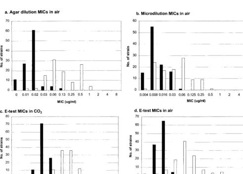
[image:2.612.57.550.364.716.2]FIG. 1. Histograms of telithromycin MICs for erythromycin-susceptible (solid bars) and -resistant (open bars) strains.
TABLE 1. Telithromycin MICs with the three methods tested against 216 strains
Method and strain typea
MIC (g/ml)
Range 50% 90%
Agar dilution, air
S ⱕ0.004–0.125 0.016 0.016
R 0.008–1.0 0.125 0.5
Microdilution, air
S ⱕ0.004–0.06 0.008 0.03
R ⱕ0.004–0.5 0.03 0.125
E test, air
S ⱕ0.004–0.06 0.016 0.016
R 0.008–1.0 0.06 0.25
E test, CO2
S 0.016–0.125 0.03 0.06
R 0.03–8.0 0.25 0.5
aStrains tested were either susceptible (S) or resistant (R) to erythromycin.
on May 15, 2020 by guest
http://jcm.asm.org/
The quality control strainsStaphylococcus aureusATCC 29213 andS. pneu-moniae ATCC 49619 were included in each run. Inoculum checks were per-formed for each strain.
Microdilution MICs.Telithromycin MICs were determined by the method
recommended by the NCCLS (19), using cation-adjusted Mueller-Hinton broth (Difco Laboratories) supplemented with 5% lysed defibrinated horse blood. Suspensions with a turbidity equivalent to that of a 0.5 McFarland standard were prepared by suspending growth from blood agar plates in 2 ml of sterile saline. Suspensions were further diluted 1:10 to obtain a final inoculum of 5⫻105 CFU/ml. Trays were incubated overnight in ambient air at 35°C. Standard quality control strains and inoculum checks (as above) were included.
E test MICs.Mueller-Hinton plates supplemented with 5% sheep blood (BBL
Microbiology Systems, Cockeysville, Md.) were inoculated with a 0.5 McFarland suspension harvested from overnight growth on plates, and telithromycin E test strips (AB Biodisk) were placed on each plate (7, 15). After overnight incubation at 35°C, the MIC was read where the ellipse of growth inhibition intersected the strip. E test MICs were determined both in air and in CO2. Standard quality control strains (see above) were used with each run.
Disk diffusion.Disk diffusion was performed by standard NCCLS
methodol-ogy (19) using Mueller-Hinton plates supplemented with 5% sheep blood (source as above), inoculated with a 0.5 McFarland suspension; 15-g telithro-mycin disks (BBL Microbiology Systems) were placed on the plates. After over-night incubation in both air and 5% CO2at 35°C, the diameters of zones of inhibition were measured with calipers. Standard quality control strains (see above) were used with each run.
Interpretation of results.For telithromycin, provisional breakpoints ofⱕ1.0
g/ml for susceptible, 2.0g/ml for intermediate, andⱖ4.0g/ml for resistant results were used. Essential agreement was defined as the MIC by one method being within 1 log2dilution of the MIC by agar dilution (taken as the reference method). Interpretative category discrepancies were defined as very major dis-crepancies when the reference method showed resistance and the comparative method showed susceptibility; major discrepancies occurred when the reference method showed susceptibility and the comparative method showed resistance;
and minor discrepancies occurred when an intermediate result was obtained with one method and a resistant or susceptible result was obtained with the other (7).
RESULTS
Of the 216 isolates included in this study, 110 were erythro-mycin and clindaerythro-mycin susceptible by disk diffusion testing, with erythromycin MICs being ⱕ0.12g/ml by agar and mi-crodilution. The remaining 106 isolates were erythromycin re-sistant (erythromycin MICs,ⱖ0.5g/ml), with 32 being clin-damycin susceptible and positive formefEgene products. All remaining 74 macrolide-resistant strains were positive forerm gene products.
The telithromycin MIC results for the four susceptibility testing methods are presented in Table 1 and Fig. 1. The MICs at which 50% and 90% of the strains are inhibited (MIC50s and MIC90s, respectively) for the erythromycin-susceptible strains varied between 0.008 and 0.016 g/ml and 0.016 and 0.03
g/ml, respectively, for methods using incubation in air. By comparison, the telithromycin MIC50s and MIC90s in air of erythromycin-resistant strains were 0.03 to 0.125 and 0.125 to 0.5g/ml, respectively. By the E test method with incubation in CO2, telithromycin MIC50s and MIC90s were 0.03 and 0.06 and for erythromycin-susceptible strains, respectively, and 0.25 and 0.5g/ml for erythromycin-resistant strains, respectively. Table 2 presents agar dilution results for erythromycin-susceptible and -resistant strains. As can be seen, telithromycin MIC50s and MIC90s rose from 0.016 and 0.016g/ml for the erythro-mycin-susceptible group to 0.06 and 0.5g/ml for the erythro-mycin-resistant group, compared to erythromycin MICs of 0.03 and 0.06g/ml and⬎64 and⬎64g/ml for the erythromycin-susceptible and -resistant groups, respectively.
[image:3.612.316.548.71.255.2]The disk diffusion results are shown in Fig. 2. Mean telithro-mycin disk diffusion zone diameters were 26.7 mm in CO2, compared with 32.9 mm in air. For erythromycin-susceptible and -resistant groups, mean zone diameters were 28.5 and 24.9 mm, respectively, in CO2, compared to 34.9 and 30.0 mm, respectively, in air. For incubation in CO2, 215 of 216 strains were susceptible at a zone diameter breakpoint of ⱖ19 mm, while all strains were susceptible in air at a zone diameter breakpoint of ⱖ22 mm. A scatter plot of telithromycin disk diffusion zones in CO2versus agar dilution MICs is presented in Fig. 3.
FIG. 2. Disk diffusion histograms with incubation in air (a) and CO2(b) for erythromycin-susceptible (solid bars) and -resistant (open bars) strains.
[image:3.612.52.293.99.156.2]FIG. 3. Scatter plot comparison of disk diffusion zone diameters in CO2with agar dilution MICs in air.
TABLE 2. Comparison of agar dilution MIC results for erythromycin-susceptible (n⫽110) and
erythromycin-resistant (n⫽106) strains
Drug Susceptible strains Resistant strains
Range 50% 90% Range 50% 90%
Telithromycin 0.004–0.125 0.016 0.016 0.008–1.0 0.06 0.5 Erythromycin 0.008–0.125 0.03 0.06 0.5–⬎64.0 ⬎64.0 ⬎64.0
on May 15, 2020 by guest
http://jcm.asm.org/
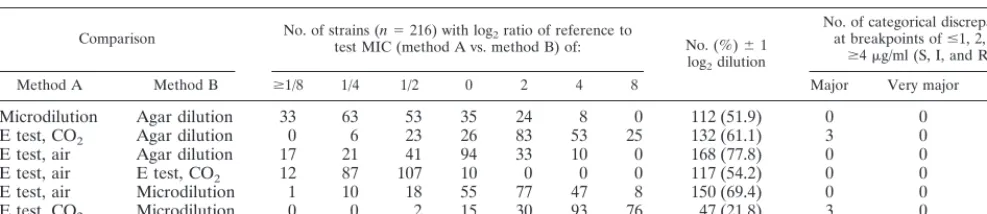
[image:3.612.53.293.475.706.2]Categorical discrepancies with the four methods are pre-sented in Table 3. The number of strains with essential agree-ment varied between 47 (21.8%) and 168 (77.8%), depending upon which methods were being compared and whether CO2 was used as opposed to air. Minor discrepancies were obtained with the E test incubated in CO2 for one strain, and major discrepancies for three strains were obtained with the E test compared with agar and microdilution.
The effect of incubation in CO2on the susceptibility results showed that incubation of E tests in CO2 resulted in MICs approximately 1 dilution higher and zone diameters 4 to 5 mm narrower than those obtained with incubation in air (Table 1; Fig. 1 and 2). Additionally, although all strains were suscepti-ble to telithromycin by agar dilution, microdilution, and E test incubated in air, four strains gave telithromycin MICs between 2 and 8 g/ml when E tests were incubated in CO2, yielding one minor and three major discrepancies with the breakpoints used, irrespective of which susceptibility method was used as the reference. The MICs and zones for erythromycin-resistant strains occurred in clusters different from those seen with erythromycin-susceptible strains. The telithromycin MICs for erythromycin-resistant strains were similar irrespective of mac-rolide resistance mechanism.
DISCUSSION
The results of this study confirm the excellent antipneumo-coccal activity of telithromycin irrespective of erythromycin susceptibility, as reported previously (1). Clustering of MICs depended on the erythromycin susceptibility of the strains (21). The agar and microdilution methods, both with incubation in air, produced similar MIC values. When E tests were incu-bated in air, excellent correlation with the results of the agar and microdilution methods was obtained. However, when E tests were incubated in CO2, MICs rose, generally by 1 dilu-tion. With a preliminary susceptible breakpoint ofⱕ1.0g/ml, all strains were telithromycin susceptible by the three MIC methods in air; however, incubation of E tests in CO2resulted in four strains for which telithromycin MICs were increased to between 2 and 4g/ml.
Disk diffusion tests showed that, with the exception of one strain, zone diameters in CO2 of ⱖ19 mm corresponded to telithromycin MICs ofⱕ1.0g/ml. In air, disk diffusion zone diameters ofⱖ22 mm corresponded with telithromycin MICs ofⱕ1.0g/ml in all 216 strains tested. NCCLS recommends incubation ofS. pneumoniaein air for agar dilution and mi-crodilution but in CO2for disk diffusion, as is also the recom-mendation of the manufacturer of the E test. In previous studies, we have reported results similar to this CO2effect on telithromycin susceptibilities with other macrolides (9, 24). Be-cause of the higher MICs observed with incubation in CO2,
there is a need to determine macrolide and ketolide MICs in air as well as in increased CO2(as used in the current study) to allow reliable testing by the E test method in the clinical laboratory.
In summary, our results show that (i) telithromycin is active against erythromycin-susceptible and -resistant strains irre-spective of macrolide resistance mechanism, although MICs for erythromycin-resistant strains are higher; (ii) susceptibility to telithromycin may be reliably tested by agar, microdilution, E test, and disk diffusion; and (iii) disk diffusion tests gave narrower zones and E tests gave higher MICs when plates were incubated in CO2than when incubated in air, but at a prelim-inary breakpoint ofⱕ1g/ml and zone diameter ofⱖ19 mm (CO2), all strains but one were telithromycin susceptible irre-spective of incubation atmosphere or method. More informa-tion from clinical or animal data is required before breakpoints can be firmly established and recommended.
ACKNOWLEDGMENT
This study was supported by a grant from Hoechst Marion Roussel Anti-infectives.
REFERENCES
1.Agouridas, C., A. Bonnefoy, and J. F. Chantot.1997. Antibacterial activity of
RU 64004 (HMR 3004), a novel ketolide derivative active against respiratory pathogens. Antimicrob. Agents Chemother.41:2149–2158.
2.Appelbaum, P. C.1992. Antimicrobial resistance in Streptococcus
pneu-moniae—an overview. Clin. Infect. Dis.15:77–83.
3.Arthur, M., C. Molinas, C. Mabilat, and P. Courvalin.1990. Detection of
erythromycin resistance by the polymerase chain reaction in conserved re-gions ofermrRNA methylase genes. Antimicrob. Agents Chemother.34:
2024–2026.
4.Block, S.1997. Causative pathogens, antibiotic resistance and therapeutic
considerations in acute otitis media. Pediatr. Infect. Dis. J.16:449–456.
5.Breiman, R. F., J. C. Butler, F. C. Tenover, J. A. Elliott, and R. R. Facklam.
1994. Emergence of drug-resistant pneumococcal infections in the United States. JAMA271:1831–1835.
6.Bryskier, A., C. Agouridas, and J.-F. Chantot.1996. Ketolides: new
semi-synthetic 14-membered ring macrolides, p. 39–50.InS. H. Zinner, L. S. Young, J. F. Acar, and H. C. Neu (ed.), Expanding indications for the new macrolides, azalides and streptogramins. Marcel Dekker, New York, N.Y.
7.Clark, C. L., M. R. Jacobs, and P. C. Appelbaum.1998. Antipneumococcal
activities of levofloxacin and clarithromycin as determined by agar dilution, microdilution, E-test, and disk diffusion methodologies. J. Clin. Microbiol.
36:3579–3584.
8.Doern, G. V., A. Brueggemann, H. P. Holley, and A. M. Rauch.1996.
Anti-microbial resistance ofStreptococcus pneumoniaerecovered from outpatients in the United States during the winter months of 1994 to 1995: results of a 30-center national surveillance study. Antimicrob. Agents Chemother.40:
1208–1213.
9.Fasola, E. L., S. Bajaksouzian, P. C. Appelbaum, and M. R. Jacobs.1997.
Variation in erythromycin and clindamycin susceptibilities ofStreptococcus pneumoniaeby four test methods. Antimicrob. Agents Chemother.41:129– 134.
10. Friedland, I. R., and G. S. Istre.1992. Management of penicillin-resistant
pneumococcal infections. Pediatr. Infect. Dis. J.11:433–435.
[image:4.612.56.550.84.191.2]11. Friedland, I. R., and G. H. McCracken, Jr.1994. Management of infections
TABLE 3. Comparison of results of telithromycin susceptibility testing by four methods
Comparison No. of strains (n⫽216) with log2ratio of reference to
test MIC (method A vs. method B) of: No. (%)⫾1 log2dilution
No. of categorical discrepancies at breakpoints ofⱕ1, 2, and
ⱖ4g/ml (S, I, and R)a Method A Method B ⱖ1/8 1/4 1/2 0 2 4 8 Major Very major Minor
Microdilution Agar dilution 33 63 53 35 24 8 0 112 (51.9) 0 0 0
E test, CO2 Agar dilution 0 6 23 26 83 53 25 132 (61.1) 3 0 1
E test, air Agar dilution 17 21 41 94 33 10 0 168 (77.8) 0 0 0
E test, air E test, CO2 12 87 107 10 0 0 0 117 (54.2) 0 0 0
E test, air Microdilution 1 10 18 55 77 47 8 150 (69.4) 0 0 0
E test, CO2 Microdilution 0 0 2 15 30 93 76 47 (21.8) 3 0 1
aS, I, and R, susceptible, intermediate, and resistant.
on May 15, 2020 by guest
http://jcm.asm.org/
caused by antibiotic-resistantStreptococcus pneumoniae. N. Engl. J. Med.
331:377–382.
12. Jacobs, M. R.1992. Treatment and diagnosis of infections caused by
drug-resistantStreptococcus pneumoniae. Clin. Infect. Dis.15:119–127.
13.Jacobs, M. R., S. Bajaksouzian, A. Zilles, G. Lin, G. A. Pankuch, and P. C.
Appelbaum.1999. Susceptibilities of Streptococcus pneumoniaeand
Hae-mophilus influenzaeto 10 oral antimicrobial agents based on pharmacody-namic parameters: 1997 U.S. surveillance study. Antimicrob. Agents Che-mother.43:1901–1908.
14. Jones, R. N., and D. J. Biedenbach.1997. Antimicrobial activity of
RU-66647, a new ketolide. Diagn. Microbiol. Infect. Dis.27:7–12.
15. Jones, R. N., M. E. Erwin, and J. L. Croco.1996. Critical appraisal of E test
for the detection of fluoroquinolone resistance. J. Antimicrob. Chemother.
38:21–25.
16. McDougal, L. K., R. Facklam, M. Reeves, S. Hunter, J. M. Swenson, B. C.
Hill, and F. C. Tenover.1992. Analysis of multiply antimicrobial-resistant
isolates ofStreptococcus pneumoniaefrom the United States. Antimicrob. Agents Chemother.36:2176–2184.
17. Munoz, R., J. M. Musser, M. Crain, D. E. Briles, A. Marton, A. J. Parkinson,
U. Sorensen, and A. Tomasz.1992. Geographic distribution of
penicillin-resistant clones ofStreptococcus pneumoniae: characterization by penicillin-binding protein profile, surface protein A typing, and multilocus enzyme analysis. Clin. Infect. Dis.15:112–118.
18. National Committee for Clinical Laboratory Standards.1997. Methods for
dilution antimicrobial susceptibility tests for bacteria that grow aerobically.
NCCLS publication no. M7-A4. National Committee for Clinical Laboratory Standards, Wayne, Pa.
19. National Committee for Clinical Laboratory Standards.1997. Performance
standards for antimicrobial disk susceptibility tests. NCCLS publication no. M2-A6. National Committee for Clinical Laboratory Standards, Wayne, Pa.
20. Pankuch, G. A., S. A. Jueneman, T. A. Davies, M. R. Jacobs, and P. C.
Appelbaum.1998. In vitro selection of resistance to four -lactams and
azithromycin inStreptococcus pneumoniae. Antimicrob. Agents Chemother.
42:2914–2918.
21. Pankuch, G. A., M. A. Visalli, M. R. Jacobs, and P. C. Appelbaum.1998.
Susceptibilities of penicillin- and erythromycin-susceptible and -resistant pneumococci to HMR 3647 (RU 66647), a new ketolide, compared with susceptibilities to 17 other agents. Antimicrob. Agents Chemother.42:624– 630.
22. Spika, J. S., R. R. Facklam, B. D. Plikaytis, M. J. Oxtoby, and the
Pneumo-coccal Surveillance Working Group.1991. Antimicrobial resistance of
Strep-tococcus pneumoniaein the United States, 1979–1987. J. Infect. Dis.163:
1273–1278.
23. Sutcliffe, J., T. Grebe, A. Tait-Kamradt, and L. Wondrack.1996. Detection
of erythromycin-resistant determinants by PCR. Antimicrob. Agents Che-mother.40:2562–2566.
24. Visalli, M. A., M. R. Jacobs, and P. C. Appelbaum.1997. Susceptibility of
penicillin-susceptible and -resistant pneumococci to dirithromycin compared with susceptibilities to erythromycin, azithromycin, clarithromycin, roxithro-mycin, and clindamycin. Antimicrob. Agents Chemother.41:1867–1870.