0095-1137/06/$08.00⫹0 doi:10.1128/JCM.44.4.1495–1501.2006
Copyright © 2006, American Society for Microbiology. All Rights Reserved.
Differentiation of
Escherichia coli
Pathotypes by Oligonucleotide
Spotted Array†
Raghavan U. M. Palaniappan,
1Yu Zhang,
1David Chiu,
1Alfonso Torres,
1Chobi DebRoy,
2Thomas S. Whittam,
3and Yung-Fu Chang
1*
Department of Population Medicine and Diagnostic Sciences, College of Veterinary Medicine, Cornell University, Ithaca, New York 148531; Gastroenteric Disease Center, Department of Veterinary Science, The Pennsylvania State University,
University Park, Pennsylvania 168022; and Microbial Evolution Laboratory, National Food Safety and
Toxicology Center, Michigan State University, East Lansing, Michigan 488243
Received 27 July 2005/Returned for modification 28 September 2005/Accepted 11 January 2006
To accurately determine the pathotypes of Escherichia coli strains, a comprehensive assessment of each
strain that targets multiple genes is required. A new approach to the identification and characterization ofE.
colipathotypes was developed by constructing gene-specific probes (70-mers) for not only the virulence genes
associated with eachE. colipathotype but also the O157-, CFT073-, and K-12-specific and common genes of
each pathotype. Analysis of oligonucleotide probes with reference and clinical isolates ofE. colipathotypes
indicated that the array could differentiate the pathotypes on the basis of their virulence and specific gene
patterns. Probes targeting common genes ofE. coli were present in all the reference and clinical strains.
Salmonella entericasubsp.enterica-specific genes andSalmonellacore genes were used as negative controls. The
entireE. colipathotype showed reactivity to only 4 of the 81Salmonella-specific gene probes. Characterization
of the genetic and virulence profiles of a single strain by using probes for virulence factors and specific and
common genes in the spotted array is an ideal diagnostic tool for determination ofE. colipathotypes and could
also have a significant impact on the epidemiological analysis ofE. coliinfections.
Escherichia coliis a normal commensal gram-negative rod-shaped bacterium that lives inside the intestinal tracts of hu-mans and warm-blooded animals. However, some E. coli
strains cause urinary tract infections, bacteremia, and bacterium-related diarrhea and are also the main cause of neonatal men-ingitis in human and animals. In 1999, the Centers for Disease Control and Prevention estimated that there were 269,060 cases of gastroenteritis caused byE. coliin the United States alone (15). PathogenicE. colistrains can be distinguished from their nonpathogenic counterparts by the presence of virulence genes, which code for adherence and colonization, invasion, cell surface molecules, secretion, transport, and siderophore formation (9). These virulence genes are generally organized as large blocks in chromosomes, plasmids, or phages and are often transmissible betweenE. colistrains. Based on the type of virulence factor present and host clinical symptoms,E. coli
strains are categorized into pathotypes: (i) enteropathogenic
E. coli(EPEC), which causes diarrhea in children and animals; (ii) enterohemorrhagicE. coli (EHEC), which is responsible for hemorrhagic colitis and hemolytic-uremic syndrome; (iii) enterotoxigenicE. coli(ETEC), which causes traveler’s diar-rhea and porcine and bovine diardiar-rhea; (iv) enteroaggregative
E. coli(EAEC), which causes persistent diarrhea in humans, and diffusely adherentE. coli(DAEC), a subclass of enteroaggre-gativeE. coliwhich causes diarrhea in children; (v) enteroin-vasiveE. coli(EIEC), which causes watery diarrhea and
dys-entery; (vi) uropathogenic E. coli (UPEC), which causes urinary tract infections in humans and animals; and (vii) neo-natal meningitisE. coli(NMEC), which is responsible for men-ingitis and sepsis (12). EIEC and EAEC strains were reported to be found only in humans and not in animals. Even thoughE. coliis a significant human and animal pathogen, there is cur-rently no rapid and efficient method for identifying the differ-ent pathotypes ofE. coli.
In order to ensure public health and to monitor biological threats, a rapid, sensitive, and specific diagnostic assay is neces-sary for the identification of E. coli pathotypes. However, the bacterial genomes are extremely dynamic, and the ability of the organisms to acquire genetic elements, such as pathogenicity islands and virulence factors, from one another in the environ-ment makes it difficult to identify the pathogens (16). Currently employed diagnostic assays, such as biochemical and immunolog-ical marker assays, PCR, reverse transcription-PCR, nucleic acid hybridization assays, and other bioassays are not comprehensive because they focus on the specific detection of a single target rather than multiple indicators of the pathogen. DNA microar-rays provide the obvious method for exploring the genome at the molecular level. Screening of multiple markers makes it possible to determine the genetic and virulence profiles of a single strain or to distinguish one strain from others. Increasing the number of genetic regions examined will increase the confidence of correct identification and is especially important for E. coli, in which virulence and genetic profiles are pertinent since they may change due to lateral gene transfer.
The recently emerging DNA microarray or gene chip tech-nology allows us to comprehensively screen thousands of genes arrayed on a single glass microscopic slide, making microarrays potentially useful for the typing of bacterial pathogens.
Mi-* Corresponding author. Mailing address: Department of Popula-tion Medicine and Diagnostic Sciences, College of Veterinary Medi-cine, Cornell University, Ithaca, NY 14853. Phone: (607) 253-3675. Fax: (607) 253-3943. E-mail: [email protected].
† Supplemental material for this article may be found at http://jcm .asm.org/.
1495
on May 16, 2020 by guest
http://jcm.asm.org/
croarrays have been used for the differentiation of bacterial and viral pathogens and the identification of virulence factors (2–4, 10, 18, 20–23, 25, 26). However, a drawback with the current research is that the typing of bacterial species by the use of DNA microarrays is based purely on a few virulence genes, some of which have been shown in many studies to be shared between many pathotypes and cannot be conclusive determinants for the differentiation of pathotypes.
In this study, we used a new approach by taking advantage of the genomic sequences ofE. coliand have developed an oli-gonucleotide spotted array (70-mers) representing the known
E. coli pathotype virulence genes (those of EHEC, EPEC, UPEC, ETEC, EAEC, and EIEC), specific genes (those ofE. coliO157 EDL933,E. coliK-12 MG1655, andE. coliCFT073), common genes (those ofE. coliO157 EDL933, E. coliK-12 MG1655, and E. coli CFT073), and negative controls (core genes ofSalmonellaand dimethyl sulfoxide buffer without oli-gonucleotides). Standardization of the DNA microarray was done with reference strains ofE. coli, and then the validity of the array was assessed with known clinical pathotypes ofE. coli. The pathotype category, the virulence profile, and its relation with other categories were determined; the results indicated that the oligonucleotide DNA microarray can be widely applicable for clinical diagnosis and epidemiological surveys.
MATERIALS AND METHODS
Bacterial strains and growth conditions.All the reference strains and field isolates ofE. colipathotypes were collected in our laboratory. The sources, catego-ries, and phenotype patterns of theE. colipathotypes are listed in Table 1. All these strains were streaked onto Luria agar plates, and a single colony was selected and propagated in Luria broth at 37°C for 12 h. Genomic DNA was isolated by using a DNeasy kit (QIAGEN, Valencia, Calif.).
Construction of virulence, common, and specific gene probes. The DNA sequences of the virulence factor genes from eachE. colipathotype were ob-tained from the NCBI database, and the probes (70-mers) were designed by using Arrayoligoselector (http://arrayoligosel.sourceforge.net/). The program
op-timizes the oligonucleotide selection based upon several parameters, including uniqueness in the genome, sequence complexity, lack of self-binding, G⫹C content, and proximity to the 3⬘end of the gene. The virulence gene probes that were designed were subjected to a search of the NCBI database with the BLAST program to confirm their uniqueness to a particular pathotype. The gene name, gene accession number, source, probe sequence, and its homology to other organisms are listed in the supplemental material (Appendix S1). The pathotype-specific genes were identified based on the annotation and comparison of the genomic sequences of strains EDL933, K-12, and CFT073 (24). We randomly selected 150 pathotype-specific genes for each pathotype and designed oligonu-cleotides (70-mers). These oligonuoligonu-cleotides that were designed were again sub-jected to a search of the NCBI database with the BLAST program by use of the genome sequences of EDL933, K-12, and CFT073 prior to synthesis of the probes (data not shown). Of these, 60, 40, and 61 gene probes of EDL933, K-12, and CFT073, respectively, showed no significant homology to other organisms in the NCBI database. A list ofE. colicommon genes based onE. coliO157 EDL933,E. coliK-12 MG1655, andE. coliCFT073 were obtained as described previously (24). Of these, 48 housekeeping genes were randomly selected, and probes were designed. Core genes of Salmonellawere also included as negative controls for the microarray analysis (14). A list of the 443 gene probes obtained fromE. coliandSalmonellais provided in the supple-mental material (Appendix S1).
Microarray printing.The probes (70-mers) were synthesized (Illumina Inc.), suspended in 50% dimethyl sulfoxide, and spotted in triplicate onto Ultra-GAPS glass slides (Corning Inc., Corning, N.Y.) at the Cornell Microarray Core Facility (www.bigredspots.cornell.edu). Autoblank was also used as a negative control.
DNA preparation and labeling.The genomic DNA of theE. colipathotypes was prepared according to the manufacturer’s instructions by using DNeasy kits (QIAGEN). The harvested genomic DNA was digested with Sau3AI (New England Biolab, Beverly, Mass.) and was purified by using a QIAquick PCR purification kit (QIAGEN). The purified fragments were labeled according to the protocol of P. Brown (http://cmgm.stanford.edu/pbrown/protocols/4_genomic.html). The purified fragments were mixed with 15g of random hexamers (Amersham, Piscataway, N.J.), boiled for 5 min, and immediately cooled on ice. Deoxynucleo-side triphosphates (6 nmol each of dATP, dGTP, and dTTP and 3 nmol of dCTP [Amersham]), 10 U of Klenow enzyme (New England Biolabs), and 3 nmol of Cy3-dCTP (Amersham) were added, and the mixture was incubated for 2 h at 37°C. The labeled probes were purified and concentrated with Microcon YM-30 (Millipore) to a volume of 12l or less. To the concentrated probe, 1l of 10 mg/ml salmon sperm DNA, 1l of 4 mg/ml yeast tRNA, 3.5l of 20⫻SSC (1⫻
[image:2.585.47.539.81.299.2]SSC is 0.15 M NaCl plus 0.015 M sodium citrate), and 1.2l of 5% sodium dodecyl sulfate (SDS) were added, and the mixture was made up to 20l with TABLE 1. Sources, categories, and genotypes of the strains used in this studya
E. colistrain Pathotype Source O type H type Genotype or phenotype
K-12 MG1655 Nonpathogenic U U U
O42 EAEC Human (child) 44 18
J96 UPEC U 4 U
CFT073 UPEC U 6 1
E2348/69 EPEC Human (child) 127 6
EDL933 EHEC Food (hamburger) 157 7
Dec1A (C2816) EPEC Human (infant) 55 6 ␣-Intimin
Dec1B (C2814) EPEC Human 55 6 ␣-Intimin
0143 (C2822) EIEC Cantaloupe Neg 45 IpaH
2-3555 (C2834) EIEC Standard strain (WHO) 143 U IpaH
1.2887 (C2828) UPEC Dog (lung) 4 Neg CNF1
1.2922 (C2832) UPEC Dog 6 Neg CNF1
2.2943 (C2824) UPEC Dog (colon) 4 U CNF1
4.0532 (C2836) ETEC Cow (feces) U U Sta
4.0663 (C2838) ETEC Pig (small intestine) U U LT Sta STb K88
4.0954 (C2842) ETEC Goat U U Sta, STb
99.0489 (C2852) EAEC Human U U EAEC
99.0490 (C2854) EAEC Human U U EAEC
99.1781 (C2856) EHEC Human 157 7 SLT1 SLT2eaeHlyA
99.1782 (C2858) EHEC Human 157 7 SLT1 SLT2eaeHlyA
99.1783 (C2860) EHEC Human 157 7 SLT1 SLT2eaeHlyA
a
Abbreviations: U, unknown; Neg, negative.
on May 16, 2020 by guest
http://jcm.asm.org/
water. The hybridization mixture was boiled for 2 min, and the denatured probe was kept at 37°C for 30 min prior to hybridization.
Microarray experiments and data analysis.The microarray chip contained oligonucleotides (70-mers) representing open reading frames (ORFs) of the virulence genes from eachE. colipathotype (EHEC, EPEC, UPEC, ETEC, EAEC, and EIEC), specific genes (those ofE. coliO157 EDL933,E. coliK-12 MG1655, andE. coliCFT073), common genes (those ofE. coliO157 EDL933,
E. coliK-12 MG1655, andE. coliCFT073), and negative controls (core genes of
Salmonellaand autoblanks). Each slide had triplicate spots of each ORF. Im-mediately before use, the slide was incubated in prehybrization solution (5⫻ SSC, 0.1% SDS) at 55°C for 1 h and was then rinsed with 1⫻SSC, 0.2⫻SSC, and finally, Milli-Q water. The slides were dried either by blowing nitrogen gas on them or by centrifugation (600 rpm for 5 min). Denatured probe was slowly applied to the slide through the edges of a LifterSlip coverslip (Erie Scientific, Portsmouth, N.H.), the slide was immediately placed in a hybridization chamber (Corning, Elmira, N.Y.), and hybridization was allowed to occur overnight by submerging the slide in a 55°C water bath. The slides were successively washed with the following three solutions: solution I, 2⫻SSC–0.1% SDS at 60°C for 30 min; solution II, 1⫻SSC at 37°C for 10 min; solution III, 0.2⫻SSC at 37°C for 10 min; and solution IV, Milli-Q water at 37°C for 5 s. Finally, the slides were dried by centrifugation or with nitrogen gas, as described above, and scanned on a GenePix 4000A scanner (Axon Instruments, Union City, Calif.).
Each microarray experiment with reference strains was repeated five times, and the experiments with the clinical isolates of theE. colipathotypes were repeated twice. Since the array was spotted in triplicate, a single hybridization yielded three sets of data for each gene. Fluorescence data for triplicate spots on one slide were collected separately by the use of Genepix pro 6.0 software. The data were then analyzed by using Avadis 3.3 prophetic software (http://avadis .strandgenomics.com/). The pixel intensity value of each spot was corrected for the background and averaged for triplicate spots. The log value for the average pixel intensity was taken and was subjected to descriptive statistical analysis. The data were filtered in such a way that genes which showed a mean value of more than two times the standard deviation (P⬍0.05) were considered positive. Hierarchical clustering was performed by using the Pearson absolute method as the distance matrix.
RESULTS
Analysis of virulence, pathotype-specific, and common gene
probes with reference strains ofE. colipathotypes.In order to
assess the virulence, pathotype-specific, and common gene probes in the microarray, reference strains ofE. coli patho-types, which includedE. colistrains with known genomic se-quence (nonpathogenic strain K-12 MG1655, EHEC O157 EDL933, and UPEC CFT073) and well-characterized strains (EAEC O42 and EPEC E2348/69), were subjected to microar-ray analysis. As indicated in the Table 2, all the reference strains were almost 100% positive to the common gene probes of theE. colipathotypes.
E. coliK-12 did not show any signal for the virulence genes of E. coli pathotypes except afaD and ibeB of UPEC and meningitis-causingE. coli, respectively. K-12 also showed no signals for Salmonella core genes or CFTO73-specific genes excepteco-306,yijF, andeco-347. However, K-12 reacted with 100% of its specific genes, as listed in Table 2.
EHEC EDL933 showed 100% reactivity to all of its viru-lence and specific genes. Although the EPEC E2348/69 ge-nome has not been completely sequenced, some of its viru-lence phenotypes are well characterized. Typical EPEC strains are known to have a locus of enterocyte effacement (LEE) region and an EAF plasmid. The presence of tir and genes encoding the type III secretion system suggest the presence of LEE, whereas the presence ofbfpandperindicates the pres-ence of the EAF plasmid. EPEC also shares a common set of virulence factors with EHEC, which includes the genes of the LEE region; these genes were designated EHEC-EPEC
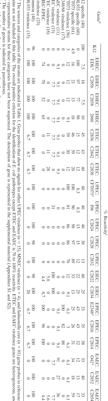
viru-TABLE 2. Percent reactivity (positive) of gene-specific probes to reference and clinical isolates of E. coli pathotypes a Gene b % Reactivity c K12 EHEC: EDL* EHEC: C2856 EHEC: C2858 EHEC: 2860 ETEC: C2836 ETEC: C2842 ETEC: C2838 UPEC: CFT073* UPEC: J96 UPEC: C2824 UPEC: C2828 UPEC: C2832 EIEC: C2822 EIEC: C2834 EPEC: E2348* EPEC: C2816 EPEC: C2814 EAEC: O42* EAEC: C2852 EAEC: C2854 K-12 specific (40) 100 7.5 18 7.5 10 45 43 40 40 40 40 38 13 40 25 33 35 38 33 40 33 EDL933 specific (60) 0 100 97 77 98 15 12 12 8.3 8.3 13 15 12 22 25 43 43 32 12 13 17 CFT073 specific (61) 4.9 16 21 13 18 46 39 20 100 90 98 97 89 33 13 25 26 31 34 18 59 CFT073 virulence (36) 3 12 12 12 9.1 39 30 12 100 81 86 86 70 12 9.1 6.1 3 6.1 6.1 6.1 6.1 E2348/68 virulence (17) 0 0000 0 0 0 0 0 0 0 0 0 0 1 0 0 8 2 8 8 0 0 0 EAEC virulence (11) 0 9.1 9.1 9.1 9.1 9.1 9.1 9.1 0 0 0 0 0 0 0 9.1 9.1 9.1 91 27 36 EIEC virulence (13) 0 0 7.7 7.7 7.7 7.7 7.7 0 0 31 0 0 0 100 100 0 0 0 7.7 0 7.7 ETEC virulence (18) 0 0000 1 1 1 1 2 8 0 0 0 0 0 5 .6 0 0 0 0 0 0 5 .6 EHEC-EPEC virulence (23) 0 74 70 52 65 0 0 0 0 0 0 0 0 4.4 8.7 100 96 100 0 0 4.4 Common (48) 96 100 100 100 100 100 100 100 100 100 100 98 100 98 100 100 100 100 100 100 100 EDL933 virulence (15) 0 100 100 100 100 6.7 6.7 6.7 0 0 0 0 0 0 20 6.7 6.7 6.7 0 0 0 a The sources and categories of the strains are indicated in Table 1. Gene probes that show no signals for other UPEC virulence ( n ⫽ 15), MNEC virulence ( n ⫽ 4), and Salmonella core ( n ⫽ 81) gene probes to reference strains are not included in this table. The overall positive criterion for the identification of E. coli pathotypes except ETEC and EAEC is 70 to 100% reactivity. ETEC and EAEC virulence genes are heterogeneous, and the representative strains for these categories have not been sequenced. The description of gene is represented in the supplemental material (Appen dixes S1 and S2). b The number of genes is indicated in parentheses. c Asterisks indicate the reference strain of each pathotype.
on May 16, 2020 by guest
http://jcm.asm.org/
[image:3.585.325.499.78.722.2]lence genes, although the probe sequences were derived from EPEC E2348/69. On the basis of these criteria, EPEC E2348/69 showed 100% reactivity to its specific genes and the EHEC-EPEC virulence gene probes, whereas EHEC EDL933 showed only 74% reactivity to the EHEC-EPEC virulence gene probes. The nonreactive probes of EDL933 include orf19,
tir,eae,espA,espB, andespD. These genes are already known to be variable on the basis of their sequences in EDL933. Since the gene probes constructed were based on the sequence of E2348/69, EDL933 was not reactive. However, the probes for thetir-2,espA2, andespB2genes, which were designed on the basis of the EDL933 genome sequence, were reactive only to EDL933 and not E2348/69, indicating the uniqueness of the probes used.
CFT073, the prototype strain of UPEC, showed 100% reac-tivity to CFT073 virulence and specific genes.
EAEC is characterized to have the following virulence genes: aaf, agg, astA, and pet (12). Our array detected astA,
aafA, aafB aggR, and pet by use of the reference strain of EAEC (O42). It also showed reactivity to all the EAEC viru-lence genes exceptaggA(91%). TheaggA-specific gene probe was derived from EAEC isolate (O3:H2) and not EAEC O42, which might be the reason for the lack of reactivity.
Salmonellacore genes were considered the negative control. These genes were derived by comparison of the genomes of different isolates ofSalmonellaand were absent fromE. coli
K-12 andE. coliO157:H7. As expected, 100% reactivity of the
Salmonellacore gene probes with isolates of Salmonella en-tericaserovar Typhimurium was observed (data not shown).E. coli pathotypes showed reactivity to 4 of 81 oligonucleotide probes specific forSalmonella. This might be because the Sal-monellacore genes established by McClelland et al. were com-pared with only twoE. coligenomes (14).
The autoblank, which consisted of spots of dimethyl sulfox-ide without any probes, was created as another negative con-trol to make sure that nonspecific hybridization to the slide was not occurring. None of the autoblanks showed a fluorescent signal.
The specificity of the array for the differentiation of refer-enceE. colipathotypes was 98%. This specificity was estimated on the basis of the numbers of true-negative spots (autoblanks) and false-negative spots (afaD,ibeB).
Validation of virulence, pathotype-specific, and common
gene probes with clinical isolates of E. coli pathotypes. All
three clinical isolates of EHEC showed 100% reactivity to EHEC-specific virulence gene probes. With regard to the EDL933-specific gene probes, two clinical isolates (C2856 and C2860) showed reactivities of 96.5 and 98%, respectively, and one clinical isolate (C2860) showed only 77% reactivity. Sim-ilar to reference strain EDL933, clinical strains C2856, C2858, and C2860 showed less reactivity to EHEC-EPEC-specific gene probes (69.5, 52, and 65%, respectively), indicating that the genomes of clinical strains are more closely related to the EDL933 genome. All the clinical strains of EHEC showed lesser reactivities to K-12-specific genes and CFT073-specific genes (Table 2).
EPEC clinical isolate C2816 was 82 and 96% positive for the EPEC and EPEC-EHEC virulence genes probes, respectively, while EPEC clinical isolate C2814 was 88 and 100% positive,
respectively, indicating that these two strains belong to the EPEC pathotype.
J96, another prototype strain of UPEC, showed 81 and 90% reactivities to CFT073 virulence genes and specific genes, re-spectively. Probes for thepapA, sfaA, fsoE,papGII, sat, and
iucD genes failed to react with the J96 strain. Probes for CFT073-specific genes kpsE, kpsD, and kpsM(K15 capsule) and probes for two hypothetical genes also showed no reactiv-ity to strain J96. The K15 capsule locus of strain J96, however, has not been characterized so far.
UPEC clinical strains C2824, C2828, and C2832 showed 86, 86, and 68% reactivities to reference strain CFT073-specific virulence gene probes, respectively, and 98, 97, and 89% reac-tivities to CFT073-specific genes, respectively. It is well known that UPEC strains exhibit greater diversity in their virulence genes, but screening with typical UPEC CFT073-specific genes indicates that these clinical isolates belong to the UPEC patho-type with a divergence in their virulence genes.
EAEC contains heterogeneous virulence markers. In com-parison with the reference strain (O42), both EAEC clinical strains showed reactivity to aggR, but only one of them was positive forastAand one of them showed reactivity toaggD,
aggB, andaggA. The clinical isolates of EAEC showed 32 to 40% and 12 to 16% reactivities to the K-12- and EDL933-specific genes, respectively.
The clinical isolates with a known phenotype of EIEC and ETEC showed 100% and 28% reactivities to the virulence gene probes for these pathotypes, respectively, in the microarray analysis. Since we used gene probes for all the toxins and various colonization factors synthesized by different ETEC iso-lates, the overall percentage of reactivity to ETEC virulence genes is low. However, ETEC isolates showed reactivity to their respective genotypes (toxins) in the array. ETEC clinical isolates showed marked differences in reactivity with ETEC virulence genes, like the EAEC isolates did (Table 2), indicat-ing that these categories contain heterogeneous virulence markers.
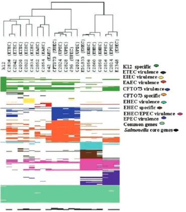
The reactivities of the individual gene probes for each pathotype are represented in the supplemental material (Ap-pendix S2). All the clinical isolates clustered with their respec-tive referenceE. colipathotypes, indicating the potential for the use of microarray analysis for differentiation of the patho-types. The diversity of pathotypes and their genetic relatedness are illustrated in Fig. 1.
DISCUSSION
Numerous bioassays targetingE. colivirulence genes have been developed for the detection and the typing of pathogenic
E. coli. The sheer number of virulence genes and the fact that the virulence genes in the pathogencity islands could poten-tially be transferred to other bacteria in the environment (16) make it hard to differentiate between differentE. coli patho-types. Using data from the completed genomic sequences of
EHEC E. coli O157 EDL933, nonpathogenic E. coli K-12
MG1655, and UPEC (E. coliCFT073) and also virulence fac-tors fromE. colipathotypes, we have developed a microarray containing not only virulence gene probes associated with each
E. colipathotype but also probes for pathotype-specific genes developed for nonpathogenic K-12, EHEC EDL933, and
on May 16, 2020 by guest
http://jcm.asm.org/
UPEC CFT073. E. coli common genes and Salmonella core genes were used as positive and negative controls, respectively. Recently, microarrays targeting multiple virulence factors have been developed for the detection of pathogenicE. coli
(1). However, the microarray analysis was evaluated as positive or negative by the color of the fluorescence intensity. Each spot in the array is subjected to variability in intensity, and the evaluation of the array by the color of the fluorescence inten-sity is difficult, especially when the fluorescence inteninten-sity is compared with the background, which may eventually lead to
[image:5.585.110.480.68.488.2]artifacts. We have calculated the background locally for each spot rather than globally for the entire image, which might have enhanced the quality of each spot. The microarray data were analyzed by taking the log value of the median without the background, and the data were further filtered statistically by taking two times the standard deviation (P⬍0.05) of the median value. PathogenicE. colistrains associated with human and animal diseases are remarkably diverse and show variabil-ity in the genes encoding surface antigens or virulence factors (5). These variable genes may not bind or may bind poorly to
FIG. 1. Dendrogram representation of virulence, specific, and common gene patterns forE. coli pathotypes. Hierarchical clustering was obtained by using Avadis software. The pathotype virulence and specific genes are indicated with different colors to denote the specificity of pathotypes in the dendrogram. Fifteen EHEC, 17 EPEC, 36 UPEC, and 11 EAEC virulence gene probes were derived from strains EDL933, E2348/69, CFT073, and O42, respectively. Twenty-three EPEC virulence gene probes were obtained from EPEC E2348/69. Sixty EHEC-specific genes, 61 UPEC-EHEC-specific genes, and 40 nonpathogenicE. coli-specific genes were from strains EDL933, CFT073, and K-12, respectively. Gene probes that show no signals for 24 UPEC-specific gene probes and also 81Salmonellacore genes (negative control) were also included in the dendrogram. The results for four nonreactive virulence gene probes of meningitis-causingE.coliare not included in the dendrogram. The diversity between EHEC and EPEC in certain regions of EHEC-EPEC is also represented as a pattern profile in the dendrogram. All the reference and clinical isolates except the ETEC and EAEC isolates contained conserved virulence and specific genes; the ETEC and EAEC isolates contained heterogeneous virulence genes. However, the uniqueness of ETEC and EAEC toxins and virulence genes is evident in the pattern profile of the dendrogram.
on May 16, 2020 by guest
http://jcm.asm.org/
the probes in the array. Seventy-mer oligonucleotide probes rather than cDNA PCR probes were used in this multiple-target assay, since oligonucleotide probes are a cost-effective alternative to cDNA PCR probes. Furthermore, the oligonucle-otide-based arrays provide a reduction in cross-hybridization and an increase in the differentiation of highly homologous regions (6, 11, 23).
The microarray’s ability to detect virulence, pathotype-spe-cific, and common genes was primarily analyzed with reference strains of E. coli pathotypes, such as nonpathogenic K-12 MG1655, EHEC O157 EDL933, EAEC O42, EPEC E2348/69, and UPEC CFT073. To validate the array further, clinical isolates representing eachE. coli pathotype were used. The results indicate that microarray analysis could potentially dif-ferentiateE. colipathotypes not only on the basis of virulence genes but also on the basis of specific and common genes. Even the potentially powerful microarray showed false-positive re-sults forafaD,ibeB, andtraT. Bekal et al. developed a microar-ray targeting virulence factors for the detection of pathogenic
E. coli, and that microarray also showed false-positive results foraagAandcdt(1).
Comparison of the LEE regions of EPEC E2348/69 and EHECE. coliO157 showed thattir,eae,espA,espB, andespD
are more diverse (7, 13). The diversity is evident in our study with EHEC reference and clinical isolates. UPEC isolates also contained heterogeneous virulence genes. UPEC prototype strain J96 showed reactivity to papC, papE, papG1, papGII,
papx,papK,papJ,papH, andpapI but not topapAorpapA2. None of the clinical isolates showed reactivity to papA or
papA2, supporting thepapAsubunit diversity in UPEC isolates (8, 19). EIEC isolates also contained different virulence genes (1, 17), but the clinical isolates of EIEC evaluated in this study had similar virulence profiles.
E. colipathotypes are categorized by the presence of viru-lence factors. The viruviru-lence factors are acquired from numer-ous sources, including bacteriophages, plasmids, and the ge-nomes of other bacteria (5). It is apparent that the virulence genes in the pathogenic island are transferable and could con-tribute to the heterogeneity ofE. colistrains (5). Therefore, it is ideal to track the pathotypes on the basis of the virulence genes. However, it would be better to include nontransferable and specific genes ofE. colipathotypes, as the categories of pathotypes keep on increasing. Screening with multiple mark-ers such as virulence, specific, and core genes ofE. colihelp us to identify the emergence of the new pathotypes and would also allow us to assess the relative genetic and virulence pro-files of a single strain in comparison with particular pathotypes. An unknown pathotype can be identified primarily by its percentage of reactivity to the virulence genes of EHEC, UPEC, EPEC, EAEC, EIEC, and ETEC. The pathotype-spe-cific genes constructed on the basis of the available genome sequences of strains EDL933, CFT073, and K-12 enhance the accuracy of identification of EHEC and UPEC. However, ETEC and EAEC are not well characterized compared to the other pathotypes, and their virulence genes are heterogeneous among isolates. Therefore, the reactivities of certain genes, especially with toxins and fimbrial types (ETECsta,stb, andlt; EAECastA and aagR) can be considered important criteria that can be used to denote the pathotype of an isolate. Al-though the clinical isolates of theE. colipathotypes showed
gene contents similar to those of reference strains, variations in gene content between pathotypes and between each strain were also observed by DNA microarray analysis. This indicates that the oligonucleotide array not only allows us to differenti-ate between pathotypes but also is useful for rapid strain char-acterization. The ability to characterize genome deletions of clinical isolates relative to the sequences of reference strains may allow us to reconstruct the phylogeny. The microarray that was developed could differentiate the major pathotypes, but the analysis of reference and clinical isolates of EHEC 2, EPEC 2, and DAEC strains may enhance its validity for wider application for the diagnosis ofE. coli infections. Since the virulence genes are heterogeneous within each category, espe-cially for ETEC, EAEC, and UPEC, the establishment of core genes of each pathotype would help us to identify the patho-type. With the completion of moreE. coligenomic sequences, the inclusion of more specific genes from each pathotype may enhance the identification of pathogenic E. coli strains and their pathotypes.
ACKNOWLEDGMENT
This project was supported with federal funds from the National Institute of Allergy and Infectious Diseases, National Institute of Health, U.S. Department of Health and Human Services, under con-tract N01-AI-30054, project ZC002-03.
REFERENCES
1.Bekal, S., R. Brousseau, L. Masson, G. Prefontaine, J. Fairbrother, and J. Harel.2003. Rapid identification ofEscherichia colipathotypes by virulence gene detection with DNA microarrays. J. Clin. Microbiol.41:2113–2125. 2.Call, D. R., M. K. Bakko, M. J. Krug, and M. C. Roberts.2003. Identifying
antimicrobial resistance genes with DNA microarrays. Antimicrob. Agents Chemother.47:3290–3295.
3.Call, D. R., F. J. Brockman, and D. P. Chandler. 2001. Detecting and genotypingEscherichia coliO157:H7 using multiplexed PCR and nucleic acid microarrays. Int. J. Food Microbiol.67:71–80.
4.Cho, J. C., and J. M. Tiedje.2001. Bacterial species determination from DNA-DNA hybridization by using genome fragments and DNA microarrays. Appl. Environ. Microbiol.67:3677–3682.
5.Donnenberg, M. S., and T. S. Whittam.2001. Pathogenesis and evolution of virulence in enteropathogenic and enterohemorrhagic Escherichia coli. J. Clin. Investig.107:539–548.
6.Dorrell, N., S. J. Hinchliffe, and B. W. Wren.2005. Comparative phylo-genomics of pathogenic bacteria by microarray analysis. Curr. Opin. Micro-biol.8:620–626.
7.Elliott, S. J., L. A. Wainwright, T. K. McDaniel, K. G. Jarvis, Y. K. Deng, L. C. Lai, B. P. McNamara, M. S. Donnenberg, and J. B. Kaper.1998. The complete sequence of the locus of enterocyte effacement (LEE) from en-teropathogenicEscherichia coliE2348/69. Mol. Microbiol.28:1–4. 8.Feria, C., J. Machado, J. D. Correia, J. Goncalves, and W. Gaastra.2001.
Virulence genes and P fimbriae PapA subunit diversity in canine and feline uropathogenicEscherichia coli. Vet. Microbiol.82:81–89.
9.Finlay, B. B., and S. Falkow.1997. Common themes in microbial pathoge-nicity revisited. Microbiol. Mol. Biol. Rev.61:136–169.
10.Fukushima, M., K. Kakinuma, H. Hayashi, H. Nagai, K. Ito, and R. Kawaguchi.2003. Detection and identification ofMycobacteriumspecies isolates by DNA microarray. J. Clin. Microbiol.41:2605–2615.
11.Hessner, M. J., V. K. Singh, X. Wang, S. Khan, M. R. Tschannen, and T. C. Zahrt.2004. Utilization of a labeled tracking oligonucleotide for visualiza-tion and quality control of spotted 70-mer arrays. BMC Genomics5:12. 12.Kaper, J. B., J. P. Nataro, and H. L. Mobley.2004. PathogenicEscherichia
coli. Nat. Rev. Microbiol.2:123–140.
13.Kresse, A. U., M. Rohde, and C. A. Guzman.1999. The EspD protein of enterohemorrhagicEscherichia coliis required for the formation of bacterial surface appendages and is incorporated in the cytoplasmic membranes of target cells. Infect. Immun.67:4834–4842.
14.McClelland, M., K. E. Sanderson, J. Spieth, S. W. Clifton, P. Latreille, L. Courtney, S. Porwollik, J. Ali, M. Dante, F. Du, S. Hou, D. Layman, S. Leonard, C. Nguyen, K. Scott, A. Holmes, N. Grewal, E. Mulvaney, E. Ryan, H. Sun, L. Florea, W. Miller, T. Stoneking, M. Nhan, R. Waterston, and R. K. Wilson.2001. Complete genome sequence ofSalmonella enterica se-rovar Typhimurium LT2. Nature413:852–856.
15.Mead, P. S., L. Slutsker, V. Dietz, L. F. McCaig, J. S. Bresee, C. Shapiro,
on May 16, 2020 by guest
http://jcm.asm.org/
P. M. Griffin, and R. V. Tauxe.1999. Food-related illness and death in the United States. Emerg. Infect. Dis.5:607–625.
16.Ochman, H., J. G. Lawrence, and E. A. Groisman.2000. Lateral gene transfer and the nature of bacterial innovation. Nature405:299–304. 17.Smajs, D., and G. M. Weinstock.2001. The iron- and temperature-regulated
cjrBCgenes ofShigellaand enteroinvasiveEscherichia colistrains code for colicin Js uptake. J. Bacteriol.183:3958–3966.
18.van Ijperen, C., P. Kuhnert, J. Frey, and J. P. Clewley.2002. Virulence typing ofEscherichia coliusing microarrays. Mol. Cell. Probes16:371–378. 19.Verdonck, F., E. Cox, and B. M. Goddeeris.2004. F4 fimbriae expressed by
porcine enterotoxigenicEscherichia coli, an example of an eccentric fimbrial system? J. Mol. Microbiol. Biotechnol.7:155–169.
20.Volokhov, D., V. Chizhikov, K. Chumakov, and A. Rasooly.2003. Micro-array-based identification of thermophilicCampylobacter jejuni,C. coli,C. lari, andC. upsaliensis. J. Clin. Microbiol.41:4071–4080.
21.Volokhov, D., A. Rasooly, K. Chumakov, and V. Chizhikov.2002. Identifi-cation ofListeriaspecies by microarray-based assay. J. Clin. Microbiol.40:
4720–4728.
22.Wang, D., L. Coscoy, M. Zylberberg, P. C. Avila, H. A. Boushey, D. Ganem,
and J. L. DeRisi.2002. Microarray-based detection and genotyping of viral pathogens. Proc. Natl. Acad. Sci. USA99:15687–15692.
23.Wang, H. Y., R. L. Malek, A. E. Kwitek, A. S. Greene, T. V. Luu, B. Behbahani, B. Frank, J. Quackenbush, and N. H. Lee.2003. Assessing unmodified 70-mer oligonucleotide probe performance on glass-slide micro-arrays. Genome Biol.4:R5.
24.Welch, R. A., V. Burland, G. Plunkett III, P. Redford, P. Roesch, D. Rasko, E. L. Buckles, S. R. Liou, A. Boutin, J. Hackett, D. Stroud, G. F. Mayhew, D. J. Rose, S. Zhou, D. C. Schwartz, N. T. Perna, H. L. Mobley, M. S. Donnenberg, and F. R. Blattner.2002. Extensive mosaic structure revealed by the complete genome sequence of uropathogenicEscherichia coli. Proc. Natl. Acad. Sci. USA99:17020–17024.
25.Wu, C. F., J. J. Valdes, W. E. Bentley, and J. W. Sekowski.2003. DNA microarray for discrimination between pathogenic O157:H7 EDL933 and non-pathogenicEscherichia colistrains. Biosens. Bioelectron.19:1–8. 26.Wu, L., D. K. Thompson, G. Li, R. A. Hurt, J. M. Tiedje, and J. Zhou.
2001. Development and evaluation of functional gene arrays for detection of selected genes in the environment. Appl. Environ. Microbiol.67:5780– 5790.