0095-1137/04/$08.00⫹0 DOI: 10.1128/JCM.42.11.4925–4930.2004
Copyright © 2004, American Society for Microbiology. All Rights Reserved.
Development of a PCR Method for Rapid Identification of
New
Streptococcus mutans
Serotype
k
Strains
Kazuhiko Nakano,
1Ryota Nomura,
1Noriko Shimizu,
1Ichiro Nakagawa,
2Shigeyuki Hamada,
2and Takashi Ooshima
1*
Departments of Pediatric Dentistry1and Oral and Molecular Microbiology,2
Osaka University Graduate School of Dentistry, Suita, Osaka, Japan
Received 19 March 2004/Returned for modification 28 June 2004/Accepted 6 July 2004
In a previous study, we isolated and characterized a new serotype kofStreptococcus mutans from human blood and oral cavities. Analysis of the genes involved in biosynthesis of the serotype-specific polysaccharide of serotypekstrains revealed that the serotypek-specific nucleotide alignment was commonly present in the 5ⴕ region of thergpFgene (350 bp from the initial sequence) compared to the reference strains, and then a method for rapid identification of serotypekstrains was developed by use of PCR with primers designed on the basis of the sequence of the variable region. PCR assays with primers specific for amplification of serotypek
strains showed a negative reaction with serotypec,e, andfstrains and a positive reaction with serotypekstrains, with the sensitivity for identification of the serotypek strains shown to range from 5 to 50 cells. Next, the frequency of positive reactions for serotypek-specific primers was surveyed with DNA taken from saliva sam-ples from 200 subjects (2 to 18 years of age), and 10 of those showed a positive reaction, which was higher than the frequency in our previous survey with a serological method. In addition, all saliva samples from subjects with serotypekstrains in our previous study were shown to be positive with the serotypek-specific primers. These results indicate that this new PCR method is effective for identification of subjects withS. mutansserotypek.
Streptococcus mutans, which is known to be a major cario-genic bacterium as well as one of the pathogens involved with bacteremia and infective endocarditis (IE), was previously clas-sified into three serotypes,c,e, andf, based on the chemical composition of serotype-specific polysaccharides (5). The se-rotype-specific polysaccharide was shown to be composed of rhamnose-glucose polymers, with a backbone of rhamnose and side chains of␣- or-linked glucosidic residues (7). The genes involved in the synthesis of serotype-specific rhamnose-glucose polymers have also been cloned and sequenced. Four rml
genes (rmlA through rmlD) are related to the synthesis of dTDP-L-rhamnose (18, 19), and the gluA gene encodes the enzyme that catalyzes the production of the immediate precur-sor of the glucose side chain donor (23). In addition, the six genes (rgpAthroughrgpF) required for synthesis of rhamnose-glucose polymers have been cloned and sequenced (15, 24). Further, thergpGgene has been implicated in the initiation of synthesis of rhamnose-glucose polymers (25).
In our previous study,S. mutansstrains with a low amount of the glucose side chain in serotype-specific polysaccharide were defined as a new serotype, serotypek, which was estimated to have a distribution in the oral cavity of approximately 2% (10). Recently, PCR methods were developed to identify serotypes
c,e, andf by using DNA extracted from saliva samples with primers constructed on the basis of the differences in the se-quences of the region downstream ofrgpFamong each of the serotypes (16). However, there is no information for serotype
kstrains regarding the genes involved in formation of the side chain of the rhamnose backbone. In the present study,
we analyzed those genes from serotypekstrains and developed a PCR method to identify subjects withS. mutansserotypek.
MATERIALS AND METHODS
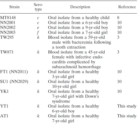
S. mutansstrains.Table 1 lists theS. mutansstrains used in this study. Blood isolates TW295 (k) and TW871 (k) (3) and orally isolated strain MT8148 (c) were selected from the stock culture collection in our laboratory (8). One hun-dred strains ofS. mutansisolated from 100 children, which included 78 serotype c, 17 serotypee, 3 serotypef, and 2 serotypek(strains FT1 and SU1) strains (NN2000 series of isolates) (10), were also analyzed, as was another serotypek strain, YK1 (10). Strains AT1 and YT1, isolated in the present study, were confirmed asS. mutansaccording to the results of a double immunodiffusion method that utilized autoclaved extracts and serotypek-specific antiserum, which has been described previously (10). The specificity of the primers used was tested against the following organisms:Streptococcus sanguinisATCC 10556, Strepto-coccus oralisATCC 10557,Streptococcus gordoniiATCC 10558,Streptococcus mitisATCC 903,Streptococcus milleriNCTC10703, andStreptococcus salivarius HHT.
Sequence analysis of genes involved in biosynthesis of serotype-specific poly-saccharide.Thergpgenes (rgpAthrough open reading frame 11 [ORF11]; total of 15,890 bp) were divided into six fragments. Each gene fragment was amplified by PCR with primers constructed on the basis of the nucleotide alignment of strain Xc (GenBank accession no. AB010970) or strain UA159 (GenBank ac-cession no. AE014133) with LATaqPolymerase (Takara Shuzo, Otsu, Japan). The PCR products were separated by electrophoresis on a 0.7% agar gel, and the amplified DNA was extracted with QIAEX (Qiagen, Du¨sseldorf, Germany). The DNA was directly cloned into a pGEM-T Easy vector (Promega, Madison, Wis.), for which the nucleotide sequence was determined by a dye-terminator reaction with a DNA sequencing system (373-18 DNA sequencer; Applied Biosystems, Foster City, Calif.) and an ABI PRISM cycle sequencing kit. Data analysis was performed with Gene Works software (IntelliGenetics, Mountain View, Calif.), and a multiple-alignment analysis was carried out with CLUSTAL W from the DDBJ (Mishima, Japan) (17). The nucleotide alignments of thergpgenes in TW295 (serotypek), TW871 (serotypek), FT1 (serotypek), and YT1 (serotype k) were compared with that of MT8148 (serotypec). Thergpgenes of strain Xc (serotypec), for which thergpgenes were originally sequenced, and those of strain UA159 (serotypec) (1), of which the complete genome has been se-quenced, were also compared with those of the serotypekstrains.
Clinical specimens.Expectorated whole saliva (approximately 1 ml in each sample) was collected from 200 children and adolescents (2 to 18 years of age;
* Corresponding author. Mailing address: Department of Pediatric Dentistry, Osaka University Graduate School of Dentistry, 1-8 Yamada-oka, Suita, Osaka 565-0871, Japan. Phone: 81 6 6879 2961. Fax: 81 6 6879 2965. E-mail: [email protected].
4925
on May 15, 2020 by guest
http://jcm.asm.org/
mean age, 7.9⫾3.6 years) who visited the Pedodontics Clinic of Osaka Univer-sity Dental Hospital in January and February 2004. Collection of the clinical specimens was carried out in accordance with the Osaka University Health Guideline for Studies Involving Human Subjects. The saliva samples were pro-cessed for the PCR assay by the method reported by Hoshino et al. (6) with some modifications. Briefly, nonstimulated whole saliva was collected in a sterile tube and kept on ice. Bacterial cells were collected in a microcentrifuge tube from 500 l of saliva at 16,000⫻gfor 5 min, then treated in a microwave oven at 500 W for 5 min, and digested inN-acetylmuramidase SG (Seikagaku Corp., Tokyo, Japan) at 50°C for 1 h. Then, 80l of nuclei lysis solution (Promega) was added and incubated at 80°C for 5 min, followed by the addition of 60l of protein precipitation solution (Promega). The proteins were removed by centrifugation at 16,000⫻g for 3 min, and the DNA was purified by phenol-chloroform extraction and ethanol precipitation. The extracted DNA was then dissolved in 50l of TE buffer (10 mM Tris-HCl, 1 mM EDTA [pH 8.0]).
Development of PCR method for identification of serotypekstrains.Table 2 lists the PCR primers used in this study. The forward primer (CEFK-F) was common for all of the serotypes and designed within the 3⬘end region ofrgpE, whereas the serotypec-,e-, andf-specific (non-serotypek) and serotypek-specific reverse primers (CEF-R and K-R, respectively) were designed within the sero-typek-specific 5⬘region of thergpFgene (350 bp from the initial sequence) (Fig. 1). The specificities of the prospective primers were tested by the program Am-plify (2), based on the DNA sequence information stored in GenBank EMBL. All of the primers were commercially synthesized (Proligo Japan, Kyoto, Japan). PCR amplification was performed in a total volume of 20l with 1l of template solution and AmpliTaqGold polymerase (Applied Biosystems) accord-ing to the manufacturer’s instructions. The PCR amplification reaction was performed in a thermal cycler (iCycler; Bio-Rad, Hercules, Calif.) with the following cycling parameters: an initial denaturation at 95°C for 4 min and then 30 cycles consisting of 95°C for 30 s, 60°C for 30 s, and 72°C for 30 s, with a final extension at 72°C for 7 min. The PCR products were subjected to electrophoresis
in a 1.5% agarose gel-Tris-acetate-EDTA buffer. The gel was stained with 0.5g of ethidium bromide per ml and photographed under UV illumination. A 100-bp DNA ladder (New England BioLabs, Beverly, Mass.) was used as the molecular size standard.
First, the sensitivity of the PCR assay was determined by using titrated cultures ofS. mutansMT8148 (serotypec), TW295 (serotypek), and FT1 (serotypek). The CEFK-F and CEF-R sets of primers were used for amplification of the genomic DNA of MT8148, and the CEFK-F and K-R sets were used for ampli-fication of that of TW295 and FT1, respectively. The detection limit for simul-taneous PCR was determined by using the known numbers of bacterial cells diluted in sterile distilled water. Genomic DNA extracted from strains MT8148 (serotypec), NN2001 (serotypec), NN2002 (serotypee), NN2003 (serotypef), TW295 (serotypek), TW871(serotypek), FT1 (serotypek), and YT1 (serotype k) was analyzed to confirm the specificity of the primers designed to classify the non-serotypek or serotypektype strains. In addition, the specificity of the primers was confirmed with genomic DNA from 100S. mutans strains (78 serotypec, 17 serotype e, 3 serotypef, and 2 serotypek strains) previously isolated (NN2000 series of isolates) (10). After analyses of the sensitivity and specificity of the present method, 200 DNA samples extracted from the saliva of the 200 subjects were analyzed.
Analysis of subjects with serotypekstrains identified previously.Saliva sam-ples were collected from two subjects from whom FT1 (serotypek) or YK1 (serotypek) had been isolated from plaque samples in our previous study. DNA from these samples was collected by the method described above and subjected to PCR with the serotypek-specific primers.
RESULTS
Nucleotide alignment ofrgpgenes fromS. mutansserotypek
strains.The nucleotide alignments ofrgpA,rgpB,rgpC,rgpD,
rgpE,rgpF, ORF7,rgpH,rgpI, ORF10, and ORF11 of TW295 (serotypek), TW871 (serotypek), FT1 (serotypek), and YT1 (serotypek) were compared with those of MT8148 (serotype
c), Xc (serotypec), and UA159 (serotype c). There were no significant differences in the putative amino acid sequences of RgpA, RgpC, RgpE, RgpH, ORF10, and ORF11 among all of the test and reference strains. On the other hand, 59 amino acids in the C-terminal region were estimated to be deleted in RgpB of strains FT1 (serotype k), YT1 (serotype k), and MT8148 (serotypec). In addition, 23 amino acids were esti-mated to be deleted in RgpD of TW871 (serotypek). As for ORF7, strains FT1 (serotypek) and YT1 (serotypek) were estimated to be composed of 603 and 675 amino acids, respec-tively, which were fewer than any of the other tested strains (803 amino acids). Further, 26 amino acids were estimated to be deleted in one-third of the N-terminal region of RgpI in strain FT1 (serotypek). The most prominent difference among the genes in each of the tested serotypekstrains commonly compared to strains MT8148 (serotypec), Xc (serotypec), and UA159 (serotypec) was identified in the 5⬘region of thergpF
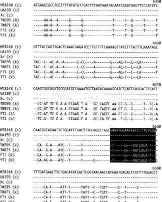
[image:2.585.301.541.69.145.2]gene (350 bp from the initial sequence) (Fig. 2). Thus, primers
[image:2.585.43.285.80.300.2]FIG. 1. Illustration of the serotypek-specific nucleotide region and location of the primers constructed for detection of serotypekstrains by PCR. A base pair (BP) scale is shown above the map. The black box indicates the serotypek-specific nucleotide alignment region in the rgpFgene. The nucleotide number corresponds to thergpFsequence in strain Xc obtained from GenBank (accession number AB010970). TABLE 1. S. mutansstrains used in the present study
Strain
Sero-type Description Reference
MT8148 c Oral isolate from a healthy child 8
NN2001 c Oral isolate from a 6-yr-old boy 10
NN2002 e Oral isolate from a 9-yr-old boy 10
NN2003 f Oral isolate from a 7-yr-old girl 10
TW295 k Blood isolate from a 59-yr-old
male with bacteremia following a tooth extraction
3
TW871 k Blood isolate from a 45-yr-old
female with infective endo-carditis complicated by subarachnoid hemorrhage
3
FT1 (NN2011) k Oral isolate from a healthy
3-yr-old girl
10
SU1 (NN2029) k Oral isolate from a healthy
10-yr-old girl
10
YK1 k Oral isolate from a healthy
7-yr-old girl with Down’s syndrome
10
YT1 k Oral isolate from a healthy
6-yr-old boy
This study
AT1 k Oral isolate from a healthy
7-yr-old girl
This study
TABLE 2. PCR primers used in the present study
Primer Sequence (5⬘–3⬘) Locationa
CEFK-Fb ATT CCC GCC GTT GGA CCA TTC C 6236–6257
CEF-Rc CCG ACA AAG ACC ATT CCA TCT C 6508–6529
K-Rd CCA ATG TGA TTC ATC CCA TCA C 6508–6529
aThe numbers correspond to the locations of the nucleotide sequences
ob-tained from GenBank accession number AB010970 (strain Xc).
bForward primer for serotypec,e,f, andk S. mutans. cReverse primer for serotypec,e, andf S. mutans. dReverse primer for serotypek S. mutans.
on May 15, 2020 by guest
http://jcm.asm.org/
[image:2.585.41.285.636.683.2]specific for the detection of serotypekstrains were constructed on the basis of those sequences.
Specificity and sensitivity of the PCR assay.The PCR assay used in the present study showed positive bands with a non-serotypek-specific set of primers with MT8148 (serotype c) and with a serotypek-specific set of primers with TW295 (se-rotypek) and FT1 (serotypek), each of which produced single bands with the expected size of 294 bp, as assessed by electro-phoresis (Fig. 3). The detection limit was determined in the presence of titrated bacterial cells, and the sensitivity of the PCR assay was found to be between 5 and 50 cells for the serotypek-specific set of primers with strains TW295 (serotype
k) and TW871 (serotypek) and between 50 and 500 cells for the non-serotypek-specific set of primers with strain MT8148 (serotypec). Non-serotype k-specific PCR products were se-lectively identified from among the serotypec,e, andfstrains (listed in Table 1), and serotypek-specific PCR products were
selectively detected from among the serotypekstrains (Fig. 4). Positive bands were identified among the S. mutans strains while the other species showed negative reactions. The speci-ficity of the primers was also confirmed with 100 strains (NN2000 series of isolates), which indicated that the PCR method utilized was valid.
Samples from subjects with a positive reaction to serotype
k-specific primers.Figure 5 shows the results from saliva sam-ples of representative subjects. Of the 200 samsam-ples, 190 showed a positive reaction to the non-serotypek-specific set of primers (specific for serotypes c, e, and f) but not to the serotype
[image:3.585.131.455.71.474.2]k-specific set, whereas 10 samples showed positive reactions to both primer sets. These 10 subjects included 3 males and 7 females, none of whom had heart disorders. In addition, saliva samples previously taken from two subjects from whom the serotypekstrains YK1 and FT1 were isolated also showed a positive reaction to the serotypek-specific primers. In addition,
FIG. 2. Multiple-sequence alignment of the serotypek-specific 5⬘region in thergpFgene compared to that of the reference strains. Only nucleotides different from MT8148 are shown. Primers used for detection of serotypekand non-serotypekstrains were constructed on the basis of the differences in nucleotide alignment (highlighted by the black box). Numbers above the nucleotides are those that appear in GenBank under accession number AB010970 (strain Xc). Serotypes are indicated in parentheses.
on May 15, 2020 by guest
http://jcm.asm.org/
the use of a double immunodiffusion method for confirmation of serotype k strains AT1 and YT1, isolated from a subject whose saliva showed a positive reaction to serotypek-specific primers, also positively reacted to the serotypek-specific prim-ers.
DISCUSSION
S. mutans is known to be a major causative bacterium of dental caries in humans and is occasionally isolated from the blood of the patients with IE (20, 21); however, its mechanism of invasion and survival in blood remains to be clarified. In a previous study (10),S. mutansserotypekstrains were shown to
be less susceptible to phagocytosis, which indicated that this newly elucidated serotype may be one of the risk factors of IE caused by the bacterium. To prevent the occurrence of IE, host risk factors, such as congenital heart failure and acquired valve replacement, have been the focus of clinical practitioners. However, it is also important to elucidate which pathogenic bacteria have a potential to cause bacteremia and the ensuing IE. Microbiological diagnoses of bacteria in blood are gener-ally based on conventional blood culturing, although molecular diagnoses have also been performed recently (4). PCR meth-ods with primers constructed based on the 16S rRNA align-ment are widely utilized for their rapid and sensitive detection of bacteremia (13). With a PCR assay, not only can the pres-ence of bacteria in blood be identified but also the bacterial species itself can be specified, even in cases of culture-negative IE (22). To identifyS. mutansby PCR, several methods have been proposed (6, 11, 14) and a PCR technique for differen-tiating between serotypesc,e, andfhas been developed (16). In the present study, we utilized a PCR method to identifyS. mutansserotypekstrains.
The frequency of subjects with serotype k in the present study was 5%, which was higher than that (2%) found when analyzing a single representative strain from a single subject by an immunodiffusion method in a previous study (10). The detection limit of the present PCR method was shown to be from 5 to 50 cells (Fig. 3), indicating that subjects with a small number of the serotypekstrain bacteria could be identified as positive. We observed positive reactions by the serotype k
blood and oral isolates, as well as negative reactions by the 98
c, e, andf serotype strains by using the present serotype k
-FIG. 3. Sensitivity of PCR assays for detection of non-serotypek and serotypekstrains ofS. mutans. The sensitivity of our PCR method was examined by using titrated cultures with 108cells per ml from
strains MT8148 (serotypec), TW295 (serotypek), and FT1 (serotype k). PCR was performed with the CEFK-F and CEF-R primer sets for MT8148 while the CEFK-F and K-R primer sets were used for TW295 and FT1. The detection limit for simultaneous PCR was determined by using the known numbers of bacterial cells diluted in sterile distilled water. The following numbers of cells were added: 5⫻104(lanes 1),
5⫻103(lanes 2), 5⫻102(lanes 3), 5⫻10 (lanes 4), and 5 (lanes 5).
[image:4.585.45.282.67.161.2]M, molecular size marker (100-bp DNA ladder).
FIG. 4. PCR assay for the detection ofS. mutansserotypekstrains. (A) The primer sets CEFK-F and CEF-R were used for the detection of serotypec,e, andfstrains. (B) The primer sets CEFK-F and K-R were used for the detection of serotypekstrains. Lane 1, MT8148 (serotypec); lane 2, NN2001 (serotypec); lane 3, NN2002 (serotypee); lane 4, NN2003 (serotype f); lane 5, TW295 (serotype k); lane 6, TW871 (serotypek); lane 7, FT1 (serotypek); lane 8, YT1 (serotype k); lane 9,S. sanguinisATCC 10556; lane 10,S. oralisATCC 10557; lane 11,S. gordoniiATCC 10558; lane 12,S. mitisATCC 903; lane 13, S. milleriNCTC10703; lane 14,S. salivariusHHT.
FIG. 5. Identification of subjects withS. mutansserotypekin saliva samples by PCR. (A) The primer sets CEFK-F and CEF-R were used for the detection of serotypec,e, andfstrains. (B) The primer sets CEFK-F and K-R were used for the detection of serotypekstrains. Lanes 1 through 5, samples from five representative subjects with a negative reaction to the serotypek-specific primers. Lanes 6 through 8, samples from three representative subjects with a positive reaction to the serotypek-specific primers. Samples in lanes 6 through 8 also reacted positively to the serotypec-,e-, andf-specific primers.
on May 15, 2020 by guest
http://jcm.asm.org/
[image:4.585.315.523.71.286.2] [image:4.585.44.284.452.630.2]specific set of primers. Further, analyses of saliva samples taken from two subjects with previously isolated serotype k
strains (FT1 and YK1) (10) revealed positive reactions to the primers, as did two other serotypekstrains (AT1 and YT1). As noted above, since serotype k strains are known to be less susceptible to phagocytosis by polymorphonuclear leukocytes (10), it is important to identify patients with serotypekstrains, especially those regarded as having a risk for IE and who are receiving medical or dental treatment. We propose that the PCR method elucidated here will be beneficial for screening subjects withS. mutansserotypekstrains.
The rgpE, rgpH, and rgpI genes have been shown to be correlated to glucose side chain formation in the serotype-specific polysaccharide ofS. mutans (12, 24), however, there were no significant differences in the nucleotide alignment of these genes found in the present serotype k strains. On the other hand, thergpB,rgpD, ORF7, andrgpIgenes were shown to be altered in one or two serotype k strains, though such alterations were not commonly detected in all of the serotype
kstrains tested. In contrast, the 5⬘region of thergpFgene (350 bp from the initial sequence) was shown to be specific for the serotypekstrains compared to the reference strains MT8148, Xc, and UA159, and we used it to develop the present PCR method for identification of serotypekstrains. It is reasonable to speculate with high probability that there is a high possibility thatrgpFitself is associated with glucose side chain formation in the serotype-specific polysaccharide ofS. mutans. In order of level of involvement, RgpA, RgpB, and RgpF, encoded by thergpA,rgpB, andrgpFgenes, respectively, were each shown to be involved in the biosynthesis of the rhamnose backbone (16, 24). Therefore, we also considered that a variation of the
rgpFgene may result in a short rhamnose backbone, causing a shortened attachment location of the glucose side chain. On the other hand, expression of thergpFgene of MT8148 in the serotypekstrains did not produce the properties of serotypes
c,e, andf(data not shown), implicating the presence of other genes involved in biosynthesis of the glucose side chain. The functions of those genes in the rhamnose backbone of the O polysaccharide ofEscherichia colihave been studied; however, the mechanisms inS. mutansremain to be elucidated in further molecular biological studies.
Our laboratory previously demonstrated that a serotype k
blood isolate of TW871 showed a lower level of sucrose-inde-pendent adherence to saliva-coated hydroxyapatite than MT8148, as well as less cariogenicity in experiments with the caries-in-ducing rat model (9). We hypothesized that an alteration of glucan-binding protein C in TW871 caused the reduction of in vitro adhesion and the caries scores in vivo. However, the caries-inducing activities of TW871 have not been discussed in terms of alteration of the serotype-specific polysaccharide compared with that in MT8148. By utilizing a mutant strain without a glucose side chain in the serotype-specific polysaccharide as well as wild-type serotype k strains, the unknown function related to dental caries may be elucidated in further studies.
ACKNOWLEDGMENTS
This study was a part of a 21st Century COE entitled “Origination of Frontier Biodentistry” at Osaka University Graduate School of Dentistry, supported by the Ministry of Education, Culture, Sports, Science, and Technology of Japan, and was supported by a
Grant-in-Aid for Scientific Research (B) 16390605 from the Japan Society for Promotion of Science.
REFERENCES
1.Ajdic, D., W. M. McShan, R. E. McLaughlin, G. Savic, J. Chang, M. B. Carson, C. Primeaux, R. Tian, S. Kenton, H. Jia, S. Lin, Y. Qian, S. Li, H. Zhu, F. Najar, H. Lai, J. White, B. A. Roe, and J. J. Ferretti.2002. Genome sequence ofStreptococcus mutansUA159, a cariogenic dental pathogen. Proc. Natl. Acad. Sci. USA99:14434–14439.
2.Engels, W. R.1993. Contributing software to internet: the Amplify program. Trends Biochem. Sci.18:448–450.
3.Fujiwara, T., K. Nakano, M. Kawaguchi, T. Ooshima, S. Sobue, S. Kawa-bata, I. Nakagawa, and S. Hamada.2001. Biochemical and genetic charac-terization of serologically untypableStreptococcus mutansstrains isolated from patients with bacteremia. Eur. J. Oral Sci.109:330–334.
4.Gauduchon, V., L. Chalabreysse, J. Etienne, M. Celard, Y. Benito, H. Lepidi, F. Thivolet-Bejui, and F. Vandenesch.2003. Molecular diagnosis of infective endocarditis by PCR amplification and direct sequencing of DNA from valve tissue. J. Clin. Microbiol.41:763–766.
5.Hamada, S., and H. D. Slade.1980. Biology, immunology, and cariogenicity ofStreptococcus mutans.Microbiol. Rev.44:331–384.
6.Hoshino, T., M. Kawaguchi, N. Shimizu, N. Hoshino, T. Ooshima, and T. Fujiwara.2004. PCR detection and identification of oral streptococci in saliva samples usinggtfgenes. Diagn. Microbiol. Infect. Dis.48:195–199. 7.Linzer, R., M. S. Reddy, and M. J. Levine.1986. Immunochemical aspects of
serotype carbohydrate antigens ofStreptococcus mutans, p. 29–38. InS. Hamada, S. M. Michalek, H. Kiyono, L. Manaker, and J. R. McGhee (ed.), Molecular microbiology and immunology ofStreptococcus mutans.Elsevier Science Publishers, Amsterdam, The Netherlands.
8.Minami, T., T. Fujiwara, T. Ooshima, Y. Nakajima, and S. Hamada.1990. Interaction of structural isomers of sucrose in the reaction between sucrose and glucosyltransferases from mutans streptococci. Oral Microbiol. Immu-nol.5:189–194.
9.Nakano, K., M. Matsumura, M. Kawaguchi, T. Fujiwara, S. Sobue, I. Na-kagawa, S. Hamada, and T. Ooshima.2002. Attenuation of glucan-binding protein C reduces the cariogenicity of Streptococcus mutans: analysis of strains isolated from human blood. J. Dent. Res.81:376–379.
10.Nakano, K., R. Nomura, I. Nakagawa, S. Hamada, and T. Ooshima.2004. Demonstration ofStreptococcusmutans with a cell wall polysaccharide spe-cific to a new serotype,k, in the human oral cavity. J. Clin. Microbiol.
42:198–202.
11.Oho, T., Y. Yamashita, Y. Shimazaki, M. Kushiyama, and T. Koga.2000. Simple and rapid detection ofStreptococcus mutansandStreptococcus sob-rinusin human saliva by polymerase chain reaction. Oral Microbiol. Immu-nol.15:258–262.
12.Ozaki, K., Y. Shibata, Y. Yamashita, Y. Nakano, H. Tsuda, and T. Koga.
2002. A novel mechanism for glucose side-chain formation in rhamnose-glucose polysaccharide synthesis. FEBS Lett.532:159–163.
13.Rothman, R. E., M. D. Majmudar, G. D. Kelen, G. Madico, C. A. Gaydos, T. Walker, and T. C. Quinn. 2002. Detection of bacteremia in emergency department patients at risk for infective endocarditis using universal 16S rRNA primers in a decontaminated polymerase chain reaction assay. J. In-fect. Dis.186:1677–1681.
14.Sato, T., J. P. Hu, K. Ohki, M. Yamaura, J. Washio, J. Matsuyama, and N. Takahashi.2003. Identification of mutans streptococci by restriction frag-ment length polymorphism analysis of polymerase chain reaction-amplified 16S ribosomal RNA genes. Oral Microbiol. Immunol.18:323–326. 15.Shibata, Y., Y. Yamashita, K. Ozaki, Y. Nakano, and T. Koga.2002.
Expres-sion and characterization of streptococcalrgpgenes required for rhamnan synthesis inEscherichia coli.Infect. Immun.70:2891–2898.
16.Shibata, Y., K. Ozaki, M. Seki, T. Kawato, H. Tanaka, Y. Nakano, and Y. Yamashita.2003. Analysis of loci required for determination of serotype antigenicity inStreptococcus mutansand its clinical utilization. J. Clin. Mi-crobiol.41:4107–4112.
17.Thompson, J. D., D. G. Higgins, and T. J. Gibson.1994. CLUSTAL W: improving the sensitivity of progressive multiple sequence alignment through sequence weighting, position-specific gap penalties and weight matrix choice. Nucleic Acids Res.22:4673–4680.
18.Tsukioka, Y., Y. Yamashita, Y. Nakano, T. Oho, and T. Koga.1997. Identi-fication of a fourth gene involved in dTDP-rhamnose synthesis in Strepto-coccus mutans.J. Bacteriol.179:4411–4414.
19.Tsukioka, Y., Y. Yamashita, T. Oho, Y. Nakano, and T. Koga.1997. Biolog-ical function of dTDP-rhamnose synthesis pathway inStreptococcus mutans. J. Bacteriol.179:1126–1134.
20.Ullman, R. F., S. J. Miller, M. J. Strampfer, and B. A. Cunha.1988. Strep-tococcus mutansendocarditis: report of three cases and review of the liter-ature. Heart Lung17:209–212.
21.Vose, J. M., P. W. Smith, M. Henry, and D. Colan.1987. Recurrent Strep-tococcus mutansendocarditis. Am. J. Med.82:630–632.
22.Wilck, M. B., Y. Wu, J. G. Howe, J. Y. Crouch, and S. C. Edberg.2001. Endocarditis caused by culture-negative organisms visible by Brown and
on May 15, 2020 by guest
http://jcm.asm.org/
Brenn staining: utility of PCR and DNA sequencing for diagnosis. J. Clin. Microbiol.39:2025–2027.
23.Yamashita, Y., Y. Tsukioka, Y. Nakano, K. Tomihisa, T. Oho, and T. Koga.
1998. Biological functions of UDP-glucose synthesis inStreptococcus mutans. Microbiology144:1235–1245.
24.Yamashita, Y., Y. Tsukioka, K. Tomihisa, Y. Nakano, and T. Koga.1998.
Genes involved in cell wall localization and side chain formation of rham-nose-glucose polysaccharide inStreptococcus mutans.J. Bacteriol.180:5803– 5807.
25.Yamashita, Y., Y. Shibata, Y. Nakano, Y. Tsuda, N. Kido, M. Ohta, and T. Koga.1999. A novel gene required for rhamnose-glucose polysaccharide synthesis inStreptococcus mutans.J. Bacteriol.181:6556–6559.