Copyright © 2004, American Society for Microbiology. All Rights Reserved.
Development of a Multiplex PCR for the Detection of
asa1
,
gelE
,
cylA
,
esp
, and
hyl
Genes in Enterococci and Survey for Virulence
Determinants among European Hospital Isolates of
Enterococcus faecium
Vanessa Vankerckhoven,
1* Tim Van Autgaerden,
1Carl Vael,
1Christine Lammens,
1Sabine Chapelle,
1Rosaria Rossi,
2Daniela Jabes,
2and Herman Goossens
1,3University of Antwerp, Antwerp, Belgium1; Vicuron Pharmaceuticals, Gerenzano, Italy2; and University of
Leiden, Leiden, The Netherlands3
Received 3 October 2003/Returned for modification 7 January 2004/Accepted 17 July 2004
A multiplex PCR for the simultaneous detection of five virulence genes (asa1, gelE,cylA, esp, andhyl) in enterococci was developed. The presence of these genes was investigated in 153 clinical and 118 fecal
Entero-coccus faecium isolates from inpatients at an increased risk of developing infections (such as patients in
intensive care units and hematology wards) from 13 hospitals in eight European countries. Of the 271 E.
faeciumisolates, 135 were vancomycin resistantE. faecium(VREF) isolates and 136 were vancomycin
suscep-tibleE. faecium(VSEF) isolates. Susceptibilities to ampicillin, gentamicin, streptomycin, vancomycin, teico-planin, ramoteico-planin, quinupristin-dalfopristin, and linezolid were tested by the microdilution method. Overall, the prevalence ofespwas significantly higher (Pⴝ0.03) in clinical VREF isolates (92%) than in fecal VREF isolates (73%). In Italy, the prevalence ofespwas significantly higher (Pⴝ0.02) in VREF isolates (91%) than in VSEF isolates (68%), whereas in the United Kingdom,hylwas significantly more prevalent (Pⴝ0.01) in VREF isolates (71%) than in VSEF isolates (29%). No significant differences were found for the other countries. Pulsed-field gel electrophoresis was used to check the clonality among the strains tested and showed the spread of two center-specific (esp-positive) VREF clones in Italy and one center-specific (hyl-positive) clone in the United Kingdom. These clones were resistant to ampicillin, gentamicin, and streptomycin. The multiplex PCR reported in this study is a convenient and rapid method for the simultaneous detection of the virulence genes asa1,gelE,cylA,esp, andhylin enterococci. Molecular analysis showed the intrahospital spread ofesp-positive VREF clones (in Italy) andhyl-positive VREF clones (in the United Kingdom); the role ofhylremains to be elucidated.
Enterococci form part of the normal flora of both the human and the animal gastrointestinal tract. However, vancomycin-resistant enterococci have emerged as a major cause of noso-comial infections (28). Several virulence factors have been described in enterococci, for instance, aggregation substance (14), gelatinase (39), cytolysin (4), enterococcal surface protein (38), and, very recently, hyaluronidase (35). The first four virulence factors are found inEnterococcus faecalis, while the fourth and fifth virulence factors are specific forEnterococcus
faecium.
Aggregation substance, encoded byasa1, which is carried on a plasmid, is a pheromone-inducible protein that enables the conjugative transfer of sex pheromone gene-containing plas-mids through the clumping of one Enterococcus to another (14). As a virulence factor, aggregation substance increases bacterial adherence to renal tubular cells (26) and heart en-docardial cells (18), augments internalization by intestinal ep-ithelial cells (30), and has been shown to increase the valvular vegetation mass in an animal model of endocarditis (3).
Gelatinase, encoded by the chromosomalgelE, is an
extra-cellular zinc endopeptidase that hydrolyzes collagen, gelatin, and small peptides (39) and that has been shown to exacerbate endocarditis in an animal model (17).
The production of cytolysin has also been shown to signifi-cantly worsen the severity of endocarditis (3) and endoph-thalmitis (24) in animal models as well as to contribute to the severity of enterococcal disease in humans (22). Cytolysin genes are carried on a plasmid or are integrated into the bacterial chromosome (23). Cytolysin consists of two compo-nents, lysin (L) and activator (A). The cytolysin operon con-sists of five genes, of which cylL1, cylL2, cylM, and cylBare relevant to the expression of component L, whereas cylA is necessary for the expression of component A (15, 21).
The enterococcal surface protein, encoded by the chromo-somalesp, has an interesting structure that includes a central core consisting of distinct tandem repeat units. This central repeat region serves as a retractable arm, extending the N-terminal globular domain through the cell wall to the surface, which might facilitate immune evasion in case of immune de-ficiency (38). Enterococcal surface protein is associated with increased virulence (38), colonization and persistence in the urinary tract (37), and biofilm formation (41). A variant esp
gene has recently been identified as a marker of highly prev-alent vancomycin-resistant E. faecium(VREF) clones among hospitalized patients (44). However, theespgene has also been * Corresponding author. Mailing address: Laboratory of Medical
Microbiology, University of Antwerp, Universiteitsplein 1, 2610 Wil-rijk, Belgium. Phone and fax: 32 3 820 26 63. E-mail: vanessa [email protected].
4473
on May 15, 2020 by guest
http://jcm.asm.org/
detected in vancomycin-susceptibleE. faecium(VSEF) isolates (45).
Recently, another virulence factor, hyaluronidase, was de-scribed inE. faecium(35). TheE. faeciumhyaluronidase, en-coded by the chromosomalhyl, shows homology to the hyalu-ronidases previously described in Streptococcus pyogenes,
Staphylococcus aureus, and Streptococcus pneumoniae (20),
which are believed to contribute to invasion of the nasophar-ynx and pneumococcal pneumonia (2, 33).
Multiplex PCR is a rapid and convenient assay that allows simultaneous amplification of more than one locus in the same reaction and is used in both clinical and research laboratories (19). The purpose of this study was to develop a multiplex PCR for the detection of five potential virulence factors in entero-cocci and to investigate their presence inE. faecium isolates from eight European countries. Any possible correlation be-tween these potential virulence factors, antibiotic resistance, and clonality was also explored.
(This work was presented in part at the 43rd Interscience Conference on Antimicrobial Agents and Chemotherapy, Chi-cago, Ill., 14 to 17 September 2003 [V. Vankerckhoven, T. Van Autgaerden, C. Vael, L. Lammens, S. Chapelle, R. Rossi, D. Jabes, and H. Goossens, Abstr. 43rd Intersci. Conf. Antimi-crob. Agents Chemother., poster B-816, 2003].)
MATERIALS AND METHODS
Bacterial isolates.A total of 271E. faeciumisolates (153 clinical isolates and 118 fecal isolates) collected in 2001 from inpatients at increased risk of the development of infections (such as patients in intensive care units [ICUs] and hematology wards) from 13 hospitals in eight European countries (12 centers: Austria,n⫽1; Belgium,n⫽1; France,n⫽2; Germany,n⫽1; Italy,n⫽3; The Netherlands,n⫽1; Spain,n⫽2; United Kingdom,n⫽1) were included in this study. In total, 135E. faeciumisolates were VREF, whereas 136 were VSEF. Of the 271 strains, 115 (96 VREF isolates and 19 VSEF isolates) were isolated in Italy; 42 (28 VREF isolates and 14 VSEF isolates) were isolated in the United Kingdom; and 114 (11 VREF isolates and 103 VSEF isolates) were isolated in Austria, Belgium, France, Greece, Spain, and The Netherlands. The isolates have been identified in a previous study on ramoplanin susceptibility (16).
The reference strains used for the multiplex PCR wereE. faecalisMMH594 (a generous gift from N. Shankar, Department of Medicinal Chemistry and Phar-maceutics, University of Oklahoma Health Sciences Center, Oklahoma City) (38) was used as a positive control strain for the detection ofasa1,gelE,cylA, and
esp;E. faeciumC68 and C38 (both strains were generous gifts from L. Rice, Research and Medical Services, Louis Stokes Cleveland Veterans Affairs Med-ical Center, and Department of Medicine, Case Western Reserve University,
Cleveland, Ohio) (35) were used as positive and negative control strains, respec-tively, for the detection ofhyl(both controls were also positive foresp); andE. faecalis217, an endocarditis isolate from The Netherlands, was used as a negative control for the detection of the virulence genes tested.
Furthermore, the multiplex PCR protocol was validated with 50E. faecalis
strains (35 clinical and 15 fecal strains),E. faecalisJH2-7 andE. faecalisOG1X (both strains were a generous gift from B. Murray, Center for Infectious Dis-eases, Department of Internal Medicine and Department of Microbiology and Molecular Genetics, University of Texas, Houston) (4),E. faecalisFA2-2 (a generous gift from N. Shankar) (38),E. faeciumE-470 (a generous gift from R. Willems, University Medical Center Utrecht, Utrecht, The Netherlands) (44), and fiveE. faeciumstrains for which the virulence genes present were unknown to us (the five strains were sent by G. Werner from the Robert Koch Institute, Wernigerode, Germany, for use as quality control strains).
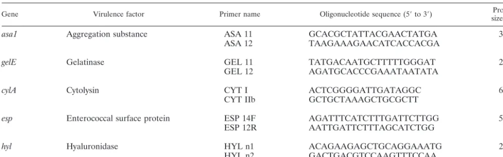
Oligonucleotide primers.The five oligonucleotide primer pairs (Eurogentec, Seraing, Belgium) used to amplify the genesasa1,gelE,cylA,esp, andhyland the expected amplicon sizes are listed in Table 1. Primers were based on published primer pairs forcylA(4) andesp(44), while primers for the detection ofasa1
(GenBank accession number X17214) (14),gelE(GenBank accession number M37185) (39), and hyl(GenBank accession number AF544400) (35) were developed by using Primer3 (http://www.genome.wi.mit.edu/cgi-bin/primer /primer3_www.cgi). Primers were designed so that the PCR products were suffi-ciently different in size to be distinguishable by agarose gel electrophoresis. Primer specificity was checked by a search with the BLAST program, available through the National Center for Biotechnology Information (http://www.ncbi.nlm.nih.gov/).
Multiplex PCR.All cultures were grown on Columbia agar (Becton Dickinson, Sparks, Md.) supplemented with 5% defibrinated horse blood and were incu-bated at 37°C. The template DNA was prepared by suspending one loopful of bacterial cells from an overnight culture in 1 ml of Milli Q water. The bacterial suspensions were heated for 5 min at 95°C and centrifuged to remove the debris. PCR was performed in a GeneAmp PCR System 9600 (Perkin-Elmer, Wellesley, Mass.). Each 50-l PCR mixture consisted of 5l of bacterial suspension; a 0.1 M concentration (each) of primers specific forasa1,gelE, andhyl; a 0.2M concentration (each) of primers specific forcylAandesp; 25l of HotStarTaq master mixture (Qiagen, Hilden, Germany), which consisted of 2.5 U of Hot-StarTaq DNA polymerase, 1.5 mM MgCl2, and 200M deoxynucleoside triphos-phates; and an additional 1.0 mM MgCl2. An initial activation step at 95°C for 15 min, during which the HotStarTaq DNA polymerase is activated, was followed by 30 cycles of denaturation (94°C for 1 min), annealing (56°C for 1 min), and extension (72°C for 1 min), followed by one cycle consisting of 10 min at 72°C. After amplification, 25l of the amplicon was mixed with 5l of gel loading buffer (50% glycerol, 0.8 mg of bromophenol blue per ml) and electrophoresed in a 1.5% pronarose D1 gel (SphaeroQ, Burgos, Spain) for 1 h at 150 V in 0.5⫻ TBE (Tris-borate-EDTA) containing 0.05 mg of ethidium bromide per liter (positive and negative controls were included in each set of amplifications). A 100-bp DNA ladder (Invitrogen, Merelbeke, Belgium) was used as a molecular size marker.
PFGE.The clonal distribution among VREF isolates was studied as described previously (16) by pulsed-field gel electrophoresis (PFGE) by the method of Descheemaeker et al. (7). Briefly, bacterial cells from an overnight culture were imbedded in low-melting-point preparative agarose (Bio-Rad Laboratories, Naz-TABLE 1. PCR primers and products for the detection of virulence genes
Gene Virulence factor Primer name Oligonucleotide sequence (5⬘to 3⬘) size (bp)Product
asa1 Aggregation substance ASA 11 GCACGCTATTACGAACTATGA 375
ASA 12 TAAGAAAGAACATCACCACGA
gelE Gelatinase GEL 11 TATGACAATGCTTTTTGGGAT 213
GEL 12 AGATGCACCCGAAATAATATA
cylA Cytolysin CYT I ACTCGGGGATTGATAGGC 688
CYT IIb GCTGCTAAAGCTGCGCTT
esp Enterococcal surface protein ESP 14F AGATTTCATCTTTGATTCTTGG 510
ESP 12R AATTGATTCTTTAGCATCTGG
hyl Hyaluronidase HYL n1 ACAGAAGAGCTGCAGGAAATG 276
HYL n2 GACTGACGTCCAAGTTTCCAA
on May 15, 2020 by guest
http://jcm.asm.org/
[image:2.603.43.542.81.237.2]areth, Belgium). After cell wall and protein digestion, the plugs were digested overnight with 30 U of SmaI (MBI Fermentas, St. Leon-Rot, Germany) at 25°C. PFGE was performed with a 1% agarose gel by using a CHEF Mapper apparatus (Bio-Rad Laboratories) in 0.5⫻TBE buffer at 14°C and 6 V/cm. For separation, a linearly ramped switching time from 5 to 35 s was applied for 24 h. The gels were stained with ethidium bromide to detect the DNA band profiles, and the image was digitized with a Gel Doc 1000 system (Bio-Rad Laboratories). Con-version, normalization, and further analysis of the DNA band patterns were performed with GelCompar software (version 4.0b; Applied Maths, Kortrijk, Belgium), as described previously (34). The similarity between PFGE patterns was evaluated by use of the Dice coefficient and was observed visually by the detection of a maximum of three clearly visible bands.
Antimicrobial susceptibility testing.The antimicrobial susceptibilities of all strains were tested as described previously (16). They were tested for their susceptibilities to ampicillin (Sigma Chemical Co., St. Louis, Mo.), gentamicin (Sigma), streptomycin (Sigma), vancomycin (Sigma), teicoplanin (Gruppo Lep-etit, Milan, Italy), ramoplanin (Vicuron Pharmaceuticals, Gerenzano, Italy), quinupristin-dalfopristin (Synercid; commercial preparation of Aventis, Milan, Italy), and linezolid (Zyvox; commercial preparation of Pharmacia, Milan, Italy) by the microdilution method, according to the guidelines of NCCLS (29). The genes responsible for resistance to vancomycin (vanA,vanB,vanC1, andvanC2) were detected by PCR, as described previously (9).
Statistical analysis.Chi-square analysis of contingency tables and Fisher’s exact test were used for statistical analysis. APvalue⬍0.05 was considered statistically significant.
RESULTS
Development of multiplex PCR. Template DNA was pre-pared by using cell suspensions of only a few colonies since thicker cell suspensions, as described by other investigators (25, 31), increased the amplicon intensity, which has also been noticed by others (13, 32). Although at first the primer con-centrations used for the detection of each gene were the same, increasing the primer concentrations forcylAand esphelped visualize the expected PCR products much more consistently. Moreover, a higher concentration of Mg2⫹ was needed to
optimize the intensities of the band patterns generated. As shown in Fig. 1, the PCR product of the expected size for each of the four control strains was observed. TheE. faecalis
positive control strain, strain MMH594 (Fig. 1, lane 2), yielded four gene-specific products (asa1,gelE,cylA, andesp), while no products were generated from theE. faecalisnegative control
strain, strain 217 (Fig. 1, lane 3). The E. faecium positive control strain forhyl, strain C68 (Fig. 1, lane 4), yielded two gene-specific products (espandhyl), while onlyespwas gener-ated from theE. faeciumnegative control strain forhyl, strain C38 (Fig. 1, lane 5). Each multiplex PCR assay was performed with a negative control containing all the reagents but no DNA template (Fig. 1, lane 6).
The 53E. faecalisstrains contained one, two, three, all, or none of the virulence genes tested for in this study, which was identical to the results generated by uniplex PCRs for each of the virulence genes tested for. Upon comparison of the two primer sets specific forespreported so far (38, 44), only those described by Willems et al. (44) could consistently detectespin two of sixE. faeciumisolates.
Multiplex PCR survey for virulence factors amongE. fae-cium isolates.We tested 271E. faecium strains for the pres-ence of five virulpres-ence factors. The genesasa1,gelE, andcylA
were not detected. Of all 271 (VREF and VSEF) strains, 176 (65%) were positive forespand 45 (17%) were positive forhyl. The prevalence of esp was significantly higher (P ⫽ 0.04) among clinical VREF isolates (24 of 26 [92%]) than among fecal VREF isolates (80 of 109 [73%]). The esp gene was significantly more prevalent (P⬍ 0.0001) among the VREF isolates than among the VSEF isolates: 104 of 135 (77%) VREF isolates versus 72 of 136 (53%) VSEF isolates. A sig-nificant difference in the prevalence of esp (P ⫽ 0.02) was found among the 115 Italian isolates (96 VREF isolates and 19 VSEF isolates): 87 of 96 (91%) VREF isolates versus 13 of 19 (68%) VSEF isolates possessed this gene. No significant dif-ference in the prevalence ofespwas observed between United Kingdom VSEF isolates and VREF isolates. Theespgene was significantly more common (P ⬍ 0.0001) among ampicillin-resistant VREF isolates (93 of 109 [85%]) than among ampi-cillin-susceptible VREF isolates (11 of 26 [42%]). Among the
esp-positive VREF isolates, ampicillin resistance was seen in 93 of 104 (89%), of which 82 of 87 (94%) were Italianesp-positive VREF isolates and 9 of 15 (60%) were esp-positive United Kingdom VREF isolates.
The hyl gene was found in 7 of 26 (27%) of the clinical VREF isolates, whereas it was found in 15 of 109 (14%) of the fecal VREF isolates (P⫽0.1). No significant difference in the prevalence of hyl was seen between the Italian VSEF and VREF isolates. A significant difference (P⫽0.01) in the prev-alence of hyl was observed among the 42 United Kingdom isolates (28 VREF isolates and 14 VSEF isolates): 20 of 28 (71%) VREF isolates versus 4 of 14 (29%) VSEF isolates were found to be positive for this gene; however, no significant difference in the prevalence of hylwas observed between the Italian VREF and VSEF isolates. The hyl gene was signifi-cantly more common (P⬍0.0001) among ampicillin-resistant VREF isolates (16 of 109 [15%]) than among ampicillin-sus-ceptible VREF isolates (6 of 26 [2%]). Ampicillin resistance was seen in 16 of 22 (73%) of thehyl-positive VREF isolates, of which the only Italianhyl-positive VREF isolates and 14 of 20 (70%) United Kingdom hyl-positive VREF isolates were resistant.
Of the 114 isolates (11 VREF isolates and 103 VSEF iso-lates) collected from Austria, Belgium, France, Greece, Spain, and The Netherlands,espwas detected in 51 (45%) isolates (2 of 11 [18%] VREF isolates and 49 of 103 [48%] VSEF isolates) FIG. 1. Multiplex PCR of control strains. Lane 1, molecular
marker (100 bp); lane 2, E. faecalis MMH594 (positive control for
asa1,gelE,cylA, andesp); lane 3,E. faecalis217 (negative control for
asa1,gelE,cylA, andesp); lane 4,E. faeciumC68 (positive control for
hyl; also positive foresp); lane 5,E. faeciumC38 (negative control for
hyl; positive foresp); lane 6, negative control (no DNA added).
on May 15, 2020 by guest
http://jcm.asm.org/
andhylwas present in 19 (17%) isolates (1 of 11 [9%] VREF isolates and 18 of 103 [18%] VSEF isolates).
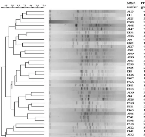
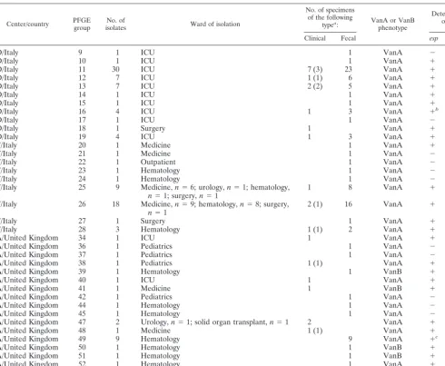
PFGE typing, susceptibility testing, and the presence of virulence determinants. PFGE groups were defined as de-scribed previously (40). A dendrogram is shown in Fig. 2. PFGE revealed the spread of two center-specificesp-positive VREF clones in Italy (referred to as centers D and F below) and one center-specifichyl-positive VREF clone in the United Kingdom (referred to as center A below); these clones were resistant to ampicillin, gentamicin, and streptomycin. In center D in Italy, among the 58 VREF isolates collected, PFGE revealed 11 groups, all of which were of the VanA phenotype (Table 2). Except for the isolates belonging to PFGE groups 9 and 17, all isolates were positive for esp, of which group 11 represented the largest number of isolates. In center F in Italy, among the 36 VREF isolates collected, PFGE revealed 9 groups, all of which were of the VanA phenotype (Table 2). The isolates of PFGE group 20 were positive forespandhyl, while the isolates of PFGE groups 25, 26, 27, and 28, of which group 26 contained the largest number of isolates, were posi-tive only foresp. The other isolates (PFGE groups 21, 22, 23, and 24) did not show the presence of any virulence factors. All of the Italian isolates were highly resistant to ampicillin, gen-tamicin, and streptomycin. In center A in the United Kingdom, among the 28 VREF isolates collected, 24 of the VanA phe-notype and 4 of the VanB phephe-notype, PFGE revealed 17 groups, of which group 49 contained the largest number of isolates (Table 2). The isolates of PFGE groups 34, 39, 40, 41, 47, 48, 49, 50, 51, and 52 were positive for espand hyl; the isolates of PFGE group 37 were positive only forhyl; and the isolates of PFGE groups 38 and 53 were positive only foresp. No virulence factors were detected in the other groups (groups 36, 42, 44, and 45). All the United Kingdom isolates were resistant to ampicillin, and of these, 19 and 21 isolates also showed resistance to gentamicin and streptomycin, respec-tively. One Italian strain of PFGE group 16 and one United
Kingdom strain of PFGE group 49 showedesppositivity, while the other isolates of the same group did not (Table 2). Finally, one strain was resistant to linezolid (8 mg/liter); this strain was isolated in the United Kingdom, belonged to PFGE group 49, and was positive for hyl. Ramoplanin was active against all strains of VREF, with an MIC at which 90% of isolates are inhibited of 0.5 mg/liter for the clinical isolates.
DISCUSSION
A multiplex PCR developed for the simultaneous detection of enterococcal genes that encode for aggregation substance (asa1), gelatinase (gelE), cytolysin (cylA), enterococcal surface protein (esp), and hyaluronidase (hyl) has not been described before. The multiplex protocol for these five genes provides a reliable and rapid alternative to phenotypic testing and uniplex PCRs. Moreover, the use of 5l of a heat-treated bacterial suspension as the DNA template is a time-saving step com-pared to the amount of time required for DNA preparation, increasing the feasibility of the technique.
We surveyed EuropeanE. faeciumisolates for the presence of these genes. The asa1, gelE, and cylAgenes were not de-tected in any of the 271E. faeciumisolates, which is in agree-ment with the results reported by other investigators who also testedE. faeciumstrains for the presence of one or more of these genes (4, 8, 10, 12, 36). However, Eaton and Gasson (10) found onegelE-positiveE. faeciumisolate without phenotypic gelatinase activity, and Elsner et al. (12) found asa1 among 13% of clinicalE. faeciumisolates, but they used hybridization. Theespgene was detected in 65% ofE. faeciumisolates, in accordance with the findings of other studies (10, 11), which identified the esp gene in about 80% of E. faecium strains. However, this is in contrast to the findings of Shankar et al. (38), who reported the absence of espinE. faecium. We de-tected the esp gene in a significantly higher number (P ⬍
0.0001) of VREF strains (77%) than VSEF strains (53%). Previous studies on the incidence ofespin VREF and VSEF are contradictory: some studies (44) indicated a higher preva-lence ofespamong VREF strains than among VSEF strains, other studies showed the opposite (5, 8, 27), whereas again in others an equal distribution of theespgene was found among VREF and VSEF strains (35, 45). Interestingly, we found that 91% of the Italian VREF strains harbored theespgene. This is in contrast to the results of Baldassarri et al. (1), who found this gene in 33% of clinical VSEF isolates. The prevalence of
espin the present study was significantly higher (P⫽0.04) in the clinical VREF isolates (92%) than in the fecal VREF isolates (73%). This is in agreement with the results of Rice et al. (35), who also reported a higher prevalence ofespamong clinical isolates compared with that among fecal isolates. These results suggest a possible role of espin the pathogenicity of enterococci.
Analysis of the clonalityE. faeciumstrains harboring theesp
gene showed that 5 of 9 clonal types (56%) from center D in Italy, 8 of 11 clonal types (73%) from center F in Italy, and 11 of 19 clonal types (58%) from center A in the United Kingdom wereesppositive. The clonal distribution ofE. faeciumstrains harboringesphas also been reported by Coque et al. (5, 6), while Willems et al. (44) described a larger number of esp -FIG. 2. Dendrogram of PFGE patterns.
on May 15, 2020 by guest
http://jcm.asm.org/
[image:4.603.43.284.70.297.2]positive clones and, thus, reported a distribution ofespmore heterogeneous than that found in our study.
Deviating results were found for one Italian strain of PFGE group 16 and one United Kingdom strain of group 49. Both strains were found to beesppositive, while other strains be-longing to the same group wereespnegative. This difference might be explained by the fact that the restriction enzyme used for PFGE, SmaI, does not recognize restriction sites in theesp
gene. Moreover, SmaI generates segments of kilobase pairs, while the multiplex PCR generates segments of base pairs, and because of the difference in the lengths of the fragments gen-erated by PFGE and PCR, it is unlikely that the virulence gene will be observed by PFGE. Our findings could be further ex-plored by using a second restriction enzyme, which might be able to detect the differences. According to Waar et al. (43),E.
faecalisisolates are clonal if they reveal a similarity ofⱖ90% by
amplified fragment length polymorphism analysis and an iden-tical pattern of virulence factors. On the basis of these conclu-sions, the clones reported in our study may have to be reclas-sified in a different PFGE cluster.
The hyl gene was detected in 17% of the 271E. faecium
isolates collected in eight European countries, which is in con-trast to the findings of Rice et al. (35), who detectedhylin only 3% of the European clinical isolates. We found thehylgene among 16% of the 135 VREF isolates and 17% of the 136 VSEF isolates. Rice et al. (35) found the hyl gene only in European VREF isolates and in none of the European VSEF isolates included in their study. Moreover, our study showed that the hyl gene is even more prevalent among the United Kingdom VREF isolates (71%) than among the U.S. VREF isolates (39%) described by Rice et al. (35).
[image:5.603.49.540.80.483.2]The esp and hyl genes were significantly more common TABLE 2. Vancomycin-resistantE. faeciumstrains in centers D and F in Italy and center A in the United Kingdom
Center/country PFGEgroup isolatesNo. of Ward of isolation
No. of specimens of the following
typea: VanA or VanB
phenotype
Detection of:
Clinical Fecal esp hyl
D/Italy 9 1 ICU 1 VanA ⫺ ⫺
D/Italy 10 1 ICU 1 VanA ⫹ ⫺
D/Italy 11 30 ICU 7 (3) 23 VanA ⫹ ⫺
D/Italy 12 7 ICU 1 (1) 6 VanA ⫹ ⫺
D/Italy 13 7 ICU 2 (2) 5 VanA ⫹ ⫺
D/Italy 14 1 ICU 1 VanA ⫹ ⫺
D/Italy 15 1 ICU 1 VanA ⫹ ⫺
D/Italy 16 4 ICU 1 3 VanA ⫹b ⫺
D/Italy 17 1 ICU 1 VanA ⫺ ⫺
D/Italy 18 1 Surgery 1 VanA ⫹ ⫺
D/Italy 19 4 ICU 1 3 VanA ⫹ ⫺
F/Italy 20 1 Medicine 1 VanA ⫹ ⫹
F/Italy 21 1 Medicine 1 VanA ⫺ ⫺
F/Italy 22 1 Outpatient 1 VanA ⫺ ⫺
F/Italy 23 1 Hematology 1 VanA ⫺ ⫺
F/Italy 24 1 Hematology 1 VanA ⫺ ⫺
F/Italy 25 9 Medicine,n⫽6; urology,n⫽1; hematology,
n⫽1; surgery,n⫽1 1 8 VanA ⫹ ⫺
F/Italy 26 18 Medicine,n⫽9; hematology,n⫽8; surgery,
n⫽1 2 (1) 16 VanA ⫹ ⫺
F/Italy 27 1 Surgery 1 VanA ⫹ ⫺
F/Italy 28 3 Hematology 1 (1) 2 VanA ⫹ ⫺
A/United Kingdom 34 1 ICU 1 VanA ⫹ ⫹
A/United Kingdom 36 1 Pediatrics 1 VanA ⫺ ⫺
A/United Kingdom 37 1 Pediatrics 1 VanA ⫺ ⫹
A/United Kingdom 38 1 Pediatrics 1 (1) VanA ⫹ ⫺
A/United Kingdom 39 1 Hematology 1 VanB ⫹ ⫹
A/United Kingdom 40 1 ICU 1 VanA ⫹ ⫹
A/United Kingdom 41 1 Medicine 1 VanB ⫹ ⫹
A/United Kingdom 42 1 Pediatrics 1 VanA ⫺ ⫺
A/United Kingdom 44 1 Hematology 1 VanA ⫺ ⫺
A/United Kingdom 45 1 Hematology 1 VanA ⫺ ⫺
A/United Kingdom 47 2 Urology,n⫽1; solid organ transplant,n⫽1 2 VanA ⫹ ⫹
A/United Kingdom 48 1 Medicine 1 (1) VanA ⫹ ⫹
A/United Kingdom 49 9 Hematology 9 VanA ⫹c ⫹
A/United Kingdom 50 1 Hematology 1 VanB ⫹ ⫹
A/United Kingdom 51 1 Hematology 1 VanB ⫹ ⫹
A/United Kingdom 52 1 Hematology 1 VanA ⫹ ⫹
A/United Kingdom 53 3 Hematology 3 VanA ⫹ ⫺
aValues in parentheses are the number of isolates from blood. bOne of four isolates.
cOne of nine isolates.
on May 15, 2020 by guest
http://jcm.asm.org/
among ampicillin-resistant VREF isolates than among ampi-cillin-susceptible VREF isolates, which is in accordance with the findings of other studies (5, 6, 27, 42).
Finally, we found that 34 of 45 (76%) hyl-positive strains were alsoesppositive, which is in accordance with the findings of Rice et al. (35), who also described the combined presence ofhylandespin⬎90% of the strains that they tested. On the contrary, Coque et al. (6) found only 4% of their isolates to be positive forespandhyl.
In conclusion, the multiplex PCR developed and described herein is a convenient and rapid method for the simultaneous detection of five potential virulence genes,asa1,gelE,cylA,esp, and hyl, in enterococci. Molecular analysis showed the intra-hospital spread ofesp-positive VREF clones (in Italy) and of
hyl-positive VREF clones (in the United Kingdom); the role of
hylremains to be elucidated.
ACKNOWLEDGMENT
This work was supported by a grant from the European Commission (QLRT-2001-01273) and Vicuron Pharmaceuticals.
REFERENCES
1. Baldassarri, L., L. Bertuccini, M. G. Ammendolia, G. Gherardi, and R. Creti.2001. Variantespgene in vancomycin-sensitiveEnterococcus faecium. Lancet357:1802.
2. Berry, A. M., and J. C. Paton.2000. Additive attenuation of virulence of
Streptococcus pneumoniaeby mutation of the genes encoding pneumolysin and other putative pneumococcal virulence proteins. Infect. Immun.68:133– 140.
3. Chow, J. W., L. A. Thal, M. B. Perri, J. A. Vazquez, S. M. Donabedian, D. B. Clewell, and M. J. Zervos.1993. Plasmid-associated hemolysin and aggre-gation substance production contribute to virulence in experimental entero-coccal endocarditis. Antimicrob. Agents Chemother.37:2474–2477. 4. Coque, T. M., J. E. Patterson, J. M. Steckelberg, and B. E. Murray.1995.
Incidence of hemolysin, gelatinase, and aggregation substance among en-terococci isolated from patients with endocarditis and other infections and from feces of hospitalized and community-based persons. J. Infect. Dis.
171:1223–1229.
5. Coque, T. M., R. Willems, R. Canton, R. Del Campo, and F. Baquero.2002. High occurrence ofespamong ampicillin-resistant and vancomycin-suscep-tibleEnterococcus faeciumclones from hospitalized patients. J. Antimicrob. Chemother.50:1035–1038.
6. Coque, T. M., R. Willems, J. Fortu´n, J. Top, S. Diz, R. Canton, E. Loza, and F. Baquero.2003. Population structure of SpanishEnterococcus faecium
isolates causing bacteremia: evolution, diversity of isolates with variable antibiotic resistance and virulence profiles and clinical outcomes, abstr. K-1111.InAbstracts of the 43rd Interscience Conference on Antimicrobial Agents and Chemotherapy. American Society for Microbiology, Washing-ton, D.C.
7. Descheemaeker, P., C. Lammens, B. Pot, P. Vandamme, and H. Goossens.
1997. Evaluation of arbitrarily primed PCR analysis and pulsed-field gel electrophoresis of large genomic DNA fragments for identification of en-terococci important in human medicine. Int. J. Syst. Bacteriol.47:555–561. 8. Dupre, I., S. Zanetti, A. M. Schito, G. Fadda, and L. A. Sechi.2003. Inci-dence of virulence determinants in clinicalEnterococcus faeciumand Entero-coccus faecalisisolates collected in Sardinia (Italy). J. Med. Microbiol.52:
491–498.
9. Dutka-Malen, S., S. Evers, and P. Courvalin.1995. Detection of glycopep-tide resistance genotypes and identification to the species level of clinically relevant enterococci by PCR. J. Clin. Microbiol.33:1434.
10. Eaton, T. J., and M. J. Gasson.2001. Molecular screening ofEnterococcus
virulence determinants and potential for genetic exchange between food and medical isolates. Appl. Environ. Microbiol.67:1628–1635.
11. Eaton, T. J., and M. J. Gasson.2002. A variant enterococcal surface protein Esp(fm) inEnterococcus faecium; distribution among food, commensal, medical, and environmental isolates. FEMS Microbiol. Lett.216:269–275. 12. Elsner, H. A., I. Sobottka, D. Mack, M. Claussen, R. Laufs, and R. Wirth.
2000. Virulence factors ofEnterococcus faecalisandEnterococcus faecium
blood culture isolates. Eur. J. Clin. Microbiol. Infect. Dis.19:39–42. 13. Feng, P., and S. R. Monday.2000. Multiplex PCR for detection of trait and
virulence factors in enterohemorrhagicEscherichia coliserotypes. Mol. Cell. Probes14:333–337.
14. Galli, D., F. Lottspeich, and R. Wirth.1990. Sequence analysis of Entero-coccus faecalisaggregation substance encoded by the sex pheromone plasmid pAD1. Mol. Microbiol.4:895–904.
15. Gilmore, M. S., R. A. Segarra, and M. C. Booth.1990. An HlyB-type function is required for expression of theEnterococcus faecalishemolysin/bacteriocin. Infect. Immun.58:3914–3923.
16. Goossens, H., D. Jabes, R. Rossi, C. Lammens, G. Privitera, and P. Cour-valin.2003. European survey of vancomycin-resistant enterococci in at-risk hospital wards and in vitro susceptibility testing of ramoplanin against these isolates. J. Antimicrob. Chemother.51(Suppl. 3):iii5–iii12.
17. Gutschik, E., S. Moller, and N. Christensen.1979. Experimental endocar-ditis in rabbits. 3. Significance of the proteolytic capacity of the infecting strains ofStreptococcus faecalis. Acta Pathol. Microbiol. Scand. Sect. B87:
353–362.
18. Guzman, C. A., C. Pruzzo, G. LiPira, and L. Calegari.1989. Role of adher-ence in pathogenesis ofEnterococcus faecalisurinary tract infection and endocarditis. Infect. Immun.57:1834–1838.
19. Henegariu, O., N. A. Heerema, S. R. Dlouhy, G. H. Vance, and P. H. Vogt.
1997. Multiplex PCR: critical parameters and step-by-step protocol. Bio-Techniques23:504–511.
20. Hynes, W. L., and S. L. Walton.2000. Hyaluronidases of gram-positive bacteria. FEMS Microbiol. Lett.183:201–207.
21. Ike, Y., D. B. Clewell, R. A. Segarra, and M. S. Gilmore.1990. Genetic analysis of the pAD1 hemolysin/bacteriocin determinant inEnterococcus faecalis: Tn917insertional mutagenesis and cloning. J. Bacteriol.172:155– 163.
22. Ike, Y., H. Hashimoto, and D. B. Clewell.1987. High incidence of hemolysin production byEnterococcus(Streptococcus)faecalisstrains associated with human parenteral infections. J. Clin. Microbiol.25:1524–1528.
23. Jett, B. D., M. M. Huycke, and M. S. Gilmore.1994. Virulence of entero-cocci. Clin. Microbiol. Rev.7:462–478.
24. Jett, B. D., H. G. Jensen, R. E. Nordquist, and M. S. Gilmore.1992. Con-tribution of the pAD1-encoded cytolysin to the severity of experimental
Enterococcus faecalisendophthalmitis. Infect. Immun.60:2445–2452. 25. Kariyama, R., H. Kuman, A. M. Hammerum, F. M. Aarestrup, and L. B.
Jensen.2001. Identification of a Tn1546-like (type 2) element in vancomy-cin-resistantEnterococcus faeciumisolated from hospitalized patients in Ja-pan. Antimicrob. Agents Chemother.45:992–993.
26. Kreft, B., R. Marre, U. Schramm, and R. Wirth.1992. Aggregation sub-stance ofEnterococcus faecalismediates adhesion to cultured renal tubular cells. Infect. Immun.60:25–30.
27. Leavis, H. L., R. Willems, J. Top, E. Spalburg, E. M. Mascini, A. C. Fluit, A. Hoepelman, A. J. de Neeling, and M. J. Bonten.2003. Epidemic and non-epidemic multidrug-resistant Enterococcus faecium. Emerg. Infect. Dis.
9:1108–1115.
28. Moellering, R. C., Jr.1992. Emergence ofEnterococcus as a significant pathogen. Clin. Infect. Dis.14:1173–1176.
29. National Committee for Clinical Laboratory Standards.2002. Performance standards for antimicrobial susceptibility testing; twelfth informational sup-plement, M100-S12, vol. 22, no. 11. National Committee for Clinical Labo-ratory Standards, Wayne, Pa.
30. Olmsted, S. B., G. M. Dunny, S. L. Erlandsen, and C. L. Wells.1994. A plasmid-encoded surface protein onEnterococcus faecalisaugments its in-ternalization by cultured intestinal epithelial cells. J. Infect. Dis.170:1549– 1556.
31. Pass, M. A., R. Odedra, and R. M. Batt.2000. Multiplex PCRs for identifi-cation ofEscherichia colivirulence genes. J. Clin. Microbiol.38:2001–2004. 32. Patel, R., J. R. Uhl, P. Kohner, M. K. Hopkins, and F. R. Cockerill, III.1997. Multiplex PCR detection ofvanA,vanB,vanC-1, and vanC-2/3genes in enterococci. J. Clin. Microbiol.35:703–707.
33. Polissi, A., A. Pontiggia, G. Feger, M. Altieri, H. Mottl, L. Ferrari, and D. Simon.1998. Large-scale identification of virulence genes fromStreptococcus pneumoniae. Infect. Immun.66:5620–5629.
34. Pot, B., P. Vandamme, and K. Kersters.1994. Analysis of electrophoretic whole-organism protein fringerprints, p. 493–521.InM. Goodfellow and A. G. O’Donnell (ed.), Modern microbial methods. Chemical methods in prokaryotic systematics. John Wiley & Sons Ltd., Chichester, United King-dom.
35. Rice, L. B., L. Carias, S. Rudin, C. Vael, H. Goossens, C. Konstabel, I. Klare, S. R. Nallapareddy, W. Huang, and B. E. Murray.2003. A potential viru-lence gene, hylEfm, predominates inEnterococcus faeciumof clinical origin. J. Infect. Dis.187:508–512.
36. Semedo, T., S. M. Almeida, P. Martins, M. F. Silva Lopes, J. J. Figueiredo Marques, R. Tenreiro, and M. T. Barreto Crespo.2003. Comparative study using type strains and clinical and food isolates to examine hemolytic activity and occurrence of thecyloperon in enterococci. J. Clin. Microbiol.41:2569– 2576.
37. Shankar, N., C. V. Lockatell, A. S. Baghdayan, C. Drachenberg, M. S. Gilmore, and D. E. Johnson.2001. Role ofEnterococcus faecalissurface protein Esp in the pathogenesis of ascending urinary tract infection. Infect. Immun.69:4366–4372.
38. Shankar, V., A. S. Baghdayan, M. M. Huycke, G. Lindahl, and M. S. Gil-more.1999. Infection-derivedEnterococcus faecalisstrains are enriched in
esp, a gene encoding a novel surface protein. Infect. Immun.67:193–200. 39. Su, Y. A., M. C. Sulavik, P. He, K. K. Makinen, P. L. Makinen, S. Fiedler,
on May 15, 2020 by guest
http://jcm.asm.org/
R. Wirth, and D. B. Clewell.1991. Nucleotide sequence of the gelatinase gene (gelE) fromEnterococcus faecalissubsp.liquefaciens. Infect. Immun.
59:415–420.
40. Tenover, F. C., R. D. Arbeit, R. V. Goering, P. A. Mickelsen, B. E. Murray, D. H. Persing, and B. Swaminathan.1995. Interpreting chromosomal DNA restriction patterns produced by pulsed-field gel electrophoresis: criteria for bacterial strain typing. J. Clin. Microbiol.33:2233–2239.
41. Toledo-Arana, A., J. Valle, C. Solano, M. J. Arrizubieta, C. Cucarella, M. Lamata, B. Amorena, J. Leiva, J. R. Penades, and I. Lasa.2001. The en-terococcal surface protein, Esp, is involved inEnterococcus faecalisbiofilm formation. Appl. Environ. Microbiol.67:4538–4545.
42. Vael, C., V. Vankerckhoven, S. Chapelle, and H. Goossens.2001. Detection ofespgene in non-epidemic vancomycin-sensitiveEnterococcus faecium iso-lates in association with ampicillin resistance, abstr. 967.InAbstracts of the
41st Interscience Conference on Antimicrobial Agents and Chemotherapy. American Society for Microbiology, Washington, D.C.
43. Waar, K., A. B. Muscholl-Silberhorn, R. J. Willems, M. J. Slooff, H. J. Harmsen, and J. E. Degener.2002. Genogrouping and incidence of virulence factors ofEnterococcus faecalisin liver transplant patients differ from blood culture and fecal isolates. J. Infect. Dis.185:1121–1127.
44. Willems, R. J., W. Homan, J. Top, M. Santen-Verheuvel, D. Tribe, X. Man-zioros, C. Gaillard, C. M. Vandenbroucke-Grauls, E. M. Mascini, E. van Kregten, J. D. van Embden, and M. J. Bonten.2001. Variantespgene as a marker of a distinct genetic lineage of vancomycin-resistantEnterococcus faeciumspreading in hospitals. Lancet357:853–855.
45. Woodford, N., M. Soltani, and K. J. Hardy.2001. Frequency of espin
Enterococcus faeciumisolates. Lancet358:584.