Introduction
Hemodynamic assessment is one of the cornerstones of critical care medicine, as hemodynamic alterations may become life-threatening in minutes. Assuring normal hemodynamic values is therefore mandatory to allow one to buy time for patient healing. Th e problem arises when we have to defi ne normal hemodynamics in critically ill patients. If we are able to defi ne normal hemodynamics, the target values for therapy will follow. In this brief opinion paper we will limit ourselves to the macro dynamics and we will discuss the determinants of hemo-dynamic impairment, the limits of normal, impaired and
failing hemodynamics, and the volume therapy to be applied.
Determinants of hemodynamic impairment
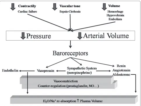
We believe the best approach to this issue is the one proposed by Schrier [1], who – based on a series of studies [2,3] – fi nally introduced a unifying hypothesis to explain the hemodynamic impairment associated with diff erent diseases or syndromes. His approach, slightly modifi ed, is presented in Figure 1, which summarizes the hemodynamic determinants, the neuro-endocrinal signal-ing and the body response. In this framework we may fi rst consider which variables are independent. Th ese variables belong to three categories: the heart (con-tractility and heart rate), the vascular tone and, fi nally, the intravascular volume. In a given disease or syndrome one or more of these variables may be aff ected. As an example, acute myo cardial infarction is paradigmatic of the problems relative to heart contractility and/or rate. A primary altera tion of the vascular tone is typical of
Abstract
Assessment and monitoring of hemodynamics is a cornerstone in critically ill patients as hemodynamic alteration may become life-threatening in a few minutes. Defi ning normal values in critically ill patients is not easy, because ‘normality’ is usually referred to healthy subjects at rest. Defi ning ‘adequate’ hemodynamics is easier, which embeds whatever pressure and fl ow set is suffi cient to maintain the aerobic metabolism. We will refer to the unifying hypothesis proposed by Schrier several years ago. Accordingly, the alteration of three independent variables – heart (contractility and rate), vascular tone and intravascular volume – may lead to underfi lling of the arterial tree, associated with reduced (as during myocardial infarction or hemorrhage) or expanded (sepsis or cirrhosis) plasma volume. The underfi lling is sensed by the arterial baroreceptors, which activate primarily the sympathetic nervous system and renin–angiotensin–aldosterone system, as well as vasopressin, to restore the arterial fi lling by increasing the vascular tone and retaining sodium and water. Under ‘normal’ conditions, therefore, the homeostatic system is not activated and water/sodium excretion, heart rate and oxygen extraction are in the range found in normal subjects. When arterial underfi lling occurs, the mechanisms are activated (sodium and water retention) – associated with low central venous oxygen saturation (ScvO2) if underfi lling is caused by low fl ow/hypovolemia, or with normal/high ScvO2 if associated with high fl ow/hypervolemia. Although the correction of hemodynamics should be towards the correction of the independent determinants, the usual therapy performed is volume infusion. An accepted target is ScvO2 >70%, although this ignores the arterial underfi lling associated with volume expansion/high fl ow. For large-volume resuscitation the worst solution is normal saline solution (chloride load, strong ion diff erence = 0, acidosis). To avoid changes in acid–base equilibrium the strong ion diff erence of the infused solution should be equal to the baseline bicarbonate concentration.
© 2010 BioMed Central Ltd
Supporting hemodynamics: what should we
target? What treatments should we use?
Luciano Gattinoni*
1,2and
Eleonora Carlesso
2R E V I E W
*Correspondence: gattinon@policlinico.mi.it
1Dipartimento di Anestesia, Rianimazione (Intensiva e Subintensiva) e Terapia del Dolore, Fondazione IRCCS Ca’ Granda – Ospedale Maggiore Policlinico, Via Francesco Sforza 35, 20122 Milan, Italy
Full list of author information is available at the end of the article
Gattinoni and Carlesso Critical Care 2013, 17(Suppl 1):S4 http://ccforum.com/content/17/S1/S4
cirrhosis and septic syn drome. A decreased intravascular volume is typical of hemorrhage. In critically ill patients more than one variable may be altered at the same time, as in sepsis where the impairment of heart contractility and the decrease of intravascular volume due to capillary leakage may be associated with the decrease of the primary artery vessel tone.
We believe that physicians approaching the hemo-dynamic status of a given patient should fi rst consider which independent determinants are more probably altered. Of interest to note, however, is that, among the primary determinants, the heart rate is the only one always assessed in clinical practice. Th e contractility measured by echocardio graphy is occasionally assessed while the vascular tone and the intravascular volume are not measured. One must note that the variables usually evaluated to assess hemo dynamics and volemia, such as pressures and fl ows, are dependent variables that, when altered, may recognize diff erent causes. Th e identifi cation of or, at least, the esti mate of which of the independent hemodynamic deter mi nants is altered makes the therapy
a logical conse quence. Unfortunately, independent of the altered variable, the fi rst intervention is usually volume replacement. A typical example is represented by hypotension following the induction of anesthesia. In this case the primary cause of hemodynamic impairment is
the pharmaco logically-induced decrease of the vessel
tone but the correction is usually performed by volume infusion.
[image:2.612.66.549.87.452.2]Th e baroreceptors, located in the carotid and aortic arch [4], sense the underfi lling of the arterial tree (which in a normal situation contains 15% of the intravascular blood volume). Th is underfi lling may be caused either by a decreased intrathoracic volume or cardiac output, as typically occurs during hemorrhage or heart failure, or by arterial vasodilation, as may occur in cirrhosis or sepsis. Interestingly, one must bear in mind that the concept of arterial tree underfi lling, which, in some way, recalls the concept of eff ective circulating blood volume [5,6], may co-exist either with hypovolemia or with hypervolemia, as most of the expanded blood volume may be confi ned in the venous tract, which acts as a capacitive reservoir.
Whatever the cause of the arterial tree underfi lling, the body response is similar and primarily consists of activa-tion, via baroreceptors, of the renin–angiotensin–aldo-sterone system and the nonosmotic release of vasopressin [7]. As shown in Figure 1 other factors may be activated, and a counter-regulation may occur. It is important, however, to realize that the primary body response is directed towards the integrity of the arterial circulation, by maximizing, through the kidneys, the reabsorption of salt and water, while increasing the arterial pressure. Th ere are several elements in favor of this unifying hypo-thesis, as recently reviewed [7]. Here it is suffi cient to say that diff erent diseases or syndromes in which underfi lling may occur, with or without plasma expansion, present increased renin, angiotensin and aldosterone levels as well as an increase of vasopressin (anti-diuretic hormone) despite a frequently associated hypo-osmolarity. In general, we believe that the hemodynamic problem and, possibly, the therapeutic interventions may be better understood if considered in this framework.
Normal hemodynamic, hemodynamic impairment and hemodynamic failure
Th e concept of normal hemodynamics is not easy to
defi ne in critically ill patients. In general, we believe that hemodynamics is adequate when the oxygen delivery to the tissues is suffi cient to maintain an aerobic meta-bolism. Th is may occur in critically ill patients at hemo-dynamic values greater than or lower than the values considered normal in healthy subjects at rest. As an example, in cases of decreased hemoglobin content and/ or its oxygen saturation, a frequent fi nding in the ICU, or if hypermetabolism is present, the cardiac output must be greater than normal to provide adequate oxygen transport. In contrast, when the metabolic requirements are reduced, as may occur in critically ill patients during deep sedation or paralysis, the aerobic metabolism may be satisfi ed with a hemodynamic set of values lower than those considered normal in healthy subjects. In other words, the normality of hemodynamics should not be judged considering the hemodynamic values per se, but instead the body response. When the body senses its hemodynamic set as adequate, the baroreceptors may be activated or not activated. If the easily measured variables such as heart rate, urinary output and sodium concentration in the urine remain in the range found in normal subjects, we may assume that the hemodynamics is normal and, obviously, adequate.
In the presence of abnormal hemodynamic values, either greater or lower than normal, the hemodynamics may be still adequate if it guarantees an aerobic metabo-lism. Th is metabolism is obtained by activating all of the homeostatic mechanisms described above. Th e hemo dy-namics then becomes inadequate (hemodynamic failure)
only when signs of anaerobic metabolism appear, despite the full activation of the mechanisms normally operating to maintain homeo stasis. Th e most recog niza ble and easy measurable output of such mecha nisms are water and sodium retention [1] as judged at least 6 hours after withdrawal of diuretic therapy (too often misused in intensive care). Th is response, at least in the early phase, should not be confused with kidney failure. On the contrary, the response may be a sign of the maximal response of a normal kidney activated by the sympathetic system and subjected to vasopressin. What is not usually realized is the speed of the change in sodium urine concentration when the system is activated [8]. Figure 2, as an example, presents the electro lyte changes during controlled hemorrhage. As shown, the kidney reacts to the blood volume decrease by retaining sodium earlier than signifi cant changes in mean arterial pressure may be detected.
If the underfi lling is caused by decreased blood volume or cardiac output, the water and sodium retention is generally associated with an increased tissue oxygen extraction, as indicated by a decrease in central venous oxygen saturation (ScvO2). Th is is, in our opinion, a reasonable surrogate of the mixed venous saturation
[9,10]. We may express ScvO2 as a function of its
determinants according to the following formula:
ScvO2 = SaO2 – (VO2/Q)×1/Hb
As shown, central venous saturation depends on the arterial oxygenation (SaO2), on the appropriate match between oxygen consumption/metabolic requirement (VO2) and on cardiac output (Q), as well as on the oxygen carrier (Hb). All determinants of oxygen transport, as well as the metabolic rate, may infl uence the central venous saturation, which is an extremely sensitive, although not specifi c, indicator of changes in respiratory function (SaO2), metabolism (VO2), cardiac output (Q) and oxygen carrier (Hb). Th e physiological meaning of ScvO2 may also be expressed as:
ScvO2 = 1 – VO2/DO2
Th is equation indicates that oxygen venous saturation refl ects the residual amount of oxygen in the venous side after consumption (VO2) of part of the oxygen delivered
(DO2). In normal conditions, at rest, the amount of
oxygen extracted from the oxygen delivered is about 25% (VO2/DO2). Th e ScvO2 is therefore around 75%. Th e
arbitrary recommended threshold of ScvO2 used in
several studies and in guidelines for sepsis treatment is 70% [11-14]. One must note, however, that ScvO2 lower than the threshold is not necessarily associated with anaerobic metabolism.
Gattinoni and Carlesso Critical Care 2013, 17(Suppl 1):S4 http://ccforum.com/content/17/S1/S4
As an example in healthy subjects during physical exercise, ScvO2 may decrease to 40% while maintaining aerobiosis, because in this condition cardiac output
remarkably increases. Th e most frequent reason for a
decrease in ScvO2 in the ICU is cardiac failure. In the framework of the unifying arterial tree underfi lling hypothesis, the asso cia tion between sodium/water reten-tion and low ScvO2 is a strong indicator of underfi lling due to low fl ow and/or hypovolemia, as typically ob-served during heart failure, hemorrhage and dehydration. In contrast, when the underfi lling is due to the arterial vasodilatation associated with volume expansion or elevated cardiac output, as in cirrhosis or in some phases of sepsis, ScvO2 may be higher than 70 to 75%. Th erefore, it is important to realize that even normal or higher than
normal ScvO2 may be associated with abnormalities of
hemodynamics, as assessed by activation of the renin– angiotensin–aldo sterone system and vasopressin release.
Considering water/sodium retention and hypo-ScvO2 or
hyper-ScvO2 together may therefore indicate whether the arterial tree underfi lling is primarily due to low heart contractility/hypovolemia or vasodilatation, respectively.
Obviously this is an oversimplifi ca tion of the problem, but we believe that this approach may provide a
reasonable framework when considering the hemo
dy-namic set in a given patient.
When all of the compensatory mechanisms are over-come, the hemodynamic failure is overt and its marker is the appearance of metabolic acidosis associated with increased plasma lactate concentration. We believe that the best approach to understand the relationship between metabolic acidosis and lactate in tissue hypoxia has been provided by Hochachka and Mommsen [15], who elegantly showed how lactate production and ATP hydrolysis are coupled and must be considered together. Here it is enough to say that the appearance of acidosis indicates the energy failure of a group of cells that, in the absence of correction, may result in cellular death after a few hours.
Figure 3 reports the impressive relationship between metabolic acidosis and mortality in a general population of critically ill patients. In contrast, elevated metabolic alkalosis, usually caused by diuretic therapy, is not
[image:4.612.69.551.89.405.2]asso-cia ted with increased mortality. Beyond pH, several
approaches have been proposed to assess the hemo-dynamic failure [16,17], such as base excess, lactate, decreased strong ion diff erence (SID), increased anion gap, increased venous to arterial diff erence in partial pressure of carbon dioxide (PCO2) and its ratio to the arterial–venous oxygen content. All these variables, however, are just diff erent facets of the same reality; that is, the perturbation of the acid–base equilibrium due to nonvolatile acid load, originating from suff ering cells (lactate) or dead cells (intracellular strong acid content is higher than plasma content). In mammalians, when metabolic acidosis starts due to the hemodynamic failure, the time for correction is limited before irreversible mitochondrial damages and cell death occurs.
Treatment target
Th e ultimate goal of the intervention on hemodynamics is to guarantee the maintenance of full aerobic meta
bo-lism. Th e hemodynamic correction may therefore be
considered a symptomatic treatment allowing buying time for the cure of the underlying disease. According to Figure 1, the most rational therapy to correct the hemo-dynamic alterations should be addressed to the correc-tion of the pathogenetic mechanisms; that is, heart contractility/rate, vascular tone or intravascular volume. In clinical practice, independent of the variable primarily altered, the fi rst intervention is usually the volume replace ment, according to the following sequence: fi rst, volume; second, cardioactive drugs; and third, blood.
Th e most popular hemodynamic target, at least in
sepsis, is to reach or maintain ScvO2 >70% [13,14]. Th is has been popularized by Rivers and colleagues’ study [12], in which the septic patients at entry had baseline ScvO2 <50%. Targeting ScvO2 of 70%, Rivers and colleagues obtained a signifi cant improvement of the survival rate. In contrast, in a previous study, we could not fi nd any diff erence in outcome in the same kind of patients with a similar target [11]. Th e baseline SvO2 of these patients, however, was 68% – remarkably diff erent from that in Rivers and colleagues’ study. We also found that targeting SvO2 of 70% was analogous to target a cardiac index of 2.5 l/minute/m2. Th e most plausible explanation for the
discre pancy between these studies is the time of
intervention: earlier in Rivers and colleagues’ study, in the emergency room; and later in our study, after admission to the ICU. However, one should note that all these studies focused on problems associated with a low
SvO2 state, ignoring the possible hemodynamic
derangements occurring in high SvO2 states.
Volume replacement
Fluid challenge
Volume replacement treatment requires assessment of the patient’s intravascular volume status (cardiac preload)
and the likelihood of responsiveness (that is, increase the stroke volume) to a fl uid challenge test. In fact, data suggest that about 50% of the critically ill patients positively respond to challenge tests [18-20]. Multiple tools have been suggested as indicators for fl uid adminis-tration, most of them as predictors of response and as targets [21]. Clinical signs, such as thirst, skin turgor, blood pressure, urine output, and so forth, are unreliable indexes of intravascular volume status. Similarly, cardiac fi lling pressures (central venous pressure (CVP) and
pulmo nary artery occlusion pressure) that have been
traditionally used to guide fl uid management are poor predictors [22]. CVP has been used for over 40 years to guide fl uid management, as an indicator of intravascular volume (values <8 cmH2O indicate hemodynamic impair-ment), even though this relationship has not been proven. Other techniques, based on echocardiography, such as left ventricular end-diastolic area, or based on thermo-dilution, such as global end-diastolic volume index, gave unsatisfying results [18].
[image:5.612.314.549.87.263.2]CVP has been used for decades as an indirect measure of left ventricle preload as it well approximates the right atrial pressure, the major determinant of right ventricle fi lling [23,24]. Moreover, changes in CVP in response to fl uid challenge tests have been used to predict volume responsiveness (target 8 to 12 cmH2O) [13]. However, there is increasing evidence that – due to a series of variables, such as venous tone, intrathoracic pressures, ventricular compliances and geometry variations, occur-ring in critically ill patients – the relationship between CVP and right ventricular end-diastolic volume is poor and that CVP (absolute or changes) does not correlate with volume responsiveness [19]. Similar prob lems were
Figure 3. Relationship between acidosis/alkalosis and mortality in critically ill patients. Relationship between acidosis/alkalosis (expressed as hydrogen ion concentration) and mortality in a general population of 754 critically ill patients (data recorded at entry to the ICU).
Gattinoni and Carlesso Critical Care 2013, 17(Suppl 1):S4 http://ccforum.com/content/17/S1/S4
encountered when referring to the pulmonary artery occlusion pressure [25,26].
During the past decades a number of dynamic tests have been used to dynamically monitor the changes in stroke volume after a maneuver that modifi es venous
return. Th ese methods have been found more reliable
and less invasive than static ones [24].
Heart–lung interaction during mechanical ventilation has been used to evaluate the variations in stroke volume, systolic pressure and pulse pressure. Pulse pressure variation estimated from the arterial waveform and stroke volume variations from pulse contour analysis and pulse oximeter plethysmographic waveform variations have been found to be reliable predictors of a positive
response to challenge tests [20]. Th ese hemodynamic
eff ects are due to the cyclic increase/decrease of intra-thoracic pressures during mechanical ventilation, aff ect-ing right and left ventricular preloads and afterloads. During insuffl ation, the increased intrathoracic pressures reduce right ventricular stroke volume and increase left ventricular stroke volume. After the blood pulmonary artery transit time (nearly two or three heart beats) even the left ventricular preload decreases with a consequent stroke volume decrease, which is at its minimum value during end expira tion. A ventilation-induced change in left ventricle stroke volume of 12 to 13% has been reported highly predictive of volume responsiveness [20].
Th ese methods, however, have some limitations,
including the use of tidal volume normalized on ideal body weight >8 ml/kg and the absence of either spontaneous respiratory activity or arrhythmias [27].
Other dynamic tests have been proposed as reliable methods to assess volume replacement responsiveness. Th ese include Doppler echocardiography to assess changes in aortic fl ow velocity and stroke volume [28,29] and changes in venocaval diameter during positive pressure ventilation estimated by echocardiography [30-33]. Th e end-expiratory occlusion test consists of the interruption of mechanical ventilation for 15 seconds to suppress the cyclic decrease of cardiac preload during insuffl ations. Th e procedure should increase cardiac preload, and an increase of 5% in cardiac output and arterial pulse pressure should predict fl uid responsiveness [34]. Finally, passive leg raising has been proposed as an auto
trans-fusion method independent of mechanical ventila tion
[35]. In conclusion, there is no gold standard clinically available to assess the volume status of the patient. How-ever, the combined use of diff erent methods may provide, in our opinion, an excellent assessment of the hemo-dynamic status.
Which fl uid?
Th ree kinds of fl uids are available: crystalloids, artifi cial colloids and albumin. Although a defi nitive indication of
the superiority of one fl uid compared with the others is still not available, the data obtained in the last few years have provided, to diff erent extents, some indications. In our opinion, however, discussion of the benefi ts/risks of the diff erent solutions only applies when large volumes are infused in a relatively short time. Modest infusion, such as 1 to 1.5 l over 24 hours, is likely to be clinically irrelevant. We may roughly divide the eff ects of the infusion into two main arms: eff ects due to the volume of the infusion, inde pendent of composition of the solution; and eff ects due to the quality of the infusion, dependent on the kind of and quantity of solutes present in the fl uid replacement.
In critically ill patients, the most general indication for large-volume resuscitation is the refi lling of the blood vessels (note that the volume infused is not necessarily proportionally distributed between the arterial and venous trees). Traditionally, we thought that to achieve the same intravascular volume the amount of crystalloids compared with colloids should be in a ratio of 3:1 [13,36].
Th e most recent large trials comparing colloids and
crystalloids, however, indicate that this fi gure must be corrected – the ratio between crystalloids and colloids, to obtain the same eff ect, being around 1.5:1 [37-39].
Th e primary eff ect of volume is to alter the acid–base status of the blood. Th is eff ect becomes clinically relevant when the extracellular fl uid dilution is in the order of 10% [40]. We investigated the genesis of acidosis induced by crystalloids in theory [41], in vitro [41,42]and in vivo [43]. In line with previous results [44-46], we found that dilutional acidosis occurs only when the three determi-nants of the acid–base status [47,48] – SID, PCO2 and total protein content – are unevenly diluted. If these
determinants are equally diluted, as during in vitro
experiments, whatever the com position of the solution used to dilute the plasma (from distilled water to normal saline), the pH does not change if the system is closed because the relative proportions between the pH deter-minants, equally diluted, are unmodifi ed. If the system in vitro is open (by tonometry) to restore the PCO2 to that before the dilution, acidosis occurs because the carbon dioxide (volatile acid load) content increases back to the predilution value while the SID and total protein content values remain diluted [41].
infusion are therefore edema diff used to the various organs [49] and disturbances of the acid–base equili-brium [40,50,51], depending on the electrolyte compo si-tion. With this background, normal saline is the worst approach for large-volume resuscitation; in fact, with the SID of the solution being equal to 0, acidosis is un-avoidable. Moreover, the chloride load and the relatively high osmolarity may increase the burden of the kidney with chloride-dependent constriction of the aff erent arterioles [52,53].
Abbreviations
CVP, central venous pressure; PCO2, partial pressure of carbon dioxide; SID, strong ion diff erence; ScvO2, central venous oxygen saturation.
Competing interests
The authors declare that they have no competing interests.
Author details
1Dipartimento di Anestesia, Rianimazione (Intensiva e Subintensiva) e Terapia del Dolore, Fondazione IRCCS Ca’ Granda – Ospedale Maggiore Policlinico, Via Francesco Sforza 35, 20122 Milan, Italy. 2Dipartimento di Fisiopatologia Medico-Chirurgica e dei Trapianti, Università degli Studi di Milano, via Francesco Sforza 35, 20122 Milano, Italy.
Declarations
This article has been published as part of Critical Care Volume 17 Suppl 1, 2013: Future of Critical Care Medicine. The supplement was proposed by Fresenius Kabi based on presentations from the ‘Future of critical care medicine (FCCM) 2012: Today’s practice and a look to the future’ symposium. Articles were commissioned by the journal, were independently prepared by the authors and have been peer reviewed by the journal. Publication of the supplement was supported by Fresenius Kabi.
Published: 12 March 2013
References
1. Schrier RW: Body fl uid volume regulation in health and disease: a unifying hypothesis.Ann Intern Med 1990, 113:155-159.
2. Schrier RW, Humphreys MH: Factors involved in antinatriuretic eff ects of acute constriction of the thoracic and abdominal inferior vena cava. Circ Res 1971, 29:479-489.
3. Bichet DG, Kortas C, Mettauer B, Manzini C, Marc-Aurele J, Rouleau JL, Schrier RW: Modulation of plasma and platelet vasopressin by cardiac function in patients with heart failure.Kidney Int 1986, 29:1188-1196.
4. Berl T, Cadnapaphornchai P, Harbottle JA, Schrier RW: Mechanism of suppression of vasopressin during alpha-adrenergic stimulation with norepinephrine.J Clin Invest 1974, 53:219-227.
5. PETERS JP: The role of sodium in the production of edema.N Engl J Med
1948, 239:353-362.
6. Rose BD: Regulation of the eff ective circulating volume. In Clinical Physiology of Acid–Base and Electrolyte Disorders. 3rd edition. New York: Mc Graw Hill; 1989:225-247.
7. Schrier RW, Niederberger M: Paradoxes of body fl uid volume regulation in health and disease. A unifying hypothesis.West J Med 1994, 161:393-408. 8. Caironi P, Langer T, Taccone P, Bruzzone P, De Chiara S, Vagginelli F, Caspani L,
Marenghi C, Gattinoni L: Kidney instant monitoring (K.IN.G): a new analyzer to monitor kidney function.Minerva Anestesiol 2010, 76:316-324.
9. Reinhart K, Rudolph T, Bredle DL, Hannemann L, Cain SM: Comparison of central-venous to mixed-venous oxygen saturation during changes in oxygen supply/demand.Chest 1989, 95:1216-1221.
10. Bloos F, Reinhart K: Venous oximetry.Intensive Care Med 2005, 31:911-913. 11. Gattinoni L, Brazzi L, Pelosi P, Latini R, Tognoni G, Pesenti A, Fumagalli R: A trial
of goal-oriented hemodynamic therapy in critically ill patients. SvO2 Collaborative Group.N Engl J Med 1995, 333:1025-1032.
12. Rivers E, Nguyen B, Havstad S, Ressler J, Muzzin A, Knoblich B, Peterson E, Tomlanovich M: Early goal-directed therapy in the treatment of severe sepsis and septic shock.N Engl J Med 2001, 345:1368-1377.
13. Dellinger RP, Levy MM, Carlet JM, Bion J, Parker MM, Jaeschke R, Reinhart K, Angus DC, Brun-Buisson C, Beale R, etal.: Surviving Sepsis Campaign: international guidelines for management of severe sepsis and septic shock: 2008.Intensive Care Med 2008, 34:17-60.
14. Levy MM, Dellinger RP, Townsend SR, Linde-Zwirble WT, Marshall JC, Bion J, Schorr C, Artigas A, Ramsay G, Beale R, Parker MM, Gerlach H, Reinhart K, Silva E, Harvey M, Regan S, Angus DC: The Surviving Sepsis Campaign: results of an international guideline-based performance improvement program targeting severe sepsis.Intensive Care Med 2010, 36:222-231.
15. Hochachka PW, Mommsen TP: Protons and anaerobiosis.Science 1983,
219:1391-1397.
16. Weil MH: Defi ning hemodynamic instability. In Functional Hemodynamic Monitoring. Edited by Edited by Vincent JL. Berlin: Springer Verlag; 2004:9-17. 17. Garcia X, Pinsky MR: Clinical applicability of functional hemodynamic
monitoring.Ann Intensive Care 2011, 1:35.
18. Michard F, Teboul JL: Predicting fl uid responsiveness in ICU patients: a critical analysis of the evidence.Chest 2002, 121:2000-2008. 19. Marik PE, Baram M, Vahid B: Does central venous pressure predict fl uid
responsiveness? A systematic review of the literature and the tale of seven mares.Chest 2008, 134:172-178.
20. Marik PE, Cavallazzi R, Vasu T, Hirani A: Dynamic changes in arterial waveform derived variables and fl uid responsiveness in mechanically ventilated patients: a systematic review of the literature.Crit Care Med
2009, 37:2642-2647.
21. Vincent JL, Weil MH: Fluid challenge revisited.Crit Care Med 2006,
34:1333-1337.
22. Osman D, Ridel C, Ray P, Monnet X, Anguel N, Richard C, Teboul JL: Cardiac fi lling pressures are not appropriate to predict hemodynamic response to volume challenge.Crit Care Med 2007, 35:64-68.
23. Weil MH, Shubin H: The ‘VIP’ approach to the bedside management of shock.JAMA 1969, 207:337-340.
24. Marik PE, Monnet X, Teboul JL: Hemodynamic parameters to guide fl uid therapy.Ann Intensive Care 2011, 1:1.
25. Pinsky MR: Clinical signifi cance of pulmonary artery occlusion pressure. Intensive Care Med 2003, 29:175-178.
26. Kumar A, Anel R, Bunnell E, Habet K, Zanotti S, Marshall S, Neumann A, Ali A, Cheang M, Kavinsky C, Parrillo JE: Pulmonary artery occlusion pressure and central venous pressure fail to predict ventricular fi lling volume, cardiac performance, or the response to volume infusion in normal subjects.Crit Care Med 2004, 32:691-699.
27. Cannesson M, Aboy M, Hofer CK, Rehman M: Pulse pressure variation: where are we today?J Clin Monit Comput 2011, 25:45-56.
28. Feissel M, Michard F, Mangin I, Ruyer O, Faller JP, Teboul JL: Respiratory changes in aortic blood velocity as an indicator of fl uid responsiveness in ventilated patients with septic shock.Chest 2001, 119:867-873.
29. Monnet X, Rienzo M, Osman D, Anguel N, Richard C, Pinsky MR, Teboul JL:
Esophageal Doppler monitoring predicts fl uid responsiveness in critically ill ventilated patients.Intensive Care Med 2005, 31:1195-1201.
30. Barbier C, Loubieres Y, Schmit C, Hayon J, Ricome JL, Jardin F, Vieillard-Baron A: Respiratory changes in inferior vena cava diameter are helpful in predicting fl uid responsiveness in ventilated septic patients.Intensive Care Med 2004, 30:1740-1746.
31. Feissel M, Michard F, Faller JP, Teboul JL: The respiratory variation in inferior vena cava diameter as a guide to fl uid therapy.Intensive Care Med 2004,
30:1834-1837.
32. Vieillard-Baron A, Augarde R, Prin S, Page B, Beauchet A, Jardin F: Infl uence of superior vena caval zone condition on cyclic changes in right ventricular outfl ow during respiratory support.Anesthesiology 2001, 95:1083-1088. 33. Vieillard-Baron A, Chergui K, Rabiller A, Peyrouset O, Page B, Beauchet A,
Jardin F: Superior vena caval collapsibility as a gauge of volume status in ventilated septic patients.Intensive Care Med 2004, 30:1734-1739. 34. Monnet X, Osman D, Ridel C, Lamia B, Richard C, Teboul JL: Predicting
volume responsiveness by using the end-expiratory occlusion in mechanically ventilated intensive care unit patients.Crit Care Med 2009,
37:951-956.
35. Cavallaro F, Sandroni C, Marano C, La Torre G, Mannocci A, De Waure C, Bello G, Maviglia R, Antonelli M: Diagnostic accuracy of passive leg raising for prediction of fl uid responsiveness in adults: systematic review and meta-analysis of clinical studies.Intensive Care Med 2010, 36:1475-1483. 36. Puyana JC: Resuscitation of hypovolemic shock. In Textbook of Critical Care.
5th edition. Edited by Edited by Fink M, Abraham E, Vincent JL, Kochanek PM.
Gattinoni and Carlesso Critical Care 2013, 17(Suppl 1):S4 http://ccforum.com/content/17/S1/S4
Philadelphia, PA: Saunders; 2005:1939-1940.
37. Finfer S, Bellomo R, Boyce N, French J, Myburgh J, Norton R: A comparison of albumin and saline for fl uid resuscitation in the intensive care unit.N Engl J Med 2004, 350:2247-2256.
38. Brunkhorst FM, Engel C, Bloos F, Meier-Hellmann A, Ragaller M, Weiler N, Moerer O, Gruendling M, Oppert M, Grond S, Olthoff D, Jaschinski U, John S, Rossaint R, Welte T, Schaefer M, Kern P, Kuhnt E, Kiehntopf M, Hartog C, Natanson C, Loeffl er M, Reinhart K; German Competence Network Sepsis (SepNet): Intensive insulin therapy and pentastarch resuscitation in severe sepsis.N Engl J Med 2008, 358:125-139.
39. Upadhyay M, Singhi S, Murlidharan J, Kaur N, Majumdar S: Randomized evaluation of fl uid resuscitation with crystalloid (saline) and colloid (polymer from degraded gelatin in saline) in pediatric septic shock.Indian Pediatr 2005, 42:223-231.
40. Scheingraber S, Rehm M, Sehmisch C, Finsterer U: Rapid saline infusion produces hyperchloremic acidosis in patients undergoing gynecologic surgery.Anesthesiology 1999, 90:1265-1270.
41. Gattinoni L, Carlesso E, Maiocchi G, Polli F, Cadringher P: Dilutional acidosis: where do the protons come from?Intensive Care Med 2009, 35:2033-2043. 42. Carlesso E, Maiocchi G, Tallarini F, Polli F, Valenza F, Cadringher P, Gattinoni L:
The rule regulating pH changes during crystalloid infusion.Intensive Care Med 2011, 37:461-468.
43. Langer T, Carlesso E, Protti A, Monti M, Comini B, Zani L, Andreis DT, Iapichino GE, Dondossola D, Caironi P etal.: In vivo conditioning of acid–base equilibrium by crystalloid solutions: an experimental study on pigs. Intensive Care Med 2012, 38:686-693.
44. Morgan TJ, Venkatesh B, Hall J: Crystalloid strong ion diff erence determines metabolic acid-base change during in vitro hemodilution.Crit Care Med
2002, 30:157-160.
45. Morgan TJ, Venkatesh B, Hall J: Crystalloid strong ion diff erence determines metabolic acid–base change during acute normovolaemic haemodilution. Intensive Care Med 2004, 30:1432-1437.
46. Morgan TJ, Vellaichamy M, Cowley DM, Weier SL, Venkatesh B, Jones MA:
Equivalent metabolic acidosis with four colloids and saline on ex vivo haemodilution.Anaesth Intensive Care 2009, 37:407-414.
47. Stewart PA: How to Understand Acid–Base. A Quantitative Acid–Base Primer for Biology and Medicine. New York: Elsevier; 1981.
48. Stewart PA: Stewart’s Textbook of Acid–Base. Amsterdam: Lulu.com; 2009. 49. Hartog CS, Bauer M, Reinhart K: The effi cacy and safety of colloid
resuscitation in the critically ill.Anesth Analg 2011, 112:156-164. 50. Williams EL, Hildebrand KL, McCormick SA, Bedel MJ: The eff ect of
intravenous lactated Ringer’s solution versus 0.9% sodium chloride solution on serum osmolality in human volunteers.Anesth Analg 1999,
88:999-1003.
51. O’Malley CM, Frumento RJ, Hardy MA, Benvenisty AI, Brentjens TE, Mercer JS, nett-Guerrero E: A randomized, double-blind comparison of lactated Ringer’s solution and 0.9% NaCl during renal transplantation.Anesth Analg
2005, 100:1518-1524.
52. Hansen PB, Jensen BL, Skott O: Chloride regulates aff erent arteriolar contraction in response to depolarization.Hypertension 1998,
32:1066-1070.
53. Wilcox CS: Regulation of renal blood fl ow by plasma chloride.J Clin Invest
1983, 71:726-735.
doi:10.1186/cc11502
Cite this article as: Gattinoni L, Carlesso E: Supporting hemodynamics: what should we target? What treatments should we use?Critical Care 2013,