en
t
re
v
ie
w
s
re
ports
de
p
o
si
te
d r
e
sea
rch
refer
e
e
d
re
sear
ch
interacti
o
ns
inf
ormation
Dirk Nagorsen
*‡
, Sara Deola
*
, Kina Smith
*
, Ena Wang
*
, Vladia Monsurro
*
,
Paola Zanovello
†
, Francesco M Marincola
*
and Monica C Panelli
*
Addresses: *Immunogenetics Section, Department of Transfusion Medicine, Clinical Center, National Institutes of Health, Bethesda, MD 20892-1502, USA. †Department of Oncology and Surgical Sciences, Oncology Section, University of Padova, Padova 35100, Italy. ‡Current
address: Charité - Universitätsmedizin Berlin, Campus Benjamin Franklin, Medizinische Klinik III, Hindenburgdamm 30, 12200 Berlin, Germany.
Correspondence: Monica C Panelli. E-mail: [email protected]
© 2005 Nagorsen et al.; licensee BioMed Central Ltd.
This is an Open Access article distributed under the terms of the Creative Commons Attribution License (http://creativecommons.org/licenses/by/2.0), which permits unrestricted use, distribution, and reproduction in any medium, provided the original work is properly cited.
Abstract
Background: Mononuclear phagocytes (MPs) stand at the crossroads between the induction of acute inflammation to recruit and activate immune effector cells and the downmodulation of the inflammatory process to contain collateral damage. This decision is extensively modulated by the cytokine microenvironment, which includes a broad array of cytokines whose direct effect on MPs remains largely unexplored. Therefore, we tested whether polarized responses of MPs to pathogens are related to the influence of selected cytokines or represent a mandatory molecular switch through which most cytokines operate.
Results: Circulating CD14+ MPs were exposed to bacterial lipopolysaccharide (LPS) followed by
exposure to an array of cytokines, chemokines and soluble factors involved in the immune response. Gene expression was studied by global transcript analysis. Two main classes of cytokines were identified that induced a classical or an alternative pathway of MP activation. Expression of
genes affected by NFκB activation was most predictive of the two main classes, suggesting that this
pathway is a fundamental target of cytokine regulation. As LPS itself induces a classical type of activation, the most dramatic modulation was observed toward the alternative pathway, suggesting that a broad array of cytokines may counteract the pro-inflammatory effects of bacterial components.
Conclusions: This analysis is directly informative of the primary effect of individual cytokines on the early stages of LPS stimulation and, therefore, may be most informative of the way MP maturation may be polarized at the early stages of the immune response.
Background
Resident and recruited mononuclear phagocytes (MPs) dis-play a versatile phenotype that reflects the plasticity of these cells in response to microenvironmental signals. This hetero-geneity spans a continuous spectrum that can be polarized into two extremes recently described by Mantovani et al. [1].
Pathogen stimulation exemplified, for instance, by lipopoly-saccharide (LPS) stimulation in the presence of interferon (IFN)-γ induces M1 MPs through engagement of Toll-like receptors (TLRs). M1 MPs are true antigen-presenting cells capable not only of killing invading organisms but of concom-itantly recruiting and activating immune effector cells [2,3]. Published: 21 January 2005
Genome Biology 2005, 6:R15
Received: 5 October 2004 Revised: 1 December 2004 Accepted: 22 December 2004 The electronic version of this article is the complete one and can be
phage activation accounts for a distinct phenotype with a key role in humoral immunity and tissue repair [5].
It has been suggested that the extreme dichotomy between a classical M1 and an alternative M2 polarization of macro-phage function may not take into account intermediate regu-lation by cytokines such as IL-10, transforming growth factors (TGF-α and TGF-β), macrophage colony-stimulating factor (M-CSF), IFN-α and IFN-β and tumor necrosis factor (TNF) [5]. Most important, a rigid dichotomy in MP function may not directly apply to physiological and/or pathological conditions in which these cells are exposed to an array of cytokines produced by innate or adaptive immune mecha-nisms during infection, tissue damage or in other conditions. Indeed, a comprehensive overview of the modulatory proper-ties of cytokines on the MP reaction to a pathogen is missing. In addition, little is known about the transcriptional changes occurring in MPs on exposure to pathogen components such as LPS.
A recent study analyzed the transcriptional profile induced by the exposure of circulating MP conditioned in vitro for 7 days with IL-4 and GM-CSF (immature dendritic cell, (DC)) to pathogen components [6]. Bacterial, viral and fungal compo-nents elicited distinctive pathways that were, however, largely overlapping. The predominant response of these DCs to most pathogen components encompassed a rapid upregu-lation of genes associated with the innate arm of the immune response followed by induction of adaptive immune response genes.
The response of circulating MP-derived DCs is short-lived as these cells can exhaust their production of effector molecules (cytokines and chemokines) within a few hours of LPS stimu-lation [7]. The transience of mRNA and protein expression can cause DCs to redirect the immune response in different ways at different time points. For instance, soon after stimu-lation, DCs elicit T-cell responses of the Th-1 type, whereas at later stage of activation they prime T-cell responses of the Th-2 type, suggesting that their function is strongly dependent on the timing and duration of exposure to individual and/or combined stimulatory conditions in the surrounding micro-environment. At the transcriptional level, the dual function of DCs shifted from an early pre-inflammatory phase occurring within 3 hours to a later regulatory phase occurring approxi-mately 8 hours following LPS exposure. During these evolv-ing stages of DC activation, cytokines play a dominant role in shaping the function of DCs and other immune cells, provid-ing a malleable link between the innate and adaptive immune responses [8]. In natural conditions, circulating or resident
non-conditioned circulating CD14+ MP to LPS. The results
suggested that these cells respond to LPS similarly to imma-ture CD14- DCs, with a surge in transcriptional activity that
peaks around 3 hours after stimulation and in which the acti-vation of genes associated with a classical actiacti-vation of innate immune mechanisms predominates [6].
The stringent dichotomy describing a classical activation of MPs into mature antigen-presenting cells caused by IFN-γ
and an alternative induction into macrophages induced by IL-4 and IL-13 may not apply to physiological conditions in which the microenvironment responds to pathogen exposure with a broad array of cytokine secretion. We therefore inves-tigated whether polarized responses of MPs to pathogens are extreme behaviors that can be observed in vitro by studying a few illustrative cytokines or whether they represent a manda-tory molecular switch through which most cytokines operate. Thus, we stimulated non-conditioned CD14+ MPs with LPS
for 1 hour. The MPs were then exposed to an array of different cytokines that may be expressed in distinct pathologic condi-tions by different immune-cell subsets. The 1-hour interval was empirically selected to induce a biphasic model in which the presumed modulation by cytokines occurred during an ongoing reaction to LPS. This allowed mapping of cytokines into conditional subclasses based on their effects on the glo-bal transcriptional changes responsible for MP activation and differentiation.
comm
en
t
re
v
ie
w
s
re
ports
refer
e
e
d
re
sear
ch
de
p
o
si
te
d r
e
se
a
rch
interacti
o
ns
inf
o
rmation
Results
Effect of LPS stimulation on circulating CD14+ MPs
Enriched MP preparations (about 90% CD14+) were exposed
to LPS. Total RNA extracts were obtained 4 and 9 hours after. These time points were selected to catch salient stages of the biphasic response of MP to LPS described by others [6,7]. Amplified antisense RNA (aRNA) [9] was hybridized to a cus-tom-made 17,000 (17K)-clone cDNA microarray chip enriched with genes relevant to immune function. The tran-scriptional profile of LPS-induced MPs was similar to that described by others in DCs [6]. In particular, genes associated with the innate response of CD14-, immature DCs to pathogen
components [6,10] were similarly upregulated in CD14+ MPs
(data not shown). This finding suggests that the differential expression of the LPS co-receptor CD14 between the two cell populations has a relatively minor impact on the transcrip-tional regulation of the innate immune response [11].
Kinetics of the response of CD14+ MPs to LPS and its
modulation by cytokines
Aliquots of CD14+ MPs were stimulated in parallel with LPS
and exposed 1 hour later to individual cytokines selected from a library of recombinant proteins possibly relevant to MP reg-ulation. MPs were kept in culture for 4 and 9 hours, at which times aRNA was prepared for transcriptional analysis. Unsu-pervised Eisen's clustering [12] was applied to the complete dataset (Figure 1). The kinetics of the response to LPS had the
greatest influence on the global transcriptional profile of MP induction; samples preferentially clustered according to time of stimulation rather than type of treatment. This was under-lined by the observation that MPs stimulated with LPS alone clustered with the cytokine-stimulated MPs according to the time elapsed after stimulation. In addition, a cluster contain-ing most of the samples obtained 9 hours after stimulation (9') included three control samples consisting of CD14+ MPs
not exposed to LPS or cytokines (no stimulation). These three samples were prepared at times 0, 4 and 9 hours to parallel the culture conditions used for stimulation. This finding sug-gested that the transcriptional profile of CD14+ MPs 9 hours
after LPS stimulation and 8 hours after treatment with most cytokines converges toward a less reactive metabolic state closer to that of unstimulated MPs. Several cytokines, how-ever, maintained a more active metabolic profile and after 9 hours retained a transcriptional footprint relatively close to that of samples treated for 4 hours (9"). This group included the genes for most IFN-α isoforms, IFN-β, vascular endothe-lial growth factor (VEGF), FLT-3 ligand, TGF-α, the chemok-ine RANTES (CCL5), IL-2, IL-4, IL-15 and the chemokchemok-ines MIP1α (CCL3) and MIP1β (CCL4), suggesting that these cytokines may have relatively prolonged kinetics of MP activation.
The average number of genes whose expression was increased compared to unstimulated MPs was higher (420 genes) after 4 hours than after 9 hours (265 genes). About half of the genes upregulated after 4 hours remained upregulated at 9 hours (223 genes). This is in accordance with Huang's observation [6] that transcriptional changes in DCs occur predominantly in the early phase of the response to pathogen components, with about half the genes displaying transitory expression and the other half sustained expression. For this reason, sub-sequent analyses were limited to the 4-hour time point.
Transcriptional modulation by cytokines 4 hours after LPS stimulation of CD14+ MPs
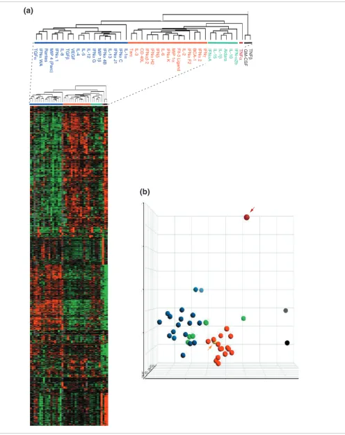
Although LPS alone strongly affected the transcriptional pro-gram of MPs, it was possible to discern the contribution of individual cytokines. This analysis was limited to samples treated for 4 hours, when the most dramatic effects on gene modulation were noted. Genes differentially expressed in samples treated with cytokines compared to those treated with LPS alone were identified. Stringent criteria were applied to select genes expressed in at least 80% of samples with a threefold or greater increase or reduction in expression over LPS in at least one of the cytokine-treated samples. This gave 2,057 genes that were deemed most relevant to the anal-ysis. Using these genes, all cytokine-treated samples were subjected to unsupervised clustering to evaluate their related-ness (Figure 2a).
Two main classes of cytokines were identified. One class (Fig-ure 2a, blue horizontal line) included IL-4, IL-13 TGF-α, TGF-β and VEGF, which are unquestionably associated with
Unsupervised clustering of LPS-stimulated CD14+ MPs exposed to distinct
cytokine treatments
Figure 1
Unsupervised clustering of LPS-stimulated CD14+ MPs exposed to distinct cytokine treatments. CD14+ MPs were stimulated in parallel with LPS and exposed after 1 h to 42 individual cytokines (see Table 1 for cytokines used). Antisense RNA obtained 4 and 9 h following LPS stimulation was hybridized to custom-made 17K cDNA arrays. Unsupervised Eisen clustering [12] was applied to the complete, unfiltered dataset of 98 experiments. Arrowheads represent control samples that did not receive any stimulation and were obtained at different time points to parallel culture conditions during stimulation (0, 4 and 9 h). Light blue and gold represent samples obtained 4 and 9 h after stimulation, respectively and black represents no stimulation. A375 is a melanoma cell line that was used for quality control alternating conventional (Cy5, red) or reciprocal (Cy3, green) labeling every 25 experiments as previously described [9]. Experiments cluster closer together according to time rather than type of stimulation, with samples obtained after 4 h clustering together (4') with the exception of few cytokines (4"). With few exceptions (9"), cytokine treatments at 9 h clustered together with non-stimulated MPs (9').
4 h 9 h
No stimulation
Figure 2 (see legend on next page)
TNF
α
TNF
β
GM-CSF
IFN-α
2b
IL-10
Aldara
IL-1
β
IL-15
IFN
α
A
IFN
γ
IFN
α
2
BCA-1
IFN
α
F2
IL-2
Flt-3 Ligand
MIP 1
α
IFN
α
K
IL-6
IFN
β
IFN
α
H2
IFN
αβ
2
CD 40L
IL-3
Tarc
IL-1
α
IFN
α
C
IFN
α
J1
IL-13
IFN
α
4B
MIP 1
β
IFN
α
G
IL-12
IL-5
IL-4
VEGF
TGF
β
IL-8
IFN
α
1
MIP 4 (Parc)
Rantes
IFN
α
WA
TGF
α
comm
en
t
re
v
ie
w
s
re
ports
refer
e
e
d
re
sear
ch
de
p
o
si
te
d r
e
se
a
rch
interacti
o
ns
inf
o
rmation
the alternative pathway of MP activation [1,5]. The second class (Figure 2a, red line) included IFN-α2, IFN-β, IFN-γ, CD40 ligand (CD40L), and FLT-3 ligand, which are generally associated with the classical pathway of MP activation [2,3]. Therefore, we considered the first cluster representative of the alternative and the second of the classical pathway of MP activation in response to LPS. As predicted by Gordon [5], few cytokines (Figure 2a, green line) did not properly belong to either class. These included IL-10, IL-1β, IL-15 and two
IFN-α isoforms. Because several of these cytokines have previ-ously been associated with the alternative pathway, we referred to this class as alternative II. Not surprisingly [5], MPs stimulated with TNF-α (Figure 2a, purple line) and, most dramatically, TNF-β and GM-CSF (Figure 2a, black line) had a totally independent effect on the transcriptional regula-tion of LPS-induced CD14+ MPs [5].
Cytokines classified according to the previous groups were tested for class prediction by applying unsupervised principal component analysis (PCA) to the global, unfiltered 17K gene dataset (Figure 2b). This analysis independently classified cytokines in two groups corresponding to the alternative (Fig-ure 2b, blue circles) and classical (Fig(Fig-ure 2b, red circles) cytokine classes. Most of the cytokines belonging to the alter-native II class (Figure 2b, green circles) grouped with the alternative group, whereas TNF-α (Figure 2b, purple circle and arrow), TNF-β and GM-CSF (Figure 2b, gray circles) remained separate. The sample treated with LPS alone (Fig-ure 2b, yellow circle and arrow) grouped with the classical cytokines, confirming the predominant pro-inflammatory effects of this bacterial product and its alignment with the classical pathway of MP activation [1,11]. This finding based on the complete dataset indicates the intrinsic bias of this study aimed at exploring the alternative modulation of the MP response to LPS.
Particular mention should be made of the erratic behavior of various IFN-α subtypes, which clustered indiscriminately between the two main cytokine classes. Interestingly, how-ever, alignment of the IFN-α protein sequences through the EMBL-EBI Clustal W database identified, with the exception of IFN-αG, a close relationship among the IFN-α subtypes that clustered with the alternative type of cytokines (data not shown). This subclassification was also supported by the
phy-logenetic relationship among interferons described by Henco [13]. This information suggests that specific domains of the IFN-α molecules may have dramatically different effects in the modulation of the MP response to pathogen [14,15]. Inter-estingly, IFN-α2, which is the one most commonly used in
clinical trials as a pro-inflammatory cytokine, clustered with the classical cytokines adjacent to IFN-γ.
Cytokine-mediated modulation of LPS-stimulated CD14+ MPs predominantly affects pathways
downstream of NFκB
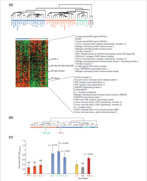
Signatures associated with several pathways of immune-cell activation were constructed by selecting genes from the global pool of 17K clones according to literature information without pre-existing information about the association of their expression to either class of cytokines. Signature genes were then subjected to supervised clustering according to the cytokine classification shown in Figure 2a. This independent process identified virtual signatures, in some cases portraying opposite transcriptional regulation by the two classes. The signature that most strongly discriminated the two classes comprised 121 genes whose expression is closely dependent on NFκB modulation [11] (Figure 3a). This is not surprising as LPS acts through engagement of Toll receptor 4 (TLR4) and CD14, with resulting activation of NFκB [3]. It would, there-fore, seem intuitive that the strongest modulation in the present experimental conditions would target this pathway. In particular, several TNF- and IL-1-related genes classically modulated by NFκB during the acute phases of the innate immune response [11] were strongly and inversely modulated by the two cytokine classes.
The same 121 genes were used for unsupervised class predic-tion by reclustering cytokine-treated samples (Figure 3b). This independent analysis segregated cytokines into two classes that with the exception of one (IFN-α2b) matched the respective original classical and alternative classification (Figure 3a, red and blue horizontal bars, respectively). Inter-estingly, the cytokines that belonged to the alternative II class clustered with the alternative cytokines (Figure 3a, green hor-izontal bars) while TNF-α (Figure 3a, purple horizontal bar), TNF-β, and GM-CSF (Figure 3a, dark gray horizontal bar) clustered separately but in proximity of the classical group. Analysis of early signaling events occurring 1 hour and 30
Definition of cytokine classes based on their modulatory effect on the response of CD14+ circulating MP to LPS
Figure 2 (see previous page)
Figure 3 (see legend on next page)
Activated p50
(OD
450nm
)
LPS No
stimulation TNF-α
NS NS NS
p = 0.049
p = 0.001 p = 0.012
NS p = 0.034
IFN-α2β TNF-α
TRAF 6
NFκB2=NFκBp100/p50B
NFκBp105/p50
NFκBp65
TNF
β
GM-CSF
TNF
α
IFN-α
2b
IL-10
Aldara
IL-1
β
IL-15
IFN
α
A
IIFN
γ
IFN
α
2
BCA-1
IFN
α
F2
IL-2
Flt-3 Ligand
MIP 1
α
IFN
α
K
IL-6
IFN
β
IFN
α
H2
IFN
αβ
2
CD 40L
IL-3
Tarc
IL-1
α
IFN
α
C
IFN
α
J1
IL-13
IFN
α
4B
MIP 1
β
IFN
α
G
IL-12
IL-5
IL-4
VEGF
TGF
β
IL-8
IFN
α
1
MIP 4 (Parc)
Rantes
IFN
α
WA
TGF
α
Tweak=Apo3/DR3 ligand (APO3L) ECSIT
Tweak=Apo3/DR3 ligand (APO3L)
Tumor necrosis factor (ligand) superfamily, member 13 Mitogen-activated protein kinase kinase 5
Mitogen-activated protein kinase 3=erk1 Toll-like receptor 7
EST, Weakly similar to A57034 transcription factor NF-kappa-B2, CD30=Ki-1 antigen=TNFR family member
Tumor necrosis factor receptor superfamily, member 12
Mitogen-activated protein kinase kinase kinase 7 interacting protein 1 Toll-like receptor 7
4-1BB ligand=TNF family member Fas (TNFRSF6) associated factor 1
Mitogen-activated protein kinase kinase kinase
Toll-like receptor 2
C1q and tumor necrosis factor related protein 6 TNF receptor-associated factor 3
TNF receptor-associated factor 3 TNFAIP3 interacting protein 2 FADD=MORT
IL-1 receptor antagonist
Mitogen-activated protein kinase kinase kinase 5=MEKK5 TNFAIP3 interacting protein 1
TRAF and TNF receptor-associated protein Tumor necrosis factor (TNF superfamily, member 2) Tumor necrosis factor (TNF superfamily, member 2) IL-1 receptor type I
TRAF1=Epstein-Barr virus-induced protein EBI6 Tumor necrosis factor, alpha-induced protein 6
0 0.2 0.4 0.6 0.8 1 1.2 1.4
IFN-γ IL-6 IL-3 IL-4 IL-13 IL-1α
(b)
comm
en
t
re
v
ie
w
s
re
ports
refer
e
e
d
re
sear
ch
de
p
o
si
te
d r
e
se
a
rch
interacti
o
ns
inf
o
rmation
minutes after LPS stimulation and, therefore, 30 minutes after the additional cytokine exposure, demonstrated significantly increased levels of the free p50 subunit of NFκB in MP whole-cell extracts treated with alternative class cytokines (IL-4 and IL-13). In addition, IL-1α and TNF-α sig-nificantly upregulated p50, whereas no significant changes were caused by classical cytokines (IFN-γ, IL-6 and IL-3). Extracts from MPs stimulated only with LPS also failed to demonstrate changes in NFκB subunit release; this is proba-bly related to the 90-minute period from stimulation that allowed a return of signaling molecules to baseline conditions (Figure 3c). The similarity of the IL-1α and TNF-α effects on p50 to those of alternative cytokines contrasts with the dra-matic differences observed on the respective transcriptional profiles, suggesting that other pathways induced by these cytokines may prevail in the conditions tested here.
Among the additional pathways tested, those mediated through STATs, Janus kinases (JAKs), and interferon regula-tory factor (IRF) did not appear consequential to the experi-mental conditions tested in this study (data not shown).
Cytokine-mediated modulation of metalloproteinase expression in LPS-stimulated CD14+ MPs
Matrix metalloproteinases (MMP) are tightly connected to MP activation. MMP released by MPs contribute to normal and pathological tissue remodeling and MP migration. In addition MMP function as regulatory proteins by promoting the activation or degradation of cytokines. Finally, MMP are susceptible to cytokine stimulation. Thirty-six MMP and MMP-related genes (filtered from a larger group of 184 MMPs, disintegrins, α-defensins, TGF-β, TNF-α, insulin-like growth factor 1 (IGF-1), epidermal growth factor (EGF), fibroblast growth factor (FGF), IL-1 and monocyte chemotac-tic protein-3 (CCL7/MCP-3) genes) [16,17] were clustered according to the alternative and classical group denomination and the 11 most representative are shown in Figure 4. The alternative group of cytokines induced the transcription of MMPs (MMP 7, 9, 10, 19), enzymes related to MMP function (disintegrin ADAM 9, pro-collagen proline dioxygenase, MEK1 kinase, serine protease inhibitor, cathepsin L) and structural proteins (gap junction connexin 26, laminin A/C). This confirms the role of alternative MP activation in promot-ing tissue remodelpromot-ing, cell-cell interactions and local control of the inflammatory process through activation (via MMP-7, 9) [18-21] or degradation (via MMP-19) of cytokine activity [17,22]. In addition, these observations suggest a role for the
cytokines in the alternative group (other than the well docu-mented IL-4 or IL-13) in polarizing MPs toward an M2 regu-latory phenotype.
Characterization of the alternative and classical groups of cytokines
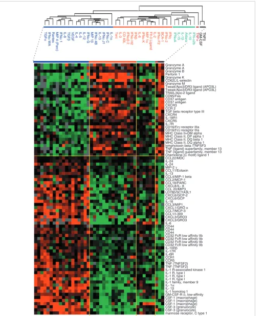
Comparison of gene-expression patterns induced by the two cytokine classes (classical and alternative) identified 2,007 genes that were differentially expressed at a less than 0.001 significance level (t-test, p2-value). Genes associated with immune function were proportionally over-represented. A selection of genes relevant to MP function is shown in Figure 5. In most cases, the pattern of expression echoed the class allocation suggested by the literature [23]. Classical cytokines induced genes responsible for the cytotoxic and migratory properties of MPs such as those for CD95, TRAIL, granzymes, perforin, CD16 (stimulatory Fcγ receptor) and CD62L. In addition, antigen presentation was enhanced as suggested by the coordinate expression of several HLA class II genes.
IFN-γ and the other classical cytokines inhibited the expression of the macrophage-derived chemokine CCL22/MDC, while upregulating the expression of CXCR3, as previously reported [23].
Alternative cytokines induced the expression of several cytokines and their respective receptors involved in the chem-otaxis and activation of neutrophils, MPs, natural killer (NK) cells, DCs, helper T lymphocytes (Th2) and B lymphocytes. Several genes known to be associated with alternative MP activation [5,23] were consistently upregulated. These included the mannose receptor, the inhibitory Fc-IIb receptor CD32, and the cell-surface molecule CD44 [24], which is associated with the disposal of inflammatory cell corpses without expansion of the inflammatory process. Several inducible chemokines were expressed in response to alterna-tive stimulation such as CCL22/MDC, supporting the emerg-ing role of this cytokine as an enhancer of polarized Th2 responses [25]. Other chemokines known to be induced by master type II cytokines and associated with the induction of Th2 responses were also induced by alternative activation; these included CCL11 (eotaxin), CCL1 (I-309), CCL2 (MCP-1) and CCL7 (MCP-3) [23]. Of interest was the relatively higher expression of IL-24, a cytokine belonging to the IL-10 family constitutively expressed by MPs [26]. Although the true role of this cytokine in inflammatory processes is not known, it is likely that its pro-apoptotic and angioregulatory properties have important roles in tissue repair and remodeling during
Effect of cytokines on the expression of genes dependent on NFκB activation
Figure 3 (see previous page)
inflammation. Finally, various chemokine receptors respon-sible for MP trafficking and localization were differentially regulated by the two cytokine groups, including in particular CCR1 and CCR5, which were induced by alternative stimula-tion, and CCR2, induced by classical stimulation.
This early transcriptional profile underlines the primary role of MPs during the acute phases of the response to pathogen as effector cells that can kill pathogens, take up antigen and migrate to local regional lymph nodes to recruit adaptive immune responses (classical activation). In contrast, alterna-tive cytokines may be produced in the microenvironment to maintain a resident MP phenotype rich in chemokine produc-tion, which can attract Th2-type immune responses while continuing pathogen clearance through retention of phago-cyte properties and promoting tissue remodeling.
Surprisingly, genes of the IL-1 family and its receptors, TNF and IL-6 were consistently upregulated by the alternative
class of cytokines, suggesting that LPS alters MP polarization with regard to these cytokines [5,10]. Particularly interesting is the alternative induction of IL-6 and its receptor, which may play a central role in mediating the transition from neu-trophils to MP recruitment during progression from acute to chronic inflammation [27].
Validation of microarray analysis by TaqMan real-time PCR
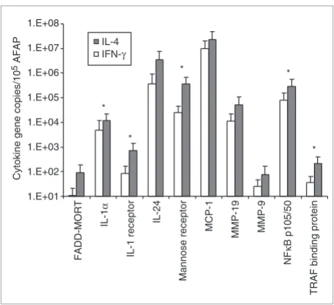
[image:8.612.56.555.86.304.2]To define the validity and accuracy of our global microarray analysis, quantitative TaqMan real-time PCR was performed on amplified RNA material isolated after stimulation of monocytes obtained from five additional normal donors with representative cytokines/soluble factors selected from the alternative and from the classical groups. Comparison of monocyte stimulation with a candidate cytokine from the alternative/M2 (IL-4) and the classical/M1 (IFN-γ) group, respectively, is shown in Figure 6. Ten genes whose expression Effect of cytokines on the expression of genes for matrix metalloproteinases (MMPs) and MMP-related genes
Figure 4
Effect of cytokines on the expression of genes for matrix metalloproteinases (MMPs) and MMP-related genes. MMP genes and MMP-related genes (such as disintegrins, IFN-γ and related genes, IFN-α, α-defensins, TGF-β, TNF-α, IGF-1, EGF, FGF, IL-1, MCP-3 - complete list available from the authors on request) were clustered according to the alternative and classical groups and filtered for 70% presence and at least one value equal to or greater than a 1.5-fold change in expression (a total of 36 genes). The clusterogram displays 11 of the most representative genes. Color coding is as Figure 2.
TNF
α
TNF
β
GM-CSF
IFN-α
2b
IL-10
Aldara
IL-1
β
IL-15
IFN
α
A
IFN
γ
IFN
α
2
BCA-1
IFN
α
F2
IL-2
Flt-3 Ligand
MIP 1
α
IFN
α
K
IL-6
IFN
β
IFN
α
H2
IFN
αβ
2
CD 40L
IL-3
Tarc
IL-1
α
IFN
α
C
IFN
α
J1
IL-13
IFN
α
4B
MIP 1
β
IFN
α
G
IL-12
IL-5
IL-4
VEGF
TGF
β
IL-8
IFN
α
1
MIP 4 (Parc)
Rantes
IFN
α
WA
TGF
α
Lamin A/C MMP 7 matrilysin MMP10 stromelysin 2
Serine protease inhibitor, Kazal type 1 Cathepsin L
Gap junction protein beta2 ( connexin 26) MMP19
MMP 9 gelatinase B type IV collagenase
Disintegrin and MMP domain 9 ( meltrin gamma=MDC9) MEK1 =Mitogen activated protein Kinase1
Procollagen proline, proline 4 dioxygenase
Selection of genes differentially expressed between classically and alternatively activated MPs based on previous annotations linking their function to MP activation
Figure 5 (see following page)
comm en t re v ie w s re ports refer e e d re sear ch de p o si te d r e se a rch interacti o ns inf o rmation
Figure 5 (see legend on previous page)
Granzyme A Granzyme A Granzyme B Perforin 1 Granzyme K CD62L/L-selectin Granzyme M
Tweak/Apo3/DR3 ligand (APO3L) Tweak/Apo3/DR3 ligand (APO3L) TRAIL/Apo-2 ligand CD95/Fas CD37 antigen CD37 antigen CXCR3 CCR 2
TGF beta receptor type III CXCR4
IL-18R1 CXCR5 IL-7R
CD16/Fcγ receptor IIIa
CD16/Fcγ receptor IIIa MHC Class II=DM alpha MHC Class II, DP alpha 1 MHC Class II, DQ beta 1 MHC Class II, DQ alpha 1 lymphotoxin beta /TNFSF3 TNF (ligand) superfamily, member 13 TNF (ligand) superfamily, member 13 Chemokine (C motif) ligand 1 CCL22/MDC
IL-24 IL-24
MIP-2 γ
CCL11/Eotaxin IL-13 CCL4/MIP-1 beta CCL2/MCP-1 CCL18/PARC CXCL8/IL- 8 CCL 20/MIP3_
LD78β/SCYA3L1
CXCL6/GCP-2 CXCL6/GCP IL-16 CCL3/MIP1_
CXCL1/GRO α
CCL7/MCP-3 CCL1/I-309 CXCL3/GRO3 CXCL3/GRO3 IL-6 CD44 CD44 CD44
CD32 FcR low affinity IIb CD32 FcR low affinity IIb CD32 FcR low affinity IIb CD32 FcR low affinity IIb
IL-10Rβ IL-17R IL-6R CCR1 CCR5 TNF (TNFSF2) TNF (TNFSF2)
IL-1 R-associated kinase 1 IL-1 R, type I
IL-1 R, type I IL-1 R, type I IL-1 family, member 9
IL- 1α
IL- 1β
IL-1 homolog 1
GM-CSF-R α, low-affinity
CSF-1 (macrophage) CSF-1 (macrophage) CSF-1 (macrophage) CSF-3 (granulocyte) CSF-3 (granulocyte) mannose receptor, C type 1
TNF α TNF β GM-CSF IFN-α 2b IL-10 Aldara IL-1 β IL-15 IFN α A IFN γ IFN α 2 BCA-1 IFN α F2 IL-2 Flt-3 Ligand MIP 1 α IFN α K IL-6 IFN β IFN α H2 IFN αβ 2 CD 40L IL-3 Tarc IL-1 α IFN α C IFN α J1 IL-13 IFN α 4B MIP 1 β IFN α G IL-12 IL-5 IL-4 VEGF TGF β IL-8 IFN α 1
MIP 4 (Parc)
higher after treatment with IL-4, as suggested by the array data. Also, the expression profiles of the other genes tested reproduced the pattern observed in the array experiments even if they did not reach statistical significance for each indi-vidual gene. This is not surprising because array experiments summarize in signature fine differences in gene expression, that often describe patterns rather than absolute significance for individual genes.
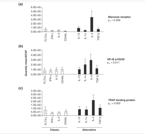
Reproducibility of the estimates of mannose receptor, NFKb-p105/50 and TRAF gene expression in MPs obtained from the same five donors was evaluated after stimulation with four cytokines/soluble factors selected from the alternative (IL-13, IL-1α, IL-4, TGF-β) and four from the classical groups (FLT-3 ligand, IFN-γ, IL-3, CD40L) (Figure 7). In all cases gene expression significantly reflected the pattern of gene expres-sion detected by microarray analysis (Figures 3, 5).
has been called the classical pathway of MP activation. It induces M1 monocytes specialized for pathogen killing and activation of innate and adaptive immune effector cells. A sec-ond, alternative, pathway induces M2-type monocytes com-mitted to clearing pathogen through internal metabolism while reducing inflammation. This process limits the collat-eral damage induced by an excessive immune response, and, upon cessation of the pathogenic stimulus, promotes tissue repair. This dichotomy is based on the study of a few cytokines deemed representative of the classical mode (LPS, IFN-γ) or the alternative mode (IL-4, IL-13) of MP activation. Other cytokines such as IL-10, TGF, M-CSF, IFN-α/β and TNF, although partially overlapping both pathways, display functional effects that diverge significantly enough that they would be inaccurately grouped in either class [5].
In this study we evaluated whether MP commitment is dependent on an early bipolar switch through which most cytokines operate. This was done by testing in parallel a library of 42 stimulatory molecules possibly present in the tis-sue microenvironment following a pathogenic insult. This modulation was tested on MPs triggered by a pathogenic stimulus, exemplified in this case by LPS. This was done on the assumption that in most circumstances resident or migra-tory MPs reaching an infected area are exposed concomi-tantly to a pathogen and to the cytokine milieu resulting from the infection. The transcriptional profile identified by this study cannot distinguish between the direct effect of each sol-uble factor analyzed and the downstream activation of tran-scriptional pathways by secondary paracrine or autocrine secretion of biological modifiers by MPs. However, the main goal of this study was to identify the overall effect on MPs of the exposure to individual cytokines. Future analyses, focused on specific cytokine patterns described here, should possibly include the addition of blocking antibodies to segre-gate secondary from primary MP responses.
The results suggest that MP activation is in most cases a bipo-lar process regulated by an internal switch through which cytokines modulate the yin and yang of the MP transcrip-tional program. In fact, most cytokines preferentially induced one or other pattern of transcriptional activation. We, there-fore, mapped most of the cytokines within a classical or an alternative classification according to their effects on CD14+,
LPS-induced MPs. In particular, it appeared that transcrip-tional programs down-stream of NFκB activation [11] were mostly associated with either class, suggesting that the NFκB system is at the center of the switch regulating MP activation/ differentiation in the conditions tested in the present study. This is not surprising as LPS signaling is mediated through
Induction of gene expression by stimulation of LPS-activated MPs with
[image:10.612.57.299.352.573.2]IFN-γ and IL-4
Figure 6
Induction of gene expression by stimulation of LPS-activated MPs with IFN-γ and IL-4. MPs obtained from PBMC from five normal donors were stimulated for 4 h with one cytokine representative of the alternative (IL-4) and one of the classical group (IFN-γ) and gene transcription measured by TaqMan real-time PCR. The relative quantification of 10 genes was calculated by normalizing the ratio of the mean copy number for each gene with the mean copy number of the reference AFAP gene in MPs from five donors. Statistically significant differences (p-value < 0.05) between the two cytokine treatments as assessed by Student's t-test are represented by an asterisk.
*
*
* *
* IL-4
IFN-γ
Cytokine gene copies/10
5 AFAP 1.E+06
1.E+05
1.E+04
1.E+03
1.E+02
1.E+01 1.E+07 1.E+08
FADD-MORT
IL-1
α
IL-1 receptor
IL-24
Mannose receptor
MCP-1
MMP-19 MMP-9
NF
κ
B p105/50
comm
en
t
re
v
ie
w
s
re
ports
refer
e
e
d
re
sear
ch
de
p
o
si
te
d r
e
se
a
rch
interacti
o
ns
inf
o
rmation
kinase (IKK)-NFκB pathway [3,11,28,29]. It appears that cytokine regulation modulates the release of the p50 subunit of NFκB, whch is in turn responsible for the downstream effects on the transcriptional program. Interestingly, reclus-tering of cytokines based on NFκB-dependent genes consoli-dated the two classes of cytokines adding TNF-α and TNF-β, IFN-α2b and GM-CSF to the classical group and the
alterna-tive type II cytokines with the alternaalterna-tive group, suggesting that NFκB may be central to MP polarization.
This information cannot of course be generalized to other conditions as the model tested is strongly biased by NFκB induction by LPS. This is underlined by the unexpected alter-native upregulation of genes associated with IL-1, TNF and IL-6 [11]. This observation suggests that in different condi-tions, cytokines may differently modulate MP activation, pos-sibly through various modulatory feedback mechanisms [5]. In addition, genes associated with the interrelated arginine and tryptophan pathways that modulate nitric oxide
[image:11.612.55.553.84.545.2]induc-Induction of gene expression by cytokines of the alternative and classical groups
Figure 7
Induction of gene expression by cytokines of the alternative and classical groups. MPs derived from PBMC from five normal donors were stimulated for 4 h with four cytokines/soluble factors selected from the alternative group (IL-13, IL-1α, IL-4, TGF-β) and four from the classical group (FLT-3 L, IFN-γ, IL-3, CD40L). Gene expression was assessed by TaqMan real-time PCR. Relative estimates of (a) mannose receptor, (b) NFκB and (c) TRAF binding protein genes were calculated by averaging the ratio of the quantity mean of each gene normalized to the reference AFAP gene in five donors. Statistical significance is expressed as Student's t-test p-value.
Mannose receptor
p1 = 0.009
Alternative Classic
NFκB p105/50
p1 = 0.011
TRAF binding protein
p1 = 0.005
Quantity mean/AFAP
0.0E+00 1.0E+05 2.0E+05 3.0E+05 4.0E+05 5.0E+05 6.0E+05
0.0E+00 1.0E+05 2.0E+05 3.0E+05 4.0E+05 5.0E+05
0.0E+00 5.0E+01 1.0E+02 1.5E+02 2.0E+02 2.5E+02 3.0E+02 3.5E+02
FLT3-L
IFN-γ
IL-3
CD40L IL-13 IL-1
α
IL-4
TGF-β
FLT3-L
IFN-γ
IL-3
CD40L IL-13 IL-1
α
IL-4
TGF-β
FLT3-L
IFN-γ
IL-3
CD40L IL-13 IL-1
α
IL-4
TGF-β
(a)
(b)
sible that cytokines may counteract this effect through modu-lation of this metabolic junction.
Cytokine and chemokine effects on MP function are tightly intertwined with the enzymatic activities of MMP. Mem-brane-bound cytokine receptors and adhesion molecules can be released from the cell surface by MMPs acting as 'shed-dases' or 'convertases'. This, in turn, can downregulate cell-surface signaling by removal of receptors, or induce paracrine activity by release of soluble proteins. Not surprisingly, IL-13 overexpression results in production of several MMPs [33]. For instance, MMP-9 activates latent TNF-α on the surface of MPs or soluble VEGF [19,34]. Furthermore, Yu and Stamenk-ovic [21] observed that gelatinase B/MMP9 bound to CD44 activates latent TGF-β stored in the pericellular matrix. MMP-7 can enhance tissue repair by facilitating migration of epithelial cells [16]. In agreement with these reports we observed that the transcription of MMP-7, MMP-9 and CD44 are coordinately induced (Figures 4, 6) by the alternative acti-vation of MPs. Conversely, the downregulation of MMP-7 and MMP-9 by the classical cytokines confirms their inhibitory effects on MMP expression. Inhibition of MMP-9 production by IFN-β and IFN-γ has been recently reported by Sanceau et al. [35], who noted that interferons regulate MMP expression through IRF/NFκB interaction. Binding of NFκB p50/p65 to the MMP-9 promoter is competitively inhibited by IFN-β and IFN-γ-induced IRF-1. Possibly, NFκB regulation of MMP pro-moters through release of p50 (Figure 3) was responsible for the transcriptional activation of MMP expression by alterna-tive cytokines observed in this study (Figure 4). These obser-vations confirm the tight specificity of the relationship between cytokine and MMP regulation, which is finely toned at several check points and strongly polarized, in these exper-imental conditions, toward an M2 phenotype.
Enhanced transcription of gap junction/connexin 26 by the alternative cytokines is also of particular interest in view of the hypothesized junctional communications among MPs or between MPs and endothelial cells [36,37]. The finding that MPs activated by alternative cytokines induce the transcrip-tion of genes for gap junctranscrip-tion components is of physiological importance and may be the missing link in the identification of factors that regulate the expression of gap junction connex-ins in MPs. In addition, it opens up the possibility that in a milieu dominated by alternative cytokines, where the ulti-mate goal is to return to homeostasis, the induction of gap junctions increases the ability to transmit or receive regula-tory signals [38] that could facilitate the return to normal housekeeping functions.
crine and paracrine modulation through the cytokine network following a primary stimulation may introduce novel on and off switches that could override the original signal. Nevertheless, this analysis is directly informative on the pri-mary effect of individual cytokines on the early stages of LPS stimulation and, therefore, may be most informative on the way MP maturation may be polarized at the early stages of the immune response. The clustering of most cytokines into two main groups suggests that their control of central switches (NFκB), or regulatory molecules (cytokines, MMPs, gap junc-tions, cytotoxic molecules, migratory markers) is essentially bimodal. This polarization program turns MPs to a 'cytotoxic' or a 'symbiotic' phenotype [18]. In physiologic conditions, this dualism is probably modulated by a multiplicity of fac-tors: the extent and duration of the environmental insult and the conditions of the resulting microenvironment. Possibly, predominant and persistent stimulation by pathogen compo-nents (such as LPS) may polarize MP towards the cytotoxic phenotype. A predominantly regulatory response is then mounted by the host, mediated by alternative cytokines that would take over at a later stage to induce a symbiotic phenotype aimed at resuming homeostasis upon pathogen clearance.
Materials and methods
MP separation and FACS staining
Peripheral blood mononuclear cells (PBMCs) from an HLA-A*0201-positive healthy caucasian male donor age 35 were collected at the Department of Transfusion Medicine, NIH. PBMCs were isolated by Ficoll gradient separation and frozen until analysis. After thawing, PBMCs were kept overnight in 175-cm2 tissue-culture flasks (Costar) in complete medium
(CM) consisting of Iscove's medium (Biofluids) supple-mented with 10% heat-inactivated human AB-serum (Gemini Bioproducts), 10 mM HEPES buffer (Cellgro; Mediatech), 0.03% l-glutamine (Biofluids), 100 U/ml penicillin/strepto-mycin (Biofluids), 10 µg/ml ciprofloxacin (Bayer), and 0.5 mg/ml amphotericin B (Biofluids). Adherent and non-adher-ent cells were gnon-adher-ently removed from the flask and cnon-adher-entrifu- centrifu-gated. MPs were separated by negative selection using the MP isolation kit and an autoMACS system (Miltenyi). Before and after separation cells were stained with anti-CD14-FITC (Becton Dickinson), and analyzed using a FACScalibur flow cytometer and CellQuest software (Becton Dickinson).
Stimulation of MPs and RNA isolation
Negatively selected CD14+ cells were washed twice with
concentra-comm
en
t
re
v
ie
w
s
re
ports
refer
e
e
d
re
sear
ch
de
p
o
si
te
d r
e
se
a
rch
interacti
o
ns
inf
o
[image:13.612.59.552.118.686.2]rmation
Table 1
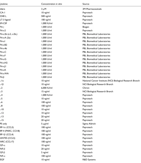
Concentration and doses of cytokines/soluble factors used for stimulation of monocytes
Cytokine Concentration in vitro Source
Aldara 3 µM 3M Pharmaceuticals
BCA-1 10 ng/ml Peprotech
CD40 L 500 ng/ml Peprotech
FLT-3 ligand 100 ng/ml Peprotech
GM-CSF 1,000 IU/ml Peprotech
IFN-γ 1,000 U/ml Biogen
IFN-α2 1,000 U/ml Peprotech
IFN-α2b (α2, α2b,) 1,000 U/ml PBL Biomedical Laboratories
IFN-αA (2a) 1,000 U/ml PBL Biomedical Laboratories
IFN-αI 1,000 U/ml PBL Biomedical Laboratories
IFN-αB2 1,000 U/ml PBL Biomedical Laboratorios
IFN-α4b 1,000 U/ml PBL Biomedical Laboratories
IFN-αC 1,000 U/ml PBL Biomedical Laboratories
IFN-αF 1,000 U/ml PBL Biomedical Laboratories
IFN-αG 1,000 U/ml PBL Biomedical Laboratories
IFN-αH2 1,000 U/ml PBL Biomedical Laboratories
IFN-αJ1 1,000 U/ml PBL Biomedical Laboratories
IFN-αK 1,000 U/ml PBL Biomedical Laboratories
IFN-αWA 1,000 U/ml PBL Biomedical Laboratories
IFN-β 1,000 U/ml PBL Biomedical Laboratories
IL-1α 10 ng/ml National Cancer Institute (NCI) Biological Research Branch
IL-1β 10 ng/ml NCI Biological Research Branch
IL-2 6,000 IU/ml Chiron
IL-3 15 ng/ml NCI Biological Research Branch
IL-4 1,000 IU/ml Peprotech
IL-5 10 ng/ml Peprotech
IL-6 100 ng/ml Peprotech
IL-8 100 ng/ml Peprotech
IL-10 10 ng/ml Peprotech
IL-12 10 ng/ml Peprotech
IL-13 20 ng/ml Peprotech
IL-15 20 ng/ml Peprotech
LPS only 5 µg/ml Sigma Aldrich
MIP-1α (CCL3) 100 ng/ml Peprotech
MIP-4 (PARC, CCl18) 100 ng/ml Peprotech
MIP-1β (CCL4) 100 ng/ml Peprotech
RANTES (CCL5) 100 ng/ml Peprotech
TARC (CCL17) 100 ng/ml Peprotech
TGF-α 10 ng/ml Peprotech
TNF-β 20 ng/ml Peprotech
TGF-β 5 ng/ml Peprotech
TNF-α 100 ng/ml Peprotech
after LPS stimulation, MPs were harvested, washed twice in PBS and lysed for RNA isolation using 700 µl RNeasy lysis buffer (Qiagen) per 25 cm2 flask, according to the
manufac-turer's protocol.
Probe preparation, amplification and hybridization to microarrays
Total RNA was isolated using RNeasy minikits (Qiagen). Amplified antisense RNA (aRNA) was prepared from total RNA (0.5-3 µg) according the protocol previously described by us [9,39]. Test samples were labeled with Cy5-dUTP (Amersham) while the reference sample (pooled normal donor PBMCs) was labeled with Cy3-dUTP. Test-reference sample pairs were mixed and co-hybridized to 17K cDNA microarrays.
Microarrays and statistical analyses
Hybridized arrays were scanned at 10-µm resolution on a GenePix 4000 scanner (Axon Instruments) at variable PMT voltage to obtain maximal signal intensities with less than 1% probe saturation. Resulting jpeg and data files were analyzed via mAdb Gateway Analysis tool [40]. Data were further ana-lyzed using Cluster and TreeView software [12] and Partek Pro software (Partek). The global gene-expression profiling of 4- and 9-h treated and untreated MP consisted of 98 experi-mental samples. Subsequent low-stringency filtering (80% gene presence across all experiments and removal of genes that did not have a log2 ≥ 1.2: 2.3 ratio in at least one of the samples) selected 10,370 genes for further analysis. Clustering of experimental samples according to Eisen et al. [12] was based on these genes. Gene ratios were average cor-rected across experimental samples and displayed according to the central method for display using a normalization factor as recommended by Ross [41].
NFκB protein activation analysis
MPs separated from peripheral blood by adherence were stimulated for 1 h with LPS and for an additional 30 min with cytokines selected from the alternative group (4, 13, IL-1α) or the classical group (IFN-γ, IL-6, IL-3). In addition, TNF-α was tested. After 90 min stimulation, cytoplasmic cell extracts were isolated using a cytoplasmic and nuclear extract kit (Active Motif), and the TransAM NFκB transcription fac-tor kit (Active Motif) was used to detect activation of NFκB subunits p50, p52, p65, c-Rel and RelB, according to the man-ufacturer's protocol.
Real-time quantitative RT-PCR
MPs obtained from PBMC of five normal caucasian donors (three males, two females, age range: 35-55 years old) were
sion of the following 10 genes: TRAF binding protein, NFκ B-p105/50, MMP9, MMP19, MCP-1, mannose receptor, IL-24, IL-1R, IL-1A and FADD-MORT. An ABI Prism 7900 HT sequence detection system with 384-well capability (Applied Biosystems) was used for detection. Primers and TaqMan probes (Biosource) were designed to span exon-intron junc-tions and to generate amplicons of less than 150 bp. TaqMan probes were labeled at the 5' end with the reporter dye mole-cule FAM (6-carboxyfluorescein; emission λmax = 518 nm)
and at the 3' end with the quencher dye molecule TAMRA (6-carboxytetramethylrhodamine; emission λmax = 582 nm). The following are the sequences for forward (f) and reverse (r) primer and probe (p) pairs:
IL-1α f: TGTATGTGACTGCCCAAGATGAA
IL-1α r: ACTACCTGTGATGGTTTTGGGTATC
IL-1α p: FAM-AGTGCTGCTGAAGGAGATGCCTG-TAMRA
IL-1 rec. f: TGTCACCGGCCAGTTGAGT
IL-1 rec. r: GCACTGGGTCATCTTCATCAATT
IL1 rec p: FAM-ACATTGCTTACTGGAAGTGGAATGGGT-CAG-TAMRA
TRAF bp f: TTGCTTACAG AGGTGTCTCAACAAG
TRAF bp r: CTCCGGATTTGTTCTGTCAGTTC
TRAF bp p: FAM-AGCAAAGTGTATTCCAGCAATGGTGT-GTCC-TAMRA
MMP9 f: TGGATCCAAAACTACTCGGAAGA
MMP9 r: GAAGGCGCGGGCAAA
MMP9 p: FAM-CGCGGGCGGTGATTGACGAC-TAMRA
MMP19 f: GACGAGCTAGCCCGAACTGA
MMP19 r: TTTGGCACTCCCGTAAACAAA
MMP19 p: FAM-TCAGCAGCTACCCCAAACCAATCAAGG-TAMRA
comm
en
t
re
v
ie
w
s
re
ports
refer
e
e
d
re
sear
ch
de
p
o
si
te
d r
e
se
a
rch
interacti
o
ns
inf
o
rmation
Mannose receptor p: FAM-ACACAAGGAAGATGGACCCT-TCTAAACCGTC-TAMRA
FADD-MORT f: GGTGGCTGACCTGGTACAAGA
FADD-MORT r: ACATGGCCCCACTCCTGTT
FADD-MORT p: FAM-TTCAGCAGGCCCGTGACCTCCA-TAMRA
NFκB p105/50 f: CTACACCGAAGCAATTGAAGTGA
NFκB p105/50 r: CAGCGAGTGGGCCTGAGA
NFκB p105/50 p: FAM-CAGGCAGCCTCCAGCCCAGTGA-TAMRA
IL-24 f: AAGAAAATGAGATGTTTTCCATCAGA
IL-24 r: CTGTTTGAATGCTCTCCGGAAT
IL-24 p: FAM-ACAGTGCACACAGGCGGTTTCTGC-TAMRA
MCP-1 f: CATGGTACTAGTGTTTTTTAGATACAGAGACTT
MCP-1 r: TAATGATTCTTGCAAAGACCCTCAA
MCP-1 p: FAM-AACCACAGTTCTACCCCTGGGATG-TAMRA
Standards for the selected genes were amplified by reverse transcriptase primer-specific amplification of 6 µg antisense RNA obtained from PBMCs stimulated in vitro with IL-2 (300 IU/ml and Flu M1 peptide) and reverse transcribed using random dN6 primers (Boehringer Mannheim). Ampli-fied cDNA standards were quantiAmpli-fied by spectrometry and the number of copies was calculated using the Oligo Calculator software [42]. Six micrograms of test antisense RNA samples were converted to cDNA using random primers and were immediately used for quantitative real-time PCR (RT-PCR). RT-PCR reactions of cDNA samples were conducted in a total volume of 20 µl, including 1 µl cDNA, 1x TaqMan Master Mix (Applied Biosystems), 2 µl of 20 µM primers and 1 µl of 12.5
µM probe. Thermal cycler parameters included 2 min at 50°C, 10 min 95°C and 40 cycles involving denaturation at 95°C for 15 sec, annealing-extension at 60°C for 1 min.
Linear regression analyses of all standard curves were 0.98 or greater. Standard curve extrapolation of copy number and quantity means were performed using the ABI Prism SDS 2.1 software (Applied Biosystems). Normalization of samples was performed by dividing the quantity mean of the gene of interest run in duplicate by the quantity mean of reference actin filament associated protein (AFAP) gene × 105 [43].
Additional data files
The following additional data are available with the online version of this article. Additional data file 1 is a spread sheet containing microarray raw data (intensity ratios of test versus reference samples) subsequently subjected to cluster analysis.
Additional data file 1
A spread sheet containing microarray raw data (intensity ratios of test versus reference samples) subsequently subjected to cluster analysis
A spread sheet containing microarray raw data (intensity ratios of test versus reference samples) subsequently subjected to cluster analysis
Click here for additional data file
Acknowledgements
D.N. was supported by a grant from Deutsche Krebshilfe.
References
1. Mantovani A, Sozzani S, Locati M, Allavena P, Sica A: Macrophage polarization: tumor-associated macrophages as a paradigm for polarized M2 mononuclear phagocytes. Trends Immunol 2002, 23:549-555.
2. Takeda K, Kaisho T, Akira S: Toll-like receptors.Annu Rev Immunol 2003, 21:335-376.
3. Anderson KV: Toll signaling pathways in the innate immune response.Curr Opin Immunol 2000, 12:13-19.
4. Mocellin S, Panelli MC, Wang E, Nagorsen D, Marincola FM: The dual role of IL-10.Trends Immunol 2003, 24:36-43.
5. Gordon S: Alternative activation of macrophages. Nat Rev Immunol 2003, 3:23-35.
6. Huang Q, Liu D, Majewski P, Schulte LC, Korn JM, Young RA, Lander ES, Hacohen N: The plasticity of dendritic cell responses to pathogens and their components.Science 2001, 294:870-875. 7. Langenkamp A, Messi M, Lanzavecchia A, Sallusto F: Kinetics of
dendritic cell activation: impact on priming of Th1, Th2 and nonpolarized T cells.Nat Immunol 2000, 1:311-316.
8. Belardelli F, Ferrantini M: Cytokines as a link between innate and adaptive antitumor immunity. Trends Immunol 2002,
23:201-208.
9. Wang E, Miller L, Ohnmacht GA, Liu E, Marincola FM: High fidelity mRNA amplification for gene profiling using cDNA microarrays.Nat Biotechnol 2000, 18:457-459.
10. Locati M, Deuschle U, Massardi ML, Martinez FO, Sironi M, Sozzani S, Bartfai T, Mantovani A: Analysis of the gene expression profile activated by the CC chemokine ligand 5/Rantes and by lipopolysaccharide in human monocytes. J Immunol 2002,
168:3557-3562.
11. Hatada EN, Krappmann D, Scheidereit C: NF-κB and the innate immune response.Curr Opin Immunol 2000, 12:52-58.
12. Eisen MB, Spellman PT, Brown PO, Botstein D: Cluster analysis and display of genome-wide expression patterns. Proc Natl Acad Sci USA 1998, 95:14863-14868.
13. Henco K, Brosius J, Fujisawa A, Fujisawa J-I, Haynes JR, Hochstadt J, Kovacic T, Pasek M, Schambock A, Schmid J, et al.: Structural rela-tionship of human interferon alpha genes and pseudogenes. J Mol Biol 1985, 185:227-260.
14. Mogensen KE, Lewerenz M, Reboul J, Lutfalla G, Uze G: The type I interferon receptor: structure, function, and evolution of a family business.J Interferon Cytokine Res 1999, 19:1069-1098. 15. Cook JR, Cleary CM, Mariano TM, Izotova L, Pestka S: Differential
responsiveness of a splice variant of the human Type I intereferon receptor to intereferons. J Biol Chem 1996,
271:13448-13453.
16. Parks WC, Shapiro SD: Matrix metalloproteinases in lung biology.Respir Res 2001, 2:10-19.
17. de Fougerolles AR, Chi-Rosso G, Bajardi A, Gotwals P, Green CD, Koteliansky VE: Global expression of extracellular matrix-integrin interactions in monocytes.Immunity 2000, 13:749-758. 18. Kreutz M, Fritsche J, Andreesen R: Macrophages in tumor biol-ogy. In The Macrophage Edited by: Burke B, Lewis CE. Oxford: Oxford University Press; 2002:457-489.
19. Haro H, Crawford HC, Fingleton B, Shinomiya K, Spengler DM, Mat-risian LM: Matrix metalloproteinase-7-dependent release of tumor necrosis factor-alpha in a model of herniated disc resorption.J Clin Invest 2000, 105:143-150.
proteolysis of insulin-like growth factor binding protein-3. Mol Biol Cell 2003, 14:4569-4580.
23. Locati M, Otero K, Schioppa T, Signorelli P, Perrier P, Baviera S, Soz-zani S, Mantovani A: The chemokine system: tuning and shap-ing by regulation of receptor expression and couplshap-ing in polarized responses.Allergy 2002, 57:972-982.
24. Vivers S, Dransfield I, Hart SP: Role of macrophage CD44 in the disposal of inflammatory cell corpses. Clin Sci (Lond) 2002,
103:441-449.
25. Mantovani A, Gray PA, Van Damme J, Sozzani S: Macrophage-derived chemokine (MDC).J Leukocyte Biol 2000, 68:400-404. 26. Conti P, Kempuraj D, Frydas S, Kandere K, Boucher W, Letourneau
R, Madhappan B, Sagimoto K, Christodoulou S, Theoharides TC: IL-10 subfamily members: IL-19, IL-20, IL-22, IL-24 and IL-26. Immunol Lett 2003, 88:171-174.
27. Kaplanski G, Marin V, Montero-Julian F, Mantovani A, Farnarier C: IL-6: a regulator of the transition from neutrophil to monocyte recruitment during inflammation. Trends Immunol 2003,
24:25-29.
28. Beutler B: Tlr4: central component of the sole mammalian LPS sensor.Curr Opin Immunol 2000, 12:20-26.
29. Guha M, Mackman N: LPS induction of gene expression in human monocytes.Cell Signal 2001, 13:85-94.
30. Alberati-Giani D, Malherbe P, Ricciardi-Castagnoli P, Kohler C, Denis-Donini S, Cesura AM: Differential regulation of indoleam-ine 2,3-dioxygenase expression by nitric oxide and inflamma-tory mediators in IFN-γ-activated murine macrophages and microglial cells.J Immunol 1997, 159:419-426.
31. Bertazzo A, Ragazzi E, Biasiolo M, Costa CVL, Allegri G: Enzyme activities involved in tryptophan metabolism along the kynurenine pathway in rabbits. Biochim Biophys Acta 2001,
1527:167-175.
32. Thomas SR, Mohr D, Stocker R: Nitric oxide inhibits indoleam-ine 2,3-dioxygenase activity in interferon-γ primed mononu-clear phagocytes.J Biol Chem 1994, 269:14457-14464.
33. Zheng T, Zhu Z, Wang Z, Homer RJ, Ma B, Riese RJ Jr, Chapman HA Jr, Shapiro SD, Elias JA: Inducible targeting of IL-13 to the adult lung causes matrix metalloproteinase- and cathepsin-dependent emphysema.J Clin Invest 2000, 106:1081-1093. 34. Munaut C, Noel A, Hougrand O, Foidart JM, Boniver J, Deprez M:
Vascular endothelial growth factor expression correlates with matrix metalloproteinases MT1-MMP, MMP-2 and MMP-9 in human glioblastomas.Int J Cancer 2003, 106:848-855. 35. Sanceau J, Boyd DD, Seiki M, Bauvois B: Interferons inhibit tumor necrosis factor-alpha-mediated matrix metalloproteinases-9 activation via interferon regulatory factor-1 binding compe-tition with NF-kappa B.J Biol Chem 2002, 277:35766-35775. 36. Lee SW, Tomasetto C, Keyomarsi K, Sager R: Transcriptional
downregulation of gap-junction proteins block junctional communication in human mammary tumor cell lines.J Cell Biol 1992, 118:1213-1221.
37. Eugenin EA, Branes MC, Berman JW, Saez JC: TNF-alpha plus IFN-gamma induce connexin43 expression and formation of gap junctions between human monocyte/macrophages that enhance physiological responses. J Immunol 2003,
170:1320-1328.
38. Alves LA, Coutinho-Silva R, Persechini PM, Spray DC, Savino W, Campos de Carvalho AC: Are there functional gap junctions or junctional hemichannels in macrophages? Blood 1996,
88:328-334.
39. Wang E, Marincola FM: Amplification of small quantities of mRNA for transcript analysis. In DNA Arrays: A Molecular Cloning Manual Edited by: Bowtell D, Sambrook J. Cold Spring Harbor, NY: Cold Spring Harbor Laboratory Press; 2002:204-213.
40. Bacon K, Baggiolini M, Broxmeyer H, Horuk R, Lindley I, Mantovani A, Maysushima K, Murphy P, Nomiyama H, Oppenheim J, et al.:
Chemokine/chemokine receptor nomenclature. J Interferon Cytokine Res 2002, 22:1067-1068.
41. mAdb Gateway Analysis tool [http://nciarray.nci.nih.gov] 42. Ross DT, Scherf U, Eisen MB, Perou CM, Rees C, Spellman P, Iyer V,