Open Access
R E S E A R C H A R T I C L E
© 2010 Visigalli et al; licensee BioMed Central Ltd. This is an Open Access article distributed under the terms of the Creative Commons Attribution License (http://creativecommons.org/licenses/by/2.0), which permits unrestricted use, distribution, and reproduction in any medium, provided the original work is properly cited.
Research article
Hind limb unloading of mice modulates gene
expression at the protein and mRNA level in
mesenchymal bone cells
Davide Visigalli
1, Antonella Strangio
2, Daniela Palmieri
1and Paola Manduca*
1Abstract
Background: We investigated the extent, modalities and reversibility of changes at cellular level in the expression of genes and proteins occurring upon Hind limb unloading (HU) in the tibiae of young C57BL/6J male mice. We focused on the effects of HU in chondrogenic, osteogenic, and marrow mesenchymal cells.
Methods: We analyzed for expression of genes and proteins at two time points after HU (7 and 14 days), and at 14 days after recovery from HU. Levels of mRNAs were tested by in situ hybridization. Protein levels were tested by
immunohistochemistry. We studied genes involved in osteogenesis (alkaline phosphatase (AP), osteocalcin (OC), bonesialoprotein (BSP), membrane type1 matrix metalloproteinase (MT1-MMP)), in extracellular matrix (ECM)
formation (procollagenases (BMP1), procollagenase enhancer proteins (PCOLCE)) and remodeling (metalloproteinase-9 (MMP(metalloproteinase-9), RECK), and in bone homeostasis (Stro-1, CXCL12, CXCR4, CD146).
Results: We report the following patterns and timing of changes in gene expression induced by HU: 1) transient or stable down modulations of differentiation-associated genes (AP, OC), genes of matrix formation, maturation and remodelling, (BMP1, PCOLCEs MMP9) in osteogenic, chondrogenic and bone marrow cells; 2) up modulation of MT1-MMP in these same cells, and uncoupling of its expression from that of AP; 3) transient down modulation of the osteoblast specific expression of BSP; 4) for genes involved in bone homeostasis, up modulation in bone marrow cells at distal epiphysis for CXCR4, down modulation of CXCL12, and transient increases in osteoblasts and marrow cells for Stro1. 14 days after limb reloading expression returned to control levels for most genes and proteins in most cell types, except AP in all cells, and CXCL12, only in bone marrow.
Conclusions: HU induces the coordinated modulation of gene expression in different mesenchymal cell types and microenvironments of tibia. HU also induces specific patterns of expression for homeostasis related genes and modulation of mRNAs and proteins for ECM deposition, maturation and remodeling which may be key factors for bone maintenance.
Background
Bone loss may be induced by immobilization of humans for long times in bed, and by hind limb unloading of rodents. HU allows ex vivo investigations into the pro-cesses and effects of weightlessness in long bones [1,2]. Effects of HU have been detected earliest on the viability of osteoblasts [3], followed by apoptosis of the osteocytes, activation of osteoclastogenesis and loss in bone mass, attributed to altered mechano-transduction by
osteo-blasts [4]. HU-induced changes in the expression of dif-ferentiation-associated genes have been reported in studies on whole bone tissue [5]. In ex-vivo experiments in rats, HU determined the decrease in number of colony forming cells from the bone marrow osteogenic compart-ment, and in AP expressing colonies [6].
The expression of differentiation-associated genes in osteoblasts and preosteoblasts is altered in simulated microgravity conditions, reproducing in vitro the condi-tion of reduced mechanical stress occurring in space flight and in HU [7-9]. In microgravity, adipocyte differ-entiation is promoted from mesenchymal precursor cells
* Correspondence: [email protected]
1 Genetics, DIBIO, University of Genoa, (Corso Europa 26), Genoa, (I-16132), Italy
[10] and collagen I synthesis is impaired in osteogenic and fibroblastic cells [7-9,11]. Osteopontin and parathy-roid hormone receptor have been associated with the modulation of bone loss in HU conditions [12,13].
Studies are needed to illustrate the changes in gene expression at the level of individual mesenchymal cell types in the bone of HU animals, and to find more infor-mation on the expression of proteins. In addition, no data are available on the effects of HU on genes governing matrix deposition and remodeling, or on genes involved in controlling the homeostasis of the different cell types in bone. Little is known about the timing of the modula-tion in different cell types for each of the responsive genes by HU. Reversibility of these changes once weight is re-applied to the limbs is poorly understood.
In this paper we report investigations to start filling these gaps and suggest that valuable information can be obtained by studying the regulation by HU of proteinases and chemokine genes and proteins at cellular levels, in association with classic osteogenic markers.
We determined by in situ hybridization and/or immu-nohistochemistry the expression of individual genes and proteins in the tibiae of 11 week old male mice C57BL/6J, after HU for 7 and 14 days. We focus our report on gene expression in chondrocytes at the growth plate, osteo-cytes, endosteum and trabecular lining osteoblasts and in bone marrow cells.
Changes in the growth plate, and then in bone mor-phology, occurred after 3, 7 and 14 days and were docu-mented. Bone loss was quantified by microcomputed tomography analysis (μCT) at 14 days. We also studied the expression of genes in tibiae of animals kept in HU for 14 days and then set on the ground for the next 14 days (re-loading). We compared same age control, and HU mice for the expression of key genes associated to the dif-ferentiation of osteoblasts (AP, OC, BSP), of genes involved in the deposition of ECM (BMP-1 and PCOLCE1 and 2) and in its remodeling (MT1-MMP, MMP-9, RECK), and of genes implied in bone homeosta-sis (Stro1, CD146, CXCR4 and CXCL12).
A decrease in AP and OC expression was previously described by examining mRNA from whole bones of HU rodents [14]. BMP1, mammalian tolloid (mTLD) and the genes for PCOLCE1 and 2, are expressed in modulated fashion during in vitro osteogenesis in rat osteoblasts (Manduca et al unpublished); the level of expression of their proteins controls the rate and amount of deposition of collagen fibers and the activation of pro-lysyl oxidase, also involved in the deposition of collagen fibrils [15,16]. PCOLCEs and BSP, known to be highly expressed by osteoblasts and osteoclasts [17], are proteins with a role in the mineralization of the ECM and PCOLCE1 is an important determinant of bone mechanical properties and of the geometry and morphology of collagen fibrils in
mammals [18,19]. PCOLCEs gene expression decreases in osteoblasts cultured in microgravity [7].
Enzymes remodeling the ECM emerged as important for the osteogenic progression [20-22]. The major MMPs family members expressed by differentiating osteoblasts, MT1-MMP, MMP-2 and -9 are developmentally regu-lated during osteogenesis in vitro [21]. In rat osteoblasts differentiating in vitro, MT1-MMP expression affects the expression of pro-MMP-2 and of AP, and the activation of pro-MMP-2 pleiotropically [22]. MMP-9 modulation is not apparently associated with that of the other MMPs. RECK is an endogenous membrane inhibitor of MMPs [23], also modulated in its expression during osteogenesis in vitro [24].
CXCR4 [25] is the receptor for Stromal cell-derived fac-tor-1 (SDF-1 or CXCL12), which is constitutively secreted by osteoblasts and bone marrow stromal cells. Dynamic levels of CXCL12 and CXCR4 expression play a key role in the homing of hematopoietic cells to the bone marrow, in the mobilization of circulating osteoblast pre-cursors and in the recruitment of bone-resorbing osteo-clasts, of osteoblasts, neutrophils, and other myeloid cells [26]. Activation of the CXCR4/CXCL12 pathway induces proliferation of hematopoietic and mesenchymal progen-itors [27]. CXCL12 also stimulates mononucleate cell fusion and TRAP activity and is a key factor in the normal homeostatic regulation of bone development and remod-eling [28]. CD146 is an endothelial, smooth muscle cell and pericyte marker [29] and Stro1 a stage- and/or lin-eage-specific stromal antigen [30].
Our results show that HU modulates the expression of the osteogenic differentiation markers AP, MT1-MMP, BSP and OC consistently in all the cells expressing these genes, even if of different differentiation lineages. Uncou-pling of the associated expression for AP and MT1-MMP occurs in cells of the osteogenic lineage.
HU modulates proteolysis related genes suggesting that pericellular proteolysis may be specifically increased. HU affects the expression of genes for matrix deposition and maturation, BMP1 and PCOLCEs, in the growth plate and in the bone cells, suggesting a lesser formation of cross linked collagen fibers and impaired bone matrix maturation.
Changes in the pattern of expression observed in cells of the distal bone marrow for genes involved in bone homeostasis, Stro1, CXCR4 and CXCL12, suggest that complex changes in bone homeostasis may occur in HU mice, apparently not involving CD146 expression.
Methods
Hindlimb unloading
11 week old C57BL/6J male mice (Charles River, Italy) were suspended for the tail in a special cage [31,32], a condition referred as HU, for 3, 7 or 14 days. Food and water were ad libitum. Control of the amount of food uptake showed no differences at any time among the groups of animals in different conditions. In each experi-ment 3 to 6 mice were HU for each time point and the same number were kept on the ground as control. Re-loading of the hind limb occurred by returning mice to standard conditions for 14 days after HU. Animal treat-ment was in accordance with the Italian Guidelines for the use of laboratory animals, which conforms with the European Community Directive published in 1986 (86/ 609/EEC).
Microcomputed Tomographic (μCT) Analysis
Whole femora (one per mouse) were examined by a μCT system (Desktop μCT 40, Scanco Medical AG, Bassers-dorf, Switzerland) at the Bone Laboratory of The Hebrew University of Jerusalem, courtesy of Prof. I. Bab and Dr. A. Bajayo, as reported previously [33]. Scans were per-formed at a 20 μm resolution in all three spatial dimen-sions and all morphometric parameters were determined using a direct 3-D approach. Three preselected regions: 1) whole bone; 2) secondary spongiosa in the distal meta-physis extending proximally 3 mm from the proximal tip of the primary spongiosa; 3) a diaphyseal segment extending 1 mm distally from the midpoint between the femoral ends. For the whole bone we determined the Apparent Volume Density (AVD) [34]. Parameters deter-mined in the metaphyseal trabecular bone included bone volume density (BV/TV), trabecular thickness (Tb.Th), trabecular number (Tb.N) and trabecular connectivity (Conn.D). Cortical thickness (Cort.Th), diaphyseal diam-eter (Dia.Dia) and the medullary cavity diamdiam-eter (Med.Dia) were determined in the mid-diaphyseal region.
Preparation for immunohistochemistry and in situ hybridization
Tibiae were dissected free of soft tissues and fixed in PAF 4% in PBS 1× a 4°C for 48 hours, decalcified with Osteo-dec (Bioptica, Italy) for 16 hours, dehydrated by succes-sive alcohol passages and xylol washing and embedded in paraffin at 58°C. Sections at the microtome were 4 μm thick and the sections in the central portion of the bone were stained with H.E. or used for immunohystochemis-try and in situ hybridization.
Immunohistochemistry
De-paraffinized sections were processed by citrate unmasking of the antigens at 95°C, peroxidase quench-ing, rinsed in PBS and incubated with the primary anti-body for 60' at room temperature, followed by rinses and
incubation for 60' with the secondary antibody (HRP polymer conjugate broad spectrum DAB kit from Zymed Laboratories inc., cat. 87-9663). Counterstaining was with Hematoxylin. Controls were run omitting the pri-mary antibody. Pripri-mary antibodies (all utilized at 1/50 dilution) were: CXCL12 polyclonal in rabbit (sc-28876, Santa Cruz), RECK polyclonal in rabbit (sc-28918, Santa Cruz), BSP polyclonal in rabbit (LF87, kindly donated by Prof. L.W. Fisher, Bethesda, USA), BMP-1 polyclonal in rabbit (sc-33200, Santa Cruz, USA, recognizing all splic-ing variants and Tll1, Tll2 protein), OC polyclonal in rab-bit (sc-30045, Santa Cruz, USA), CD146 polyclonal in rabbit (sc-28667, Santa Cruz, USA) and Stro-1 mouse ascites (kindly donated by Prof. Ranieri Cancedda, CBA, Genoa, Italy). We show exemplary micrographs from at least two experiments of HU and 2 or 3 mice for each condition from each experiment, which gave consistent results.
In situ hybridization
digestion with HCl 0.2 M and Proteinase K 10 μg/ml at 37°C, hybridization was in 3× SSC, 1 mg/ml tRNA, 10 mM DTT, Denhardt's solution 1×, 50% formamide, 1 mg/ ml denatured salmon sperm, 10% dextran sulfate and 1 ng/μl of the denaturated probe, maintained overnight at high stringency. Temperatures of annealing and strin-gency of washes were optimized for each probe [38]. The slides were washed in 2× SSC, digested with RNAse A 100 μg/ml at 37°C, incubated overnight at 4°C with anti-digoxigenin Fab fragments conjugated to alkaline phos-phatase (Boeringher-Mannheim, Germany) in buffer with 1.5 M NaCl, 0.1 M TRIS-HCl, 2 mM MgCl2, 0.3% Triton X-100, 10% fetal calf serum, pH 7.5. Color devel-opment was in buffer 0.1 M NaCl, 0.1 M TRIZMA base, 5 mM MgCl2, 10% polyvinyl alcohol 89-98 kDa (Sigma, USA), 1 mM levamisole (Sigma, USA), 0.16 mg/ml BCIP, 0.33 mg/ml NBT (Roche, Germany), pH 9.5, for 3-6 hours at 30°C [39]. Slides were counterstained with nuclear fast red 0.005% and mounted in glycerol gelatin (Sigma, USA). We show exemplary micrographs from at least two experiments of HU, and 3 mice for each condition and from each experiment, which gave consistent results.
Image acquisition
The images were captured with a Leica DMBR micro-scope mounted with a Leica camera DFC320 and acquired with Leica Firecam version 1.9.1.
Statistics
For mice weight a one-way ANOVA test was performed to define significance.
Results
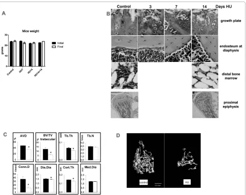
We examined 3 mice for each time point of HU in each of 2 or 3 separate experiments compared with equal num-bers of control mice. No significant differences in body weight were found in HU mice compared with age matched control mice during all the experimental peri-ods, nor with mice kept in HU for 14 days plus 14 days after reloading of hind limbs (Figure 1A). This shows lack of significant growth during the experimental time and suggests lack of stress due to the HU condition.
Figure 1B shows the morphology of growth plate, dia-physeal endosteum and bone marrow in the distal epiph-ysis of controls and in mice kept for 3, 7 or 14 days in HU. 3 days after HU, the cartilage at growth plate already shows a disrupted columnar arrangement, with loss of the connection to the morphologically unchanged trabe-cular organization underneath. Osteogenic cells, here shown lining the endosteum at diaphysis, but equally on trabeculae (not shown), became disorganized and fewer after 7 days of HU. At this time no obvious thinning of the diaphyseal bone was detected. 14 days after HU, the tra-beculae appear severely disarranged and have reduced
thickness. This observation is more evident in the micro-graph at low enlargement including the whole proximal epiphysis. Bone marrow in the distal epiphysis shows decrease in cellularity after 14 days in HU, which might be due to increase in size/number of adipocytes.
Quantitative μCT analysis shows significant HU-induced decreases in total bone volume (AVD, 26%) which is the percent mineralized tissue volume over the total bone volume defined by the external bone envelope. Also decreased are trabecular bone volume (51%), trabec-ular thickness (20%), and connectivity density (66%) at 14 days in HU. The diaphyseal diameter shows a 57 μm decrease (28.5 μm in diaphyseal radius). In the absence of a parallel significant reduction in the medullar cavity diameter, this decrease results in 16 μm thinning of the cortex (Figure 1C). Substantial cortical and trabecular bone loss, with disorganization of trabecular architec-ture, is evident after 14-days HU (Figure 1D).
We examined gene expression by in situ hybridization and protein expression by immunohistochemistry. We could not detect changes in gene expression in mice after 3 days of HU. We here report, summarized in Table 1, the changes observed in gene expression in mice after 7 and 14 days of HU in comparison with matched age controls. Expression levels in control mice remained constant at each time within the 28 day experimental period, and so for each of the figures below, we only show the data from controls at 14 days. In each of the figures images show in sequence from left to right in the panel columnar pre-hypertrophic chondrocytes, osteoblasts on endosteum and on trabeculae, in bone marrow cells in the central diaphysis and in the distal epiphysis.
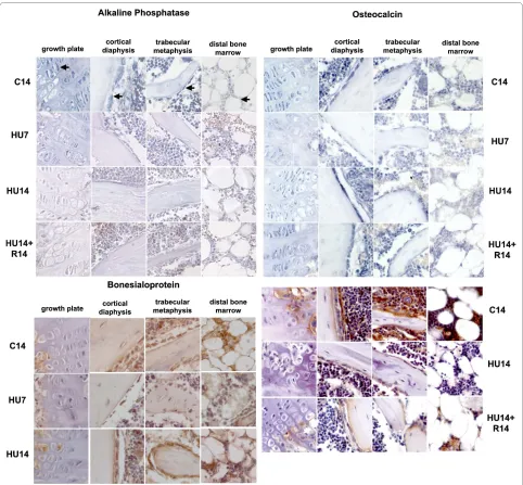
Among the genes characterizing the osteogenic pro-gression, the mRNA for AP (Figure 2, top-left) is expressed in control mice in all these cells, with lesser intensity in those in the distal epiphysis. Its amount decreases severely after 7 days of HU (HU7) and remains low at 14 days of HU (HU14) in pre-hypertrophic chon-drocytes, osteoblasts on endosteum, and a smaller decrease is detected in bone marrow cells at the distal epiphysis. The level of AP mRNA does not return to con-trol level in HU14 mice recovering on the ground for 14 days (HU14+R14).
The expression of BSP protein (Figure 2, bottom-left) is restricted to osteogenic and bone marrow cells. Com-pared to controls, the protein level transiently decreases after 7 days of HU and returns to control levels after 14 days of HU.
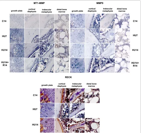
MT1-MMP expression occurs in the same cell types as those expressing AP, and its mRNA increases in HU7 and HU14 mice (Figure 3, top-left). The level of gene expres-sion decreases after recovery. All the cell types that express MT1-MMP show a similar pattern and timing of change in gene expression during HU and after recovery.
MMP-9 mRNA (Figure 3, top-right) is expressed in chondrogenic, osteogenic, bone marrow mesenchymal cells and macrophages. The expression of the transcript is
progressively decreased during the two weeks of HU, compared to control, and returns to a level similar to con-trol after recovery.
The expression of the protein for the endogenous inhibitor of MMPs, RECK (Figure 3, bottom) is observed in osteoblasts and in mesenchymal cells under the growth plate. The amount of RECK transiently decreases after 7 days of HU in all cell types, returns to control levels after 14 days of HU, and no further changes occur after recov-ery for 14 days (not shown).
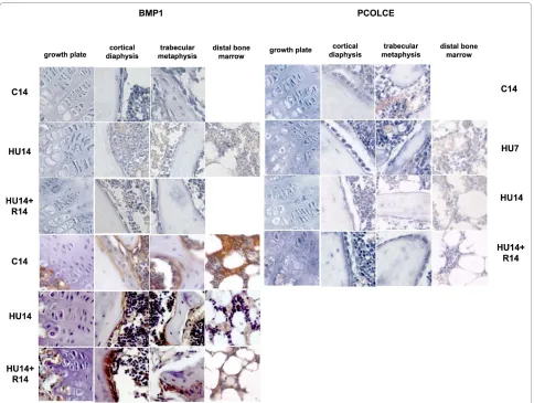
The expression of BMP1 and PCOLCE1, 2 occurs in the same cell types expressing AP. 14 days after HU is detected down modulation in the levels of expression for mRNAs (Figure 4, top-left) and proteins (Figure 4,
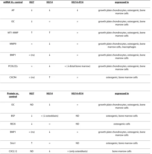
[image:5.595.60.540.93.474.2]Table 1: Gene and protein expression in HU mice and recovery versus control.
mRNA Vs. control HU7 HU14 HU14+R14 expressed in
AP | | | growth plate chondrocytes, osteogenic, bone
marrow cells
OC | = = growth plate chondrocytes, osteogenic, bone
marrow cells
MT1-MMP 9 9 = growth plate chondrocytes, osteogenic, bone
marrow cells
MMP9 = | = growth plate chondrocytes, osteogenic, bone
marrow cells, macrophages
BMP1 = (ns) | = growth plate chondrocytes, osteogenic, bone
marrow cells
PCOLCEs = | = (|distal bone marrow) growth plate chondrocytes, osteogenic, bone marrow cells
CXCR4 = (ns) 9 = osteogenic, bone marrow cells
Protein vs. control
HU7 HU14 HU14+R14 expressed in
OC ND | = growth plate chondrocytes, osteogenic, bone
marrow cells
BSP | = (|osteoblasts) ND osteogenic, bone marrow cells
RECK | = ND osteogenic cells
BMP1 = (ns) | = growth plate chondrocytes, osteogenic, bone
marrow cells
Stro1 9 = ND osteogenic, bone marrow cells
CXCL12 ND | = (only osteoblasts) bone marrow cells
Arrows pointing upwards indicate increase, pointing down indicate decrease of expression related to the corresponding control; = stays for equal to control. ND, not done; ns, not shown in the corresponding figure.
tom-left), and the mRNA level is restored after recovery for 14 days.
The modulation of expression of mRNAs for the PCOLCE 1,2 (Figure 4, top-right), is similar to that of BMP1, with a decrease detected after 14 days of HU, and returning to control level after recovery for 14 days.
Stromal protein Stro1 (Figure 5, top-left) is expressed in osteogenic and bone marrow cells and is transiently up modulated after 7 days of HU.
The CXCL12 protein is detected in controls with high-est expression in the bone marrow cells (Figure 5, top-right) and is down modulated in HU14 mice; after recov-ery for 14 days the protein is detected again in bone cells and also in the osteocytes within the trabeculae, but its expression is not restored in the cells of the distal bone marrow.
(Fig-ure 5, third panel from the top), is highly up modulated in the bone marrow at distal epiphysis after 14 days of HU and only slightly decreases after recovery.
Immunostaining for the stromal protein CD146 and in situ hybridization for VEGF (with a probe detecting the 3 forms of VEGF-A) were also done, and these genes did not show significant differences between control and HU mice (not shown).
Discussion
We report the evidences of modulation of the expression of genes and proteins in mesenchymal cells of mice tibia during HU. We also report the restoration of control gene and protein expression after re-loading of the limbs for 14 days.
Although in HU rodents the osteoblasts and mesenchy-mal osteogenic precursors were identified as the first cel-lular component responding to the lack of weight bearing with increased apoptosis [3], the effects of HU on gene expression at the cellular level in mesenchymal cells was not previously studied [5-9] and no information was available about how HU affects the expression of genes involved in the deposition, organization and remodeling of ECM.
We investigated either gene and/or protein expression, according to the availability of probes, as summarized in Table 1.
We show that HU can modulate the expression not only of genes involved in osteogenesis, but also of genes involved in ECM proteolysis and deposition, and of genes involved in the homeostasis of bone.
The modulation by HU of many genes and proteins occurs at the same time and with the same sign (increase or decrease of expression) in the cells residing in different tissue microenvironments of the long bone, with the exceptions described below for bone marrow in the distal epiphysis.
In control mice co-expression of AP, MT1-MMP, OC, MMP-9, PCOLCEs and BMP1 occurs in chondrogenic, osteogenic and bone marrow mesenchymal cells, while BSP, Stro1, VEGF-A and CD146 (these last two not shown) and CXCR4 expression are restricted to osteo-genic and bone marrow cells. RECK expression is local-ized to osteoblasts at the endosteum and under the growth plate, and CXCL12 expression to bone marrow cells.
No changes were detected after 3 days of HU but changes in gene and protein expression were evident by 7 days as decreases in the levels of AP, OC, BSP, RECK, and increases in the levels of MT1-MMP and Stro1. At this time disorganization of the trabecular arrangement at the growth plate and changes in the morphology of the osteo-blasts lining the endosteum were already evident, but no obvious thinning of the diaphyseal bone was detected.
After 14 days of HU, bone erosion was measured and decreases in the level of expression of MMP 9, BMP1 and PCOLCEs, CXCR4, CXCL12 were detected. The changes in gene expression observed after 7 days of HU persisted and/or were enhanced for some genes, (as the decrease in AP and the increase in MT1-MMP), while for other genes and proteins the level of expression reverted to that of the control (as for OC, BSP, RECK, Stro1).
Recovery on the ground for 14 days, after 14 days of HU, determined return to control level and reversal of the changes induced by HU, for expression of all the genes and proteins in chondrocytes at the growth plate and for osteoblasts and bone marrow cells in proximal and mid tibiae, with the exception of AP. This observation is in agreement with the slow recovery of AP expression after HU in rodents, in man and other mammals after space flight.
Whereas AP expression decreased and was not revers-ible by re-loading, MT1-MMP expression increased dur-ing HU and returned to control by 14 days after limb re-loading. This evidence suggests that the positive pleiotro-phism that couples MT1-MMP and AP expression during osteogenesis [22] was disrupted during HU.
In general, and with the exceptions below, synchrony and similarity in direction of the changes induced by HU and occurring upon recovery were observed in all the mesenchymal cells of different lineages and in the context of different local microenvironments. This suggests that a common mechanism for response to mechanical forces is activated in all cell types, and mediates the sensing of the changes induced by unloading, causing responses at cel-lular level. This mechanism would be independent from the different composition and characteristic of the local ECM, such as in cartilage, bone or bone marrow.
We suggest that MT1-MMP, expressed in all the cell types residing in these different ECMs, may play a key role in sensing the mechanical changes induced by HU. MT1-MMP expression is know to be modulated by mechanical forces [39], and biochemical microenviron-ment [40], and has the potential to "sense" the interaction with ECM [18], which in turn can affect the expression of other genes.
reflect the response to HU of cells in the mesenchymal compartment of bone marrow.
The decrease in procollagenase and PCOLCEs activi-ties, occurring only after 7 days of HU, may then diminish collagen fibrillogenesis and deposition of the novel ECMs of the several kinds present in the long bone, contributing to the loss of bone density.
The effects of HU on CXCR4 and CXCL12 in the tibia are specifically localized to the compartment of distal bone marrow. In general, this compartment has a distinct
response by comparison to the diaphyseal bone marrow, e.g. it is preferentially enriched in adipocytes in HU ani-mals and in other models of induced osteoporosis [41,42]. A specific pattern of HU-induced modulation was found for the CXCL12 protein, highly expressed in the bone marrow cells, which is down modulated after 14 days of HU. CXCL12 level was restored after recovery in osteoblasts and in the osteocytes within the trabeculae, but not in the bone marrow. CXCR4, the CXCL12 recep-tor, mRNA was up modulated after 14 days in HU,
[image:8.595.57.539.94.541.2]ularly in the cells of the bone marrow in the distal tibia, where it continued to be expressed at a relatively high level also after recovery. The effects of HU on cells in the bone marrow in the distal epiphysis might unbalance the CXCR4/CXCL12 signaling at least regionally. In view of the role that this signaling pathway plays in bridging hematopoiesis, osteogenesis and osteoclastogenesis, these effects might be relevant in the homeostasis of the whole bone. Since CXCL12 expression did not return to control level in the distal bone marrow after recovery, the unbalance might persist and, the restoration of bone bal-ance might lag.
In this report we have not mentioned gene expression in multinucleated cells in the bone marrow and in osteo-clasts; these are part of an ongoing study. We had prob-lems focusing on osteocytes at the microscope, possibly due to the relatively thick sections obtained from paraffin embedded whole bones, which limited our report about this cell type.
Conclusion
[image:9.595.58.540.95.549.2]To summarize, the net effects at the cellular level of HU treatment in mice were to depress the expression of genes for osteogenic progression, to uncouple the expression of
AP and MT1-MMP, to decrease generalized ECM degra-dation via down modulation of MMP-9, and to increase the pericellular proteolysis due to increased expression of MT1-MMP, in the presence of transiently decrease of the MMPs-inhibitory protein RECK in the osteogenic cells. Anabolic genes that govern collagen synthesis and depo-sition in fibrils (procollagenases and their enhancer pro-teins) were for the first time identified as targets of HU-dependent inhibition in cartilage, bone marrow and bone cells. This inhibition possibly aggravates the loss of matrix mass, already known to occur during HU, due to the reduction in collagen synthesis. The down-modula-tion of these funcdown-modula-tions may also contribute, together with the transient decrease of BSP protein in osteogenic cells, to the specific impairment of the maturation of osteo-genic matrix.
The simultaneous timing and direction of the response to HU for genes involved in the osteogenic progression and genes involved in proteolysis and deposition of the
ECM occurring in cells of different bone compartments and lineages, suggests that it might exist a common pro-cess of sensing the reduced gravity. We suggest that MT1-MMP increase is a key element in this response, able to mediate and influence further changes during HU. The response to HU for specific genes involved in homeosta-sis and localized to cells of the distal bone marrow points the possibility that HU might affect the homeostatic bal-ance of the whole bone; this point requires further studies in vitro and in vivo.
No changes were detected in CD146 protein and for VEGF-A.
Limb re-loading caused recovery of the previous pat-tern of expression with the exceptions of AP in the whole bone, and of CXCL12/CXCR4 in distal bone marrow. This suggests that, at least in the experimental time ana-lyzed, after re-loading of the limbs "all will not return as
well as before". Further investigation is particularly
[image:10.595.57.541.94.459.2]important to define if and when the expression of these
genes is recovered and how it is relevant for the re-acqui-sition of bone mass after re-loading.
Competing interests
The authors declare that they have no competing interests.
Authors' contributions
DV was the main research fellow, who performed the HU experiments, col-lected the samples of bones, prepared the molecular probes and performed and the in situ hybridization. He also contributed in writing the manuscript. AS did the histological preparation of the bones, and their sectioning. She took care of the immunohistochemistry experiments. DP contributed to the blind reading of the in situ hybridization and immunohistochemistry slides, she also participated in selecting and editing the figures and to the final editing of the manuscript. PM designed and supervised the experiments, coordinated the
project and wrote the manuscript. All authors read and approved the final manuscript.
Acknowledgements
This work was funded by an OSMA grant from ASI (Italian Space Agency) to Prof. Paola Manduca. We acknowledge the contribution of Prof. I. Bab and Dr. A. Bajayo of Bone Laboratory of The Hebrew University of Jerusalem, Jerusalem, Israel for morphometric measures of bone. We thank Prof. D. Corte Camerino and Dr. J.F. Desaphy for use of the animal facilities of the Pharmacobiology Dept. of the University of Bari and Prof. C. Tacchetti of the DIMES of the Univer-sity of Genoa for access to microscopic facilities. We are very grateful to Dr. Dai Williams for reading and copywriting the manuscript.
Author Details
1Genetics, DIBIO, University of Genoa, (Corso Europa 26), Genoa, (I-16132), Italy
[image:11.595.56.542.94.524.2]and 2DIMES, University of Genoa, (Via De Toni), Genoa, (I-16132), Italy
References
1. Wronski TJ, Morey-Holton ER: Skeletal response to simulated weightlessness: a comparison of suspension techniques. Aviat Space Environ Med 1987, 58:63-8.
2. Zhang P, Hamamura K, Yokota H: A brief review of bone adaptation to unloading. Genomics Proteomics Bioinformatics 2008, 6:4-7.
3. Aguirre JI, Plotkin LI, Stewart SA, Weinstein RS, Parfitt AM, Manolagas SC, Bellido T: Osteocyte apoptosis is induced by weightlessness in mice and precedes osteoclast recruitment and bone loss. J Bone Miner Res 2006, 21:605-15.
4. Wronski TJ, Morey-Holton ER, Doty SB, Maese AC, Walsh CC:
Histomorphometric analysis of rat skeleton following spaceflight. Am J Physiol 1987, 252:R252-5.
5. Salingcarnboriboon R, Tsuji K, Komori T, Nakashima K, Ezura Y, Noda M:
Runx2 is a target of mechanical unloading to alter osteoblastic activity and bone formation in vivo. Endocrinology 2006, 147:2296-2305. 6. Basso N, Bellows CG, Heersche JN: Effect of simulated weightlessness on
osteoprogenitor cell number and proliferation in young and adult rats. Bone 2005, 36:173-83.
7. Patel MJ, Liu W, Sykes MC, Ward NE, Risin SA, Risin D, Jo H: Identification of mechanosensitive genes in osteoblasts by comparative microarray studies using the rotating wall vessel and the random positioning machine. J Cell Biochem 2007, 101:587-99.
8. Pardo SJ, Patel MJ, Sykes MC, Platt MO, Boyd NL, Sorescu GP, Xu M, van Loon JJ, Wang MD, Jo H: Simulated microgravity using the Random Positioning Machine inhibits differentiation and alters gene expression profiles of 2T3 preosteoblasts. Am J Physiol Cell Physiol 2005,
288:C1211-21.
9. Carmeliet G, Nys G, Stockmans I, Bouillon R: Gene expression related to the differentiation of osteoblastic cells is altered by microgravity. Bone 1998, 22:139S-143S.
10. Zayzafoon M, Gathings WE, McDonald JM: Modeled microgravity inhibits osteogenic differentiation of human mesenchymal stem cells and increases adipogenesis. Endocrinology 2004, 145:2421-32. 11. Seitzer U, Bodo M, Müller PK, Açil Y, Bätge B: Microgravity and
hypergravity effects on collagen biosynthesis of human dermal fibroblasts. Cell Tissue Res 1995, 282:513-7.
12. Ishijima M, Tsuji K, Rittling SR, Yamashita T, Kurosawa H, Denhardt DT, Nifuji A, Noda M: Resistance to unloading-induced three dimensional bone loss in osteopontin-deficient mice. J Bone Miner Res 2002,
17:661-7.
13. Ono N, Nakashima K, Schipani E, Hayata T, Ezura Y, Soma K, Kronenberg HM, Noda M: Constitutively active parathyroid hormone receptor signaling in cells in osteoblastic lineage suppresses mechanical unloading-induced bone resorption. J Biol Chem 2007, 282:25509-16. 14. Ito T, Ohmori S, Kanda K, Kawano S, Murata Y, Seo H: Changes in serum
1,25-dihydroxyvitamin D3 and mRNAs for osteocalcin and alkaline phosphatase in femur unloaded by tail suspension in rats. Environ Med 1994, 38:103-6.
15. Hopkins DR, Keles S, Greenspan DS: The bone morphogenetic protein 1/ Tolloid like metalloproteinases. Matrix Biol 2007, 26:508-23.
16. Moali C, Font B, Ruggiero F, Eichenberger D, Rousselle P, Francois V, Oldberg A, Bruckner-Tuderman L, Hulmes DJ: Substrate-specific modulation of a multisubstrate proteinase. C-terminal processing of fibrillar procollagens is the only BMP-1 dependent activity to be enhanced by PCPE-1. J Biol Chem 2005, 280:24188-94.
17. Fisher LW, Whitson SW, Avioli LV, Termine JD: Matrix sialoprotein of developing bone. J Biol Chem 1983, 258:12723-7.
18. Malaval L, Wade-Guéye NM, Boudiffa M, Fei J, Zirngibl R, Chen F, Laroche N, Roux JP, Burt-Pichat B, Duboeuf F, Bolvin G, Jurdic P, Lafage-Proust MH, Amédée J, Vico L, Rossant J, Aubin JE: Bone sialoprotein plays a functional role in bone formation and osteoclastogenesis. J Exp Med 2008, 205:1145-1153.
19. Steiglitz BM, Kreider JM, Frankenburg EP, Pappano WN, Hoffman GG, Meganck JA, Liang X, Höök M, Birk DE, Goldstein SA, Greenspan DS:
Procollagen C proteinase enhancer 1 genes are important
determinants of the mechanical properties and geometry of bone and the ultrastructure of connective tissues. Mol Cell Biol 2006, 26:238-49.
20. Holmbeck K, Bianco P, Caterina J, Yamada S, Kromer M, Kuznetsov SA, Mankani M, Robey PG, Poole AR, Pidoux I, Ward JM, Birkedal-Hansen H:
MT1-MMP-deficient mice develop dwarfism, osteopenia, arthritis, and connective tissue disease due to inadeguate collagen turnover. Cell 1999, 99:81-92.
21. Filanti C, Dickson GR, Di Martino D, Ulivi V, Sanguineti C, Romano P, Palermo C, Manduca P: The expression of metalloproteinase2, 9, and -14 and of tissue inhibitors-1 and -2 is developmentally modulated during osteogenesis in vitro. J Bone Miner Res 2000, 15:2154-68. 22. Manduca P, Castagnino A, Lombardini D, Marchisio S, Soldano S, Ulivi V,
Zanotti S, Garbi C, Ferrari N, Palmieri D: Role of MT1-MMP in the osteogenic differentiation. Bone 2009, 44:251-265.
23. Noda M, Oh J, Takahashi R, Kondo S, Kitayama H, Takahashi C: RECK: a novel suppressor of malignancy linking oncogenic signaling to extracellular matrix remodeling. Cancer Metastasis Rev 2003, 22:167-175. 24. Zambuzzi WF, Yano CL, Cavagis AD, Peppelenbosch MP, Granjeiro JM,
Ferreira CV: Ascorbate-induced osteoblast differentiation recruits distinct MMP-inhibitors: RECK and TIMP-2. Mol Cell Biochem 2009,
322:143-50.
25. Sugiyama T, Kohara H, Noda M, Nagasawa T: Maintenance of the hematopoietic stem cell pool by CXCL12-CXCR4 chemokine signaling in bone marrow stromal cell niches. Immunity 2006, 25:977-988. 26. Otsuru S, Tamai K, Yamazaki T, Yoshikawa H, Kaneda Y: Circulating bone
marrow-derived osteoblast progenitor cells are recruited to the bone-forming site by the CXCR4/stromal cell-derived factor-1 pathway. Stem Cells 2008, 26:223-34.
27. Dar A, Kollet O, Lapidot T: Mutual, reciprocal SDF-1/CXCR4 interactions between hematopoietic and bone marrow stromal cells regulate human stem cell migration and development in NOD/SCID chimeric mice. Exp Hematol 2006, 34:967-75.
28. Wright LM, Maloney W, Yu X, Kindle L, Collin-Osdoby P, Osdoby P: Stromal cell-derived factor-1 binding to its chemokine receptor CXCR4 on precursor cells promotes the chemotactic recruitment, development and survival of human osteoclasts. Bone 2005, 36:840-53.
29. Shi S, Gronthos S: Perivascular niche of postnatal mesenchymal stem cells in human bone marrow and dental pulp. J Bone Miner Res 2003,
18:696-704.
30. Simmons PJ, Torok-Storb B: Identification of stromal cell precursors in human bone marrow by a novel monoclonal antibody, STRO-1. Blood 1991, 78:55-62.
31. Morey-Holton ER, Globus RK: Hindlimb unloading rodent model: technical aspects. J Appl physiol 2002, 92:1367-77.
32. Pierno S, Desaphy JF, Liantonio A, Luca De, Zarrilli A, Mastrofrancesco L, Procino G, Valenti G, Conte Camerino D: Disuse of rat muscle in vivo reduces protein kinase C activity controlling the sarcolemma chloride conductance. J Physiol 2007, 584:983-95.
33. Bajayo A, Goshen I, Feldman S, Csernus V, Iverfeldt K, Shohami E, Yirmiya R, Bab I: Central interleukin-1 receptor signaling regulates bone growth and mass. Proc Natl Acad Sci USA 2005, 102:12956-12961.
34. Alexander JM, Bab I, Fish S, Mueller R, Uchiyama T, Gronowicz G, Nahounou M, Zhao Q, White DW, Chorev M, Gazit D, Rosenblatt M:
Human parathyroid hormone 1-34 reverses bone loss in ovariectomized mice. J Bone Min Res 2001, 16:1665-1673.
35. Desbois C, Hogue DA, Karsenty G: The mouse osteocalcin gene cluster contains three genes with two separate spatial and temporal patterns of expression. J Biol Chem 1994, 269:1183-90.
36. Basyuk E, Bertrand E, Journot L: Alkaline fixation drastically improves the signal of in situ hybridization. Nucleic Acids Res 2000, 28:E46.
37. Kadkol SS, Gage WR, Pasternack GR: In situ hybridization - theory and practice. Mol Diagn 1999, 4:169-83.
38. De Block M, Debrouwer D: RNA-RNA in situ hybridization using digoxigenin-labeled probes: the use of high-molecular-weight polyvinyl alcohol in the alkaline phosphatase indoxyl-nitroblue tetrazolium reaction. Anal Biochem 1993, 215:86-9.
39. De Croos JN, Jang B, Dhaliwal SS, Grynpas MD, Pilliar RM, Kandel :
Membrane type-1 matrix metalloproteinase is induced following cyclic compression of in vitro grown bovine chondrocytes. Osteoarthritis Cartilage 2007, 15:1301-10.
40. Barbolina MV, Adley BP, Ariztia EV, Liu Y, Stack MS: Microenvironmental regulation of membrane type 1 matrix metalloproteinase activity in ovarian carcinoma cells via collagen-induced EGR1 expression. J Biol Chem 2007, 282:4924-31.
Received: 15 January 2010 Accepted: 5 July 2010 Published: 5 July 2010
This article is available from: http://www.biomedcentral.com/1471-2474/11/147 © 2010 Visigalli et al; licensee BioMed Central Ltd.
This is an Open Access article distributed under the terms of the Creative Commons Attribution License (http://creativecommons.org/licenses/by/2.0), which permits unrestricted use, distribution, and reproduction in any medium, provided the original work is properly cited.
41. Zayzafoon M, Gathings WE, McDonald JM: Modeled microgravity inhibits osteogenic differentiation of human mesenchymal stem cells and increases adipogenesis. Endocrinology 2004, 145:2421-32. 42. Marie PJ, Kaabeche K: PPAR Gamma Activity and Control of Bone Mass
in Skeletal Unloading. PPAR Res 2006:64807.
Pre-publication history
The pre-publication history for this paper can be accessed here: http://www.biomedcentral.com/1471-2474/11/147/prepub
doi: 10.1186/1471-2474-11-147
Cite this article as: Visigalli et al., Hind limb unloading of mice modulates
gene expression at the protein and mRNA level in mesenchymal bone cells