Expression of the
aa
1
bb
1 integrin, VLA-1, marks a
distinct subset of human CD4
+
memory T cells
Itamar Goldstein, … , Hong Jiang, Leonard Chess
J Clin Invest.
2003;
112(9)
:1444-1454.
https://doi.org/10.1172/JCI19607
.
The
a
1
b
1 integrin, very late antigen-1 (VLA-1), is a collagen receptor expressed in many
CD4
+T cells localizing to inflamed tissues. Here we show that the expression of VLA-1 is a
stable marker of a distinct subset of CD4
+memory T cells. Thus, in human peripheral blood
lymphocytes (PBLs), approximately 1–4% of the CD4
+T cells express VLA-1, and following
T cell receptor activation ex vivo, the percentage of VLA-1
+cells increases within the
CD45RO
+population. Importantly, the activated VLA-1
+and VLA-1
–cells can be isolated
and maintained in culture as phenotypically stable subsets. Functionally, CD4
+memory T
cells, operationally defined as the cells that divide rapidly following stimulation with a recall
antigen, are highly enriched for VLA-1
+cells. Moreover, depletion of the small fraction of
VLA-1
+cells present in CD4
+PBLs prior to stimulation significantly abrogated the
proliferative response to recall antigens. Notably, the VLA-1
+cells in fresh CD4
+PBLs are
composed of resting CD45RO
+/RA
–, CCR7
–, CD62L
+, CD25
–, and VLA-4
hicells.
Interestingly, this VLA-1
+subset is enriched for Th1-type cells, and Th1-polarizing
conditions during T cell activation favor the emergence of VLA-1
+cells. Thus, VLA-1
expression is a stable marker of a unique subset of human memory CD4
+T cells that
predominantly differentiates into Th1 cells.
Article
Aging
Find the latest version:
Introduction
The α1β1 integrin is a heterodimeric cell surface receptor composed of a β1 chain shared also by other β1 integrins and an α1 chain that contains a specific “inserted” I-domain in its extracellular por-tion responsible for collagen recognipor-tion (1, 2). The
α1β1 integrin uniquely binds to collagen IV but also binds to collagen I and weakly to laminin (3–5). The binding of very late antigen-1 (VLA-1) to these ECMs is thought to facilitate cellular migration through the laminin- and collagen IV–rich basement mem-branes, as well as to enhance migration within the collagen-rich interstitium (6). Moreover, VLA-1 interaction with collagen activates signaling path-ways that promote cell survival and progression through the cell cycle (7–9).
In T cells, VLA-1 becomes expressed only very late (more than 6 days) after T cell activation (10, 11). Fur-thermore, VLA-1/collagen interactions augment T cell receptor–mediated (TCR-mediated) proliferation and cytokine secretion (12, 13). Importantly, recent stud-ies in murine models of inflammation show that deleting the α1 integrin gene or blocking the func-tions of VLA-1 in vivo with mAb’s prevents the devel-opment of delayed-type hypersensitivity-like (DTH-like) immune responses, including those associated with graft-versus-host disease, adjuvant or Ab-induced arthritis, and hapten-Ab-induced colitis (14–18). Notably, in the wild-type untreated animals the majority of T cells (and monocytes) infiltrating the inflamed tissues express VLA-1 (15, 18).
Similarly, human T cells infiltrating inflamed tissues, particularly in Th1-mediated inflammation, are high-ly enriched for VLA-1+cells. For example, the T cells infiltrating the synovium of rheumatoid arthritis (RA) patients (12, 19), the lungs of sarcoidosis patients (20), and tuberculin purified protein derivative–induced (PPD-induced) skin blisters (11) are highly enriched in VLA-1–expressing cells. It is of interest that these VLA-1+ T cells in the inflamed tissue usually coexpress CD45RO, a marker of previously activated T cells (21, 22). In the fresh peripheral blood lymphocytes (PBLs) of normal individuals as well as patients, however, the expression of VLA-1 is restricted to only a small frac-tion of the CD45RO+T cells (21, 23). The frequent coexpression of VLA-1 with CD45RO in both inflam-matory sites and in peripheral blood, taken together
Expression of the
α
1
β
1 integrin, VLA-1, marks a distinct
subset of human CD4
+memory T cells
Itamar Goldstein,
1Shomron Ben-Horin,
1Jianfeng Li,
1Ilan Bank,
2Hong Jiang,
1and
Leonard Chess
11Department of Medicine, Columbia University, College of Physicians and Surgeons, New York, New York, USA 2Department of Medicine and Laboratory for Immunoregulation, Chaim Sheba Medical Center and Tel Aviv University,
Tel Aviv, Israel
The α1β1 integrin, very late antigen-1 (VLA-1), is a collagen receptor expressed in many CD4+T cells
localizing to inflamed tissues. Here we show that the expression of VLA-1 is a stable marker of a dis-tinct subset of CD4+memory T cells. Thus, in human peripheral blood lymphocytes (PBLs),
approx-imately 1–4% of the CD4+T cells express VLA-1, and following T cell receptor activation ex vivo, the
percentage of VLA-1+cells increases within the CD45RO+population. Importantly, the activated VLA-1+
and VLA-1–cells can be isolated and maintained in culture as phenotypically stable subsets.
Func-tionally, CD4+memory T cells, operationally defined as the cells that divide rapidly following
stim-ulation with a recall antigen, are highly enriched for VLA-1+cells. Moreover, depletion of the small
fraction of VLA-1+cells present in CD4+PBLs prior to stimulation significantly abrogated the
pro-liferative response to recall antigens. Notably, the VLA-1+cells in fresh CD4+PBLs are composed of
resting CD45RO+/RA–, CCR7–, CD62L+, CD25–, and VLA-4hicells. Interestingly, this VLA-1+subset is
enriched for Th1-type cells, and Th1-polarizing conditions during T cell activation favor the emer-gence of VLA-1+cells. Thus, VLA-1 expression is a stable marker of a unique subset of human
mem-ory CD4+T cells that predominantly differentiates into Th1 cells.
J. Clin. Invest.112:1444–1454 (2003). doi:10.1172/JCI200319607.
Received for publication July 25, 2003, and accepted in revised form August 26, 2003.
Address correspondence to: Leonard Chess, Department of Medicine, Columbia University, College of Physicians and Surgeons, 630 West 168th Street, PH8E Suite 101, New York, New York 10032, USA. Phone: (212) 305-9984;
Fax: (212) 305-4943; E-mail: [email protected].
Conflict of interest:The authors have declared that no conflict of interest exists.
with the observations that VLA-1 is expressed only in a fraction of the CD45RO cells, suggests the possibility that VLA-1 may mark a subset of memory T cells.
In this regard, precise identification of CD4+memory T cells is problematic, partly because unambiguous markers of memory have not been described (24). Thus, although CD45RO expression has been used to distin-guish naive from memory T cells, it is clear that not every T cell that expresses CD45RO is necessarily a memory cell, since virtually all recently activated T cells also express CD45RO. In this regard, CD45RO expression reflects more accurately an imprint of prior activation than a marker of memory cells. In addition, recent stud-ies give rise to the notion that the memory lineage is composed of different subsets based on the expression of other cell surface molecules, including chemokine receptors, selectins, and adhesion molecules (24–27). For example, the memory T cell pool has been subdivided based on the expression of the lymph node–homing chemokine receptor CCR7 into two major subsets. Because CCR7+cells home predominantly to lymph nodes, they have been termed the central memory sub-set. Migration of memory T cells from the lymph nodes to the blood and peripheral organs is associated with the loss of CCR7 expression and the functional differentia-tion of the memory cells into effector T cells capable of secreting cytokines and functioning as inducer and cyto-toxic cells. Thus, these CCR7–cells have been termed effector memory T cells (25).
Here we will present evidence that following CD4+T cell activation, whereas all the T cells express CD45RO, stable VLA-1 expression is observed only in a subset of the CD45RO+T cells. To determine whether this VLA-1– expressing subset represented memory cells, we defined the memory CD4+T cells functionally by their greater capacity to rapidly proliferate upon re-encounter with a recall antigen (e.g., tetanus toxoid, PPD, or mumps antigens). We used the vital dye CFSE dilution tech-nique (28, 29) to identify the memory population of cells as those that have rapidly divided following trig-gering with recall antigens. This enabled us to quanti-tate using flow cytometry not only the proportion of memory cells but also their cell surface phenotype with respect to VLA-1 and other surface markers. We found that the small fraction of VLA-1+T cells in fresh human PBLs form a major fraction within the memory cells proliferating in response to recall antigen and are pre-dominantly Th1-type memory cells. Moreover, deple-tion of the CD4+VLA-1+T cells from the PBLs marked-ly abrogates the recall response to recall antigens. Interestingly, the circulating CD4+VLA-1+memory cells are predominantly CCR7–, CD25–, VLA-4hi, and enriched for CD62L+cells.
Methods
Ab’s and reagents. The hybridoma producing the func-tion-blocking anti-human VLA-1 (1B3.1) that recog-nizes the αchain I-domain was originally generated in our laboratory at Columbia University (30).
Hybrido-ma cells producing anti-huHybrido-man VLA-1 (TS2/7) mAb were purchased from American Tissue Culture Collec-tion (Rockville, Maryland, USA). Another mAb that also recognizes the I-domain of human VLA-1 (AGF.1.3) was kindly provided by Biogen Inc. (Cam-bridge, Massachusetts, USA), and the phycoerythrin-conjugated (PE-phycoerythrin-conjugated) mAb to human VLA-1 (SR84; CD49a) was purchased from PharMingen (San Diego, California, USA). The following fluorochrome-conjugated murine anti-human mAb’s were all pur-chased from PharMingen: CD4, CD8, CD25, CD45RO, CD45RA, CD62L, CD69, VLA-2 (CD49b), VLA-4 (CD49d), anti–IFN-γ, and anti–IL-4. Isotype control mouse IgG1, IgG2a, and IgG2b (pure and fluoro-chrome-conjugated), biotinylated anti-human CCR7, and streptavidin-PE were also obtained from PharMin-gen. FITC-conjugated anti-TCR Vβ2 was purchased from Immunotech (Marseille, France). The hybridoma producing mAb’s to anti-CD3 (OKT3) was purchased from American Tissue Culture Collection. Fluorescein-conjugated F(ab′)2fragment goat anti-mouse IgG plus IgM were purchased from Jackson ImmunoResearch Laboratories (West Grove, Pennsylvania, USA).
FACS analysis. The immunostaining and subsequent flow-cytometric analysis of human lymphocytes was carried out as described previously (31). Briefly, cells were first treated with human Ig (Sigma-Aldrich, St. Louis, Missouri, USA) to reduce nonspecific Ab bind-ing to Fc receptors and subsequently were incubated with saturating concentrations of the indicated fluo-rescein-conjugated mAb for 15 minutes at 4°C. In some experiments, when pure mAb were used for the initial step, the cells were washed and then incubated for 15 minutes at 4°C with fluorescein-conjugated F(ab′)2fragments of polyclonal goat anti-mouse IgG plus IgM. Fluorescence intensity was measured on a FACScan cytofluorograph (Becton Dickinson Immunocytometry Systems, San Jose, California, USA) and analyzed using the Cellquest software (Becton Dickinson Immunocytometry Systems). Viable lym-phocytes were defined by their forward scatter/side scatter (FSC/SSC) characteristics or propidium iodide (10 µg/ml) in order to more accurately exclude dead cells from analysis. Of note, a very stringent compen-sation for interdetector spillage of fluorescence was necessary during sample acquisition due to the usual-ly low fluorescence intensity, which is characteristic of VLA-1 staining in most VLA-1+human T cells.
They were subsequently washed and incubated with anti-hapten microbeads and separated over a magnet-ic column system. The VLA-1+and VLA-1 fractions were likewise isolated using the MACS method. Briefly, cells were stained with anti–VLA-1 mAb’s (AGF.1.3), washed, incubated for 15 minutes with goat anti-mouse microbeads (Miltenyi Biotech GmbH), and then selected over magnetic columns, as above. In some experiments, the VLA-1+cells were isolated by incuba-tion with TS2/7, a mAb directed to a different epitope of VLA-1 to exclude effects unique to the AGF 1.3 mAb’s. Purity of subsets following isolation was deter-mined by immunostaining and FACS and usually was greater than 95%.
Purified T cells were plated at 1 ×10⁶to 2 ×10⁶cells/ well into 24-well plates containing 2 ×10⁶cells/well of irradiated autologous PBLs (250 Gy) in 1 ml of medi-um consisting of X-Vivo 15 (BioWhittaker Inc., Walk-ersville, Maryland, USA) supplemented with 1% heat-inactivated human AB serum, 100 U/ml penicillin, and 100 µg/ml streptomycin and maintained in a humidi-fied 37°C, 5% CO2incubator. The cells were stimulat-ed immstimulat-ediately after plating with either 0.1 µg/ml of toxic shock syndrome toxin-1 (TSST-1) superantigen (Sigma-Aldrich), 2.5 µg/ml anti-CD3 mAb’s, 3 floccu-lation units/ml (Lf/ml) tetanus toxoid (TT; setts Biological Laboratories, Worcester, Massachu-setts, USA), 5 µg/ml PPD (Parke-Davis Division, Warner Lambert Co., Morris Plains, New Jersey, USA), 5 µg/ml mumps viral antigens (BioWhittaker Inc.), or medium alone. To generate long-term T cell lines, the purified fresh CD4+ PBLs were stimulated with 0.1
µg/ml of TSST-1 and irradiated autologous APCs every 10 days. The cells were cultured in a medium consist-ing of RPMI, 10% FCS (HyClone Laboratories, Logan, Utah, USA), 2 mM L-glutamine, 100 U/ml penicillin, and 100 µg/ml streptomycin. Three days after the ini-tial stimulation, and every 3 days thereafter, the medi-um was supplemented with 50 U/ml of recombinant IL-2 (Hoffman-La Roche Inc., Nutley, New Jersey, USA). The VLA-1+and VLA-1–fractions were usually purified from the mixed cultures several days after the second stimulation and maintained as above. To further gen-erate VLA-1+and VLA-1–CD4+T cell clones, the cells were plated in 96-well U-bottom plates at a density of 3, 1, or 0.3 cells/well containing either 105irradiated autologous PBLs (250 Gy) or autologous EBV-trans-formed B cells (500 Gy) and pulsed with 0.1 µg/ml of TSST. Subsequently, proliferating wells were screened for their VLA-1 phenotype, and selected clones were expanded by repeated stimulation with TSST-1–pulsed irradiated APCs every 10 days.
Intracellular cytokine staining and Th polarization assays. T cells were activated with 20 ng/ml of phorbol 12,13-dibutyrate and 0.8 µM of ionomycin (Sigma-Aldrich) in the presence of 2 µg/ml of monensin (GolgiStop; PharMingen) for 5 hours at 37°C. Following this brief activation, cells were harvested, fixed, and permeabi-lized using the Cytofix/Cytoperm Plus kit and then
immunostained for intracellular IFN-γor IL-4 pro-duction, all in accordance with the manufacturer’s recommendations (PharMingen). All samples were then analyzed on a FACScan cytofluorograph (Becton Dickinson Immunocytometry Systems).
To induce Th1 polarization, the CD4+cells were stim-ulated with 2.5 µg/ml of anti-CD3 mAb in a medium containing 5 ng/ml recombinant human IL-12 (R&D Systems Inc., Minneapolis, Minnesota, USA). To induce Th2 polarization, the CD4+cells were activated in a medium containing 10 µg/ml of neutralizing anti–INF-γ mAb’s and 20 ng/ml of recombinant human IL-4 (both from R&D Systems Inc.). Cytokines and anti–IFN-γmAb’s were thereafter added to the cul-tures every 3 days, and cells were harvested and used for the different assays at day 8.
Analyzing T cell proliferation using the CFSE dilution method. The flow cytometric–based analysis of T cell proliferation by serial halving of the fluorescence intensity of the vital dye, CFSE, has been described pre-viously (29, 32). Briefly, T cells at 107/ml were sus-pended in 1 ml RPMI and then pulsed with 4 µM CFSE (Molecular Probes Inc., Eugene, Oregon, USA) for 15 minutes at 37°C with constant shaking. Subse-quently, the CFSE was quenched with 50% human AB serum for 1 minute, and the cells were washed twice with large volumes of RPMI medium. The desired sub-sets of cells were purified and then triggered with either a polyclonal stimulus, recall antigens, or medi-um alone, as described above. The cells were then cul-tured for 8 days without the addition of IL-2 to mini-mize the background proliferation. In experiments addressing the proliferative contribution of the VLA-1+ subset to the overall recall response, we first isolated CD4+PBLs using negative depletion, as above. Subse-quently, cells were rested overnight and then immuno-magnetically depleted of the rare VLA-1+CD4+T cells prior to antigenic triggering. To nullify the effects resulting from the separation procedure itself, we used sham depletion as our control group. The sham deple-tion consisted of incubating the cells with an isotype-matched mouse IgG (instead of the anti–α1 integrin mAb’s) followed by goat anti-mouse immunobeads. This was followed by passing the cells through an identical magnetic column as done for the experimen-tal group. The cells were harvested on day 8 and ana-lyzed for CFSE content (generation number), various surface markers, and, when indicated, for intracellular cytokines. Since some dye is lost from the parental generation and some T cells can slowly proliferate in response to soluble factors in the medium (bystander effect), we considered in the data analysis as antigen-responsive only those CD4+T cells that have under-gone more than cellular divisions.
Results
The VLA-1 receptor is expressed in vitro in a subset of CD45RO+T cells. VLA-1 is expressed in a small fraction
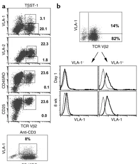
of fresh, resting PBLs (approximately 1–4%), and almost all these VLA-1+cells also coexpress CD45RO (21, 23). Following TCR activation of unselected human PBLs, an increase in the percentage of VLA-1+ cells becomes evident 1–2 weeks after activation (10). For example, in initial studies we analyzed the expres-sion of VLA-1 in fresh CD4+T cells induced to expand by direct TCR triggering using the TSST-1 superanti-gen, which exclusively triggers TCR Vβ2+cells. We fol-lowed the expression of VLA-1 in the Vβ2+T cells for up to 14 days. By day 8, as expected, most of the Vβ2+ T cells expressed the activation molecules CD45RO and CD25 (Figure 1a, upper four panels). In contrast, however, the VLA-1 molecule is expressed in only a small fraction (range: 6.8–13.5%, n = 10; Figure 1a) of the activated CD45RO+Vβ2+cells, and this expression was maximal by day 7 and stable thereafter. Further-more, in fresh CD4+T cells stimulated with anti-CD3 and maintained under similar conditions, the expres-sion of VLA-1 was also detected in only a small frac-tion (5.3–12.1%, n = 5) of the activated CD45RO+cells (Figure 1a, bottom panel). In contrast to VLA-1, the
α2β1integrin (VLA-2), which is also expressed late after activation, is expressed on more than 90% of the Vβ2+T cells.
Because VLA-1 is expressed in only a fraction of the CD45RO+T cells, we next asked whether the expression of VLA-1 is a stable phenotype of this subset of T cells. Thus, fresh CD4+T cells were stimulated twice (10 days apart) with TSST-1, and then at day 21, a time point when the majority of the cells are Vβ2+but only a frac-tion express VLA-1 (approximately 10–20%), we puri-fied the VLA-1+and VLA-1–cells from the mixed cul-tures (Figure 1b). Subsequently, the two different lines were cultured separately in IL-2–containing medium and repeatedly stimulated with TSST-1 and irradiated APCs, every 10 days. We found that the majority of cells in these two different cultures maintained their respec-tive VLA-1+or VLA-1–phenotypes for prolonged peri-ods of time (Figure 1b). Moreover, using limiting dilu-tion cloning, we derived highly purified T cell lines/clones that maintained a stable VLA-1+or VLA-1– phenotype. Taken together, these data demonstrate that VLA-1 is a stable cell surface marker of a subset of CD45RO+T cells.
VLA-1 marks a major subset within the memory lineage of CD4+T cells. As shown above, following activation VLA-1
[image:5.576.51.330.50.382.2]is expressed only in some CD45RO+CD4+T cells. Thus, we hypothesized that these VLA-1+ cells represent memory cells, operationally distinguished by their greater capacity to proliferate upon re-encounter with a recall antigen (e.g., TT, PPD, or mumps antigens). We used the CFSE dye-dilution technique to identify the
Figure 1
VLA-1 is expressed only in a subset of the activated CD45RO+CD4+T cells. (a) CD4+T cells were isolated
from PBLs as described in Methods. Subsequently, the cells were stimulated with irradiated APCs and TSST-1 (upper four panels) or anti-CD3 (bottom panel). The cultures were assayed at day 8 for the cell surface coexpression of CD4 and Vβ2, together with VLA-1, CD45RO, CD25, or VLA-2. The data shown are rep-resentative of at least ten independent experiments in different normal donors. (b) CD4+PBLs were
stimu-lated twice (every 10 days) with APCs and TSST-1. Subsequently, the cultures were purified into VLA-1–
or VLA-1+cell fractions. The cells were maintained in
memory cells, defined as the population of cells that have undergone at least two to three successive cellular divisions during the first week following recall antigen triggering. This approach enabled us to easily analyze individual cells for their cell generation number togeth-er with surface VLA-1 expression, as well as othtogeth-er sur-face molecules previously associated with memory cell subsets. We compared the response to recall antigen with the polyclonal response to superantigen, reason-ing that polyclonal stimulation would not only trigger memory cells but also indiscriminately trigger the large population of antigen-naive cells present in PBLs. We predicted that if VLA-1 marks the memory population, we should observe a significant increase in the per-centage of VLA-1–expressing cells in the cells dividing in response to recall antigens as compared with cells responding to polyclonal triggering.
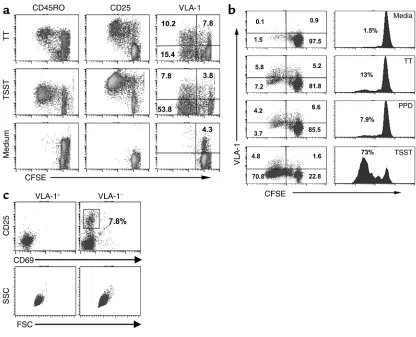
Thus, fresh CD4+PBLs from normal donors were labeled with CFSE and stimulated with TT (or other nominal antigens) or TSST-1. As shown in a
represen-tative experiment (Figure 2a), VLA-1+is expressed on the surface of more than 40% of the T cells rapidly dividing in response to TT triggering. In contrast, in T cells stimulated by TSST-1, a significantly smaller frac-tion (approximately 12%) of the rapidly dividing cells express VLA-1. In addition, the combined data from similar experiments in multiple donors (n = 11) showed a significant enrichment for VLA-1+T cells in TT-spe-cific dividing cells (range: 16.8–59.5%) and a consider-ably lower expression of VLA-1 following polyclonal triggering (range: 3.1–12.7%; P < 0.01, Student’s ttest). Furthermore, as seen in Figure 2b, the recall response to PPD in a sensitized individual was also enriched for VLA-1+cells (56%), as was the recall response to mumps virus antigens (range: 42–63%, n = 4).
Because a single T cell dividing n times will generate 2ndaughter cells, the proportion of CFSE-low cells at
[image:6.576.86.502.53.390.2]day 8 correlates directly with the original precursor fre-quency of TT-specific T cells in PBLs. Thus, as expect-ed, this proportion was different for each donor, as was Figure 2
VLA-1 expression preferentially distinguishes a large fraction of the CD4+T cells responding to recall antigens. (a) CD4+T cells from PBLs were
labeled with CFSE and plated in 24-well plates at 106cells/well containing 2 ×10⁶irradiated APCs and triggered by TT (upper panels), TSST-1
(middle panels), or medium (lower panels). The cells were harvested on day 8 and analyzed for CFSE content and the expression of VLA-1, CD25, or CD45RO. The data shown are representative of more than ten independent experiments in different donors. (b) CD4+PBLs
isolat-ed from a PPD+and TT-vaccinated individual were labeled with CFSE and triggered with TT, PPD, or TSST-1, harvested on day 8, and analyzed
for generation number and VLA-1 expression. (c) CD4+T cells from fresh PBLs were purified into VLA-1+and VLA-1–fractions and analyzed
the proportion of VLA-1+cells within the memory pop-ulation. Nevertheless, a dramatic enrichment for VLA-1+ cells following TT stimulation compared with TSST-1 stimulation was observed in all donors. Moreover, as shown in Figure 2a, the proportion of VLA-1+cells that have divided following TT stimulation (upper right panel) was much higher compared with the proportion of dividing CD45RO+T cells (upper left panel). Thus, these data are compatible with the notion that persist-ent VLA-1 expression distinguishes a T cell population highly enriched for memory cells.
As noted above, in normal individuals VLA-1 is expressed only in a small fraction of approximately 1–4% of the circulating CD45RO+CD4+T cells. More-over, these VLA-1+ cells are small cells that do not express CD25 or CD69 (Figure 2c), two molecules that usually mark all recently activated T cells. Because these findings suggest that the CD45RO+CD4+VLA-1+ PBLs represent a population of resting cells, it was of interest to determine directly their contribution to the overall response to recall antigens. Thus, we depleted
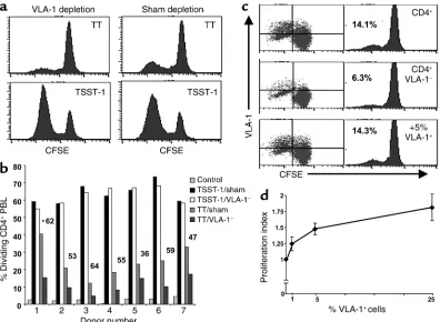
[image:7.576.91.487.312.602.2]these VLA-1+cells from fresh PBLs and in parallel sub-mitted the control group to a sham depletion proce-dure, as described in Methods. Subsequently, the VLA-1 and sham-depleted populations were labeled with CFSE, pulsed with TT or with TSST-1, and cultured for 8 days. We found that VLA-1 depletion signifi-cantly diminished the proliferative response to TT. In contrast, the depletion procedure had minimal effect on the proliferative response to TSST-1 (Figure 3a), in which a large fraction of dividing cells are likely derived from the naive Vβ2+cells in PBL. Overall, as depicted in Figure 3b, the combined data from the VLA-1 depletion experiments performed in seven nor-mal individuals demonstrate a significant reduction in the recall response to TT that ranged between 36% and 64% when compared with TSST-1 stimulation (range: 6.7–5.1%; P = 0.02, Wilcoxon signed rank test). Like-wise, VLA-1 depletion significantly reduced the response to other recall antigens, including PPD (65% reduction; n = 1) and mumps antigens (range: 50–67%; n = 4, P < 0.05).
Figure 3
Depleting VLA-1+cells from the PBLs significantly abrogates the proliferative response to TT. (aand b) CD4+T cells from fresh PBLs of
nor-mal individuals were depleted of the VLA-1+cells or sham depleted (see Methods). Subsequently, the two cell groups were labeled with CFSE
and stimulated with either TT or TSST-1 and irradiated APCs. The cells were harvested at day 8 and analyzed for CFSE dilution. (a) A rep-resentative experiment for donor 6. (b) The bar graph shows the results from seven different individuals. The Pvalue of 0.02 (Wilcoxon signed rank test) was obtained by comparing the relative reduction induced by VLA-1 depletion in the combined TT-stimulation experiments to the relative reduction measured in the TSST-stimulation experiments (asterisk indicates percentage of reduction in the TT response when com-paring sham to VLA-1 depletion). (c and d) The sham-depleted CD4+PBLs or the VLA-1–fraction with a increasing numbers of VLA-1+cells
added back (0%, 1%, 5%, and 25%) were stimulated with TT and cultured for 8 days and then assayed for cellular divisions and VLA-1 expres-sion. (c) A representative experiment in one donor. (d) Graph shows the combined results obtained from five different individuals. The pro-liferation index was calculated as the percentage of propro-liferation in a given sample divided by the percentage of propro-liferation in the pure VLA-1–
To further address the contribution of the VLA-1+ sub-set to the recall response, we asked whether adding back the VLA-1+fraction eluted from the columns to the VLA-1– fraction could restore the proliferative response. We found that adding back the VLA-1+cells results in the complete reconstitution of the recall response (Figure 3c). Moreover, as shown in Figure 3d, adding back the VLA-1+cells at increasing numbers into the VLA-1– frac-tion resulted in a dose-dependent increase (P < 0.01, n = 5, repeated measures ANOVA test) in the recall response. Interestingly, a near optimal proliferative recall response was reached at the 5% VLA-1+cell frequency, a number that approximates the upper limit of the physi-ological frequency of VLA-1+cells in the PBLs. This is further underscored by the observation that increasing the VLA-1+cell ratio above 25% had no additional incre-mental effect on the recall response (data not shown). Taken together, these results show that VLA-1 expres-sion preferentially marks a subset of memory cells.
Moreover, the circulating VLA-1+CD4+T cells are rest-ing memory cells that account for a significant fraction of the proliferative response to recall antigens.
VLA-1 expression is associated with the Th1 phenotype. It is known that the development of Th1-mediated inflam-matory responses in vivo is markedly abrogated in anti-VLA-1–treated mice and in α1–/–knockout mice (15,
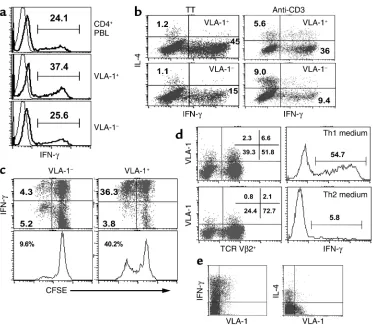
18). Moreover, as discussed above, a high percentage of the T cells infiltrating the inflammatory lesions in a variety of Th1-mediated diseases, both in mice and humans, express VLA-1. Thus, it was of interest to determine whether VLA-1 is preferentially expressed on Th1 cells. We first addressed this question in fresh CD4+PBLs purified into VLA-1+and VLA-1–fractions. Thus, the two cell populations were briefly activated with PMA/ionomycin and subsequently analyzed for intracellular production of IFN-γ and IL-4. We found that the VLA-1+fraction of CD4+T cells was somewhat enriched for cells expressing intracellular INF-γ com-Figure 4
VLA-1 expression is associated with Th1 polarization. (a) CD4+PBLs were purified into VLA-1+and VLA-1–fractions, activated with
PMA/ion-omycin for 5 hours, and analyzed for intracellular IFN-γproduction. (b) CD4+PBLs were stimulated with TT or anti-CD3; 10 days later the
cells were harvested and purified into VLA-1+and VLA-1–fractions, activated with PMA/ionomycin, and analyzed for intracellular IFN-γand
IL-4. (c) CD4+PBLs were labeled with CFSE, stimulated with TT, and 10 days later purified into VLA-1+and VLA-1–fractions. Subsequently,
the two subsets were activated and analyzed for divisions and intracellular IFN-γ. (d) CD4+PBLs were stimulated with TSST-1 in two
differ-ent environmdiffer-ents: Th1 (IL-12+) or TH2 (IL-4+and anti–IFN-γ). Ten days later cells were harvested and analyzed for VLA-1 and Vβ2 surface
[image:8.576.108.481.51.382.2]pared with the VLA-1–fraction (Figure 4a), whereas IL-4–producing cells were equally infrequent (approx-imately 2%) within the two fractions.
Next we asked whether, in fresh CD4+T cells induced to expand ex vivo by either a recall antigen (e.g., TT) or anti-CD3, the VLA-1+progeny is enriched for Th1 cells. Thus, following activation, the cells were cultured for 8 days, purified into VLA-1+or VLA-1–fractions, and ana-lyzed for production of intracellular IFN-γ and IL-4. We found that the ratio of IFN-γ+cells in the VLA-1+ frac-tion, regardless of the stimulafrac-tion, was significantly higher (threefold to fourfold) compared with the VLA-1– fraction (Figure 4b). Moreover, in CD4+T cells activat-ed by anti-CD3, the VLA-1–fraction was enriched for Th2 cells. Interestingly, following TT triggering, which induces Th1-biased responses in most normal individ-uals, we observed that within the dividing memory progeny almost all of the VLA-1+cells (more than 90%) produce IFN-γ (Figure 4c). Moreover, the VLA-1+ frac-tion contained a higher frequency of TT-specific mem-ory CD4+T cells (Figure 4c, lower panel).
These observations suggest that following TCR trig-gering, VLA-1–expression is preferentially associated with Th1 polarization. To further address this notion, we stimulated fresh CD4+PBLs with TSST-1 and con-ditioned them in either Th1-polarizing (+ IL-12) or Th2-polarizing (+ IL-4 and anti–IFN-γneutralizing mAb’s) environments. We found that culturing the cells in a Th1 environment induced an increased percentage of VLA-1–expressing cells. In contrast, the percentage of VLA-1+cells was reduced in the Th2 environment (Fig-ure 4d, left panel). In addition, as expected, the fre-quency of IFN-γ–secreting cells was significantly increased in the Th1 environment (right panel).
Next, it was of interest to ask whether VLA-1 expres-sion is also associated with the Th1 phenotype in vivo in chronically activated T cells. To address this ques-tion, we analyzed T cells isolated from fresh synovial fluids of patients with RA (n = 4) or psoriatic arthritis (PsA) (n = 2) for cytokine production, upon brief ex vivo activation. Again, we found that more than 90% of VLA-1–expressing CD4+T cells had a Th1 phenotype in all samples. Moreover, previous studies show that the synovial CD4+T cells from patients with RA are highly enriched for VLA-1+cells and usually contain 20–40% VLA-1+cells. In nonrheumatoid patients, including those with PsA, this percentage is usually lower, at 10–20% (12, 19). Taken together, these observations demonstrate that VLA-1 preferentially marks a major subset within the memory and effector Th1 population both in vitro and in situ at sites of inflammation. It should be noted, however, that VLA-1 expression does not mark all Th1-polarized cells.
Cell surface molecules coexpressed on VLA-1+and VLA-1– subsets of human CD4+cells. Because in previous studies
of both murine and human memory cells, the CCR7, CD62L, and VLA-4 molecules have been used to define subsets of memory and effector cells (25, 33, 34), we next evaluated the expression of these molecules in the
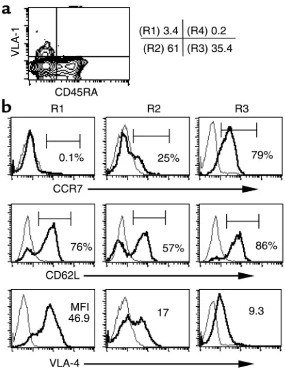
VLA-1+and VLA-1–subsets present in fresh PBLs and in the cell population that proliferates in response to recall antigens, ex vivo. We first analyzed the VLA-1+ cells present in PBLs (designated R1 in Figure 5a) and found that these cells invariably did not express the CCR7 molecule, but were enriched in CD62L+ and VLA-4hicells (Figure 5b). In this regard, the CCR7– VLA-4hiphenotype has been shown previously to be associated with tissue-localizing effector memory cells in mice (35). The high percentage of CD62L+cells in the VLA-1+population is of interest because CD62L is crucial for lymphocyte migration to lymphoid tissues (36). In contrast, the VLA-1– CD45RA–/RO+ subset (designated R2) was more heterogeneous with respect to the expression of CCR7, CD62L, and VLA-4. As expected, the naive CD45RA+T cell population was highly enriched for cells expressing CCR7 and CD62L and low levels of VLA-4.
[image:9.576.310.518.344.617.2]In addition, we attempted to analyze the differential coexpression of CCR7 and CD62L in the VLA-1+and VLA-1–CD4+memory T cells dividing in response to TT triggering. We found that the majority of the cells, regardless of their VLA-1 phenotype, downregulate
Figure 5
The VLA-1+CD4+T cells have a distinct cell surface phenotype. (a)
Fresh CD4+PBLs were analyzed for the cell surface coexpression of
CD45RA and VLA-1, and the four quadrants were identified as R1 to R4, where R1 = VLA-1+CD45RA–; R2 = VLA-1–CD45RA–; R3 = VLA-1–
CD45RA+; and R4 = VLA-1+CD45RA+. (b) Isolated CD4+T cells were
further purified into VLA-1+and VLA-1–fractions and then analyzed
for the coexpression of CD45RA together with CCR7, CD62L, or VLA-4. The histograms represent the viable CD4+T cell gate, and the data are
both CCR7 and CD62L within the first week follow-ing antigen activation. This further underscores the previous observations that the expression and down-regulation of CCR7 and CD62L in T cells is highly activation dependent.
Discussion
The studies described here provide evidence that in human CD4+T cells VLA-1 expression is highly restrict-ed to a distinct subset of memory cells with the poten-tial to mediate Th1-type immune responses in periph-eral tissues. Sevperiph-eral lines of evidence support this conclusion. First, when fresh CD4+PBLs are activated by a polyclonal trigger, even though all responding CD4+T cells stably express CD45RO, only a small per-centage expresses VLA-1, and the capacity to express VLA-1 is a stable property of this T cell subset. Second, when memory T cells are triggered by a recall antigen, VLA-1 becomes expressed on a large fraction of the rap-idly dividing cells that can be readily assayed by CFSE dilution. Third, the very small fraction (approximately 1–4%) of VLA-1+CD4+T cells found in fresh PBLs con-tribute significantly to the overall recall response. Fourth, the VLA-1+ fraction of the memory T cells dividing after recall antigen stimulation is highly enriched for Th1-polarized effector cells. Last, VLA-1+ CD4+T cells isolated from the peripheral blood are composed of small, nonactivated cells that have a dis-tinct cell surface phenotype of CCR7–, CD62L+, and VLA-4hi. Likewise, VLA-1+CD4+T cells isolated from fresh SFLs of arthritis patients lack CCR7 and CD25 expression and are predominantly Th1 cells; neverthe-less, many Th1 cells do not express VLA-1. Thus, VLA-1 expression in a subset of human memory CD4+T cells is associated with a distinct phenotype, suggesting that their main function in vivo is to mediate DTH-like (immune) responses in peripheral tissues.
The primary evidence that VLA-1 expression is a sta-ble marker of a subset of activated T cells stems from our initial observation that following polyclonal TCR triggering of CD4+PBLs, VLA-1 does not mark all acti-vated CD45RO+T cells. Moreover, VLA-1+and VLA-1– CD45RO+subsets purified from the activated T cell cultures and maintained in culture over time retain their VLA-1– or VLA-1+ phenotype. Because the CD45RO+phenotype is known to contain the memory cells, we asked whether VLA-1 expression defined a sub-set of memory cells. In this regard, as already noted, precise identification of human CD4+memory T cells is difficult, mainly because the phenotypic markers (e.g., CD45RO) used to distinguish memory T cells actually mark prior antigen activation rather than the acquisition of memory functions. Operationally, we defined memory cells as those cells that rapidly divide (more than two divisions) in response to recall antigens within the first week following recall antigen triggering ex vivo. We observed a dramatic increase in the per-centage of VLA-1+cells in this rapidly dividing popula-tion. Interestingly, following polyclonal triggering of T
cells in which both naive and memory cells are activat-ed, a much smaller percentage of VLA-1+CD45RO+ cells is observed. Taken together, these data suggest that VLA-1 is a unique marker because it preferentially distinguishes a subset within the activated CD45RO+ memory T cells. Interestingly, in human peripheral blood the depletion of VLA-1+ CD4+T cells, which account for only 1–4% of the cells, markedly reduces (36% to 64%) the proliferative recall response to TT.
Even though VLA-1 expression marks a large subset within the dividing CD45RO+CD4+memory T cells, a substantial fraction of the cells within this memory population does not express VLA-1. Thus, it was of interest to study the functions associated with the VLA-1+ and VLA-1–CD4+T cell subsets responding to recall antigens. We chose to study the profile of Th cytokine secretion by the VLA-1+and VLA-1–subsets because in humans T cells localizing to inflamed tissues in Th1 cell–driven immune responses are enriched for VLA-1+ cells (11, 12, 19, 20). Thus, we studied whether VLA-1 expression in CD4+T cells is preferentially associated with the Th1 phenotype defined in vitro by the pro-duction of IFN-γbut not IL-4. We found that following recall antigen or polyclonal triggering, the purified VLA-1+subset was highly enriched for effector Th1 cells. The VLA-1– fraction was more heterogeneous, although somewhat enriched in Th2 cells, and also contained Th1, Th0, and nonpolarized cells. Moreover, activating CD4+PBLs under different Th-polarizing conditions demonstrated that a Th1 environment favors the emergence of VLA-1+cells. Consistent with these ex vivo findings, we also observed that the great majority of VLA-1+CD4+T cells isolated from inflamed synovial fluids of patients with RA, where they form 10–40% of the population, express IFN-γbut not IL-4 (Figure 4e). It should be noted, however, that many Th1 cells in SFLs do not express VLA-1. Therefore, VLA-1 should not be considered a marker of Th1 polarization, but rather a marker of a distinct subset of antigen-expe-rienced Th1 cells.
The precise mechanism by which a Th1-biased cytokine environment favors the induction of VLA-1+ cells is unknown. In this context, several experimental observations suggest that the microenvironment in the lymph nodes, including interactions with local den-dritic cells, may influence the peripheral homing pref-erence of effector memory T cells (37). The type of anti-gen, the adjuvant used, and the route of immunization can all affect this environment. Thus, it will be of inter-est to study the influence of these parameters on the induction of VLA-1+memory T cells in vivo in animal models, where the nature of the antigen and the immu-nization conditions can be readily manipulated.
increased size and granularity usually observed in recently activated T cells (Figure 2c). These observa-tions are consistent with detailed studies in mice, show-ing that long-lived antigen-experienced T cells are usu-ally small, resting cells that do not express the activation markers CD25 or CD69 (24, 34). It is of interest that upon activation ex vivo both VLA-1+and VLA-1–CD4+PBL T cells re-express CD25 and CD69. Moreover, Lanzavecchia and colleagues have sug-gested that human memory T cells can be divided into two functionally distinct subsets based on the expres-sion of CCR7 (25), a lymph node–homing chemokine receptor (38). The CCR7+subset of memory cells that circulate through the lymphoid system was termed cen-tral memory. In contrast, the CCR7–memory T cells predominantly migrate into peripheral tissues and are enriched for effector-type cells; thus they are termed effector memory cells. We find that VLA-1 expression is associated with the CCR7–phenotype. It is important to note, however, that CCR7 expression is neither a spe-cific marker of the memory lineage nor a stable mark-er; many CCR7+memory and naive T cells differentiate into CCR7–T cells following antigen activation (25, 27). Because VLA-1 expression is a stable phenotypic marker of a subset of CCR7–CD45RO+T cells, we pro-pose that the expression of VLA-1 might more accu-rately identify the subset of memory T cells that local-ize to nonlymphoid tissues. This notion is further supported by the in vivo observations showing that VLA-1+cells are frequent among T cells isolated from inflamed tissues (e.g., SFL and PPD induced skin blis-ters). In contrast, we find that VLA-1 is only rarely expressed on CD45RO+CD4+T cells isolated from lym-phoid tissues (e.g., hyperplastic tonsils), even though many of the tonsillar T cells express molecules associ-ated with the postactivation phenotype, including CD45RO and CD25 (data not shown).
In addition to CCR7, the L-selectin molecule (CD62L) has also been suggested to distinguish central memory cells (39). The CD62L molecule mediates T cell traffick-ing to lymphoid tissues through interactions with its lig-and, the mucosal addressin cell-adhesion molecule-1, particularly expressed in the high endothelial venules (40). Therefore, CCR7+cells that migrate into lymph nodes usually express high levels of CD62L (34). CD62L, however, is also expressed in a significant percentage of CCR7–memory cells (25). Moreover, similar to CCR7, the CD62L molecule is not a stable surface marker, and usually T cells rapidly shed this receptor following cellu-lar activation (41). Interestingly, we find that the VLA-1+ subset is highly enriched for CD62L+cells. Our inter-pretation of these data is that CD62L expression is a fea-ture of resting memory T cells, as exemplified by the VLA-1+PBLs. In this context, other studies show that CD62L expression identifies the majority of T cells that proliferate to recall antigens in human PBLs (42, 43). Taken together, our results provide evidence that the VLA-1+CD62L+phenotype identifies a subset of quies-cent memory CD45RO+CD4+PBLs.
Another distinction of the circulating VLA-1+T cells is that most express high levels of VLA-4, whereas the VLA-1– fraction contains both VLA-4loand VLA-4hi cells. In this context, in a recent study Swain and col-leagues showed that in mice, anti-viral CD4+Th1 cells found in situ at the site of viral infection show a dis-tinct surface phenotype of CCR7–and VLA-4hi(35). In addition, earlier studies from the laboratories of Mori-moto and Schlossman have indicated that the expres-sion of high levels of CD29 (the β1 integrin chain shared by all VLA molecules) identifies the majority of CD45RA–PBLs with memory functions (43, 44). Taken together, these data may imply that the VLA-1+VLA-4hi cells form an important subset within the CD29hi memory population. It should be noted, however, that the CD29hipopulation also contains cells that do not express VLA-1, of which a small percentage (2–5%) express VLA-2. The stability and distinct functions of this VLA-1–VLA-2+population is unknown.
The function of the VLA-1 molecule expressed on the surface of a fraction of the memory T cells remains to be fully elucidated. For instance, because VLA-1 is the primary receptor for collagen IV (3), it is thought to facilitate the migration of lymphocytes through vascu-lar basement membranes into tissues (18). VLA-1 is also a receptor for collagen I (5), which is abundant in inter-stitial tissues, and thus VLA-1 may also facilitate the migration of lymphocytes in tissues. In addition, it is known that signaling via VLA-1 in T cells augments subthreshold TCR triggering (12, 13). In this context, studies in the α1–/–mice show that VLA-1 is the sole
collagen receptor that can activate the adaptor protein Shc pathway (7). This signaling was shown to promote cell survival and progression through the cell cycle in fibroblasts (8, 9). In addition, Meharra and colleagues (45) observed that in the α1–/–mice the α1β1integrin
is required for the survival of certain subsets of intes-tinal T cells within epithelial tissues. Moreover, we found recently that signaling via VLA-1 can induce IFN-γ production in activated Th1-type cell lines, inde-pendent of concomitant TCR retriggering (unpub-lished observation). Thus, VLA-1 functions may include inducing cellular programs that sustain Th1-mediated immunity in collagen-rich environments.
A corollary of our data is that VLA-1 expression may distinguish autoaggressive Th1 cells in human autoimmunity. This conclusion is compatible with the recent findings in mice that blockade of the VLA-1 pathway, either by genetic deletion (α1–/–) or with
Acknowledgments
We thank Israeli A. Jaffe for his assistance in obtaining the synovial fluid samples from patients and Eva Glick-man-Nir for her excellent technical assistance. This work was funded in part by NIH grants U19 AI-46132 and RO1 AI-44927.
1. Hemler, M.E., Huang, C., and Schwarz, L. 1987. The VLA protein family. Characterization of five distinct cell surface heterodimers each with a common 130,000 molecular weight β subunit. J. Biol. Chem.
262:3300–3309.
2. Kern, A., Briesewitz, R., Bank, I., and Marcantonio, E.E. 1994. The role of the I domain in ligand binding of the human integrin α1β1. J. Biol. Chem.
269:22811–22816.
3. Kern, A., and Marcantonio, E.E. 1998. Role of the I-domain in collagen binding specificity and activation of the integrins α1β1 and α2β1. J. Cell Physiol.176:634–641.
4. Riikonen, T., Vihinen, P., Potila, M., Rettig, W., and Heino, J. 1995. Anti-body against human α1β1 integrin inhibits HeLa cell adhesion to laminin and to type I, IV, and V collagens. Biochem. Biophys. Res. Commun.
209:205–212.
5. Heino, J. 2000. The collagen receptor integrins have distinct ligand recog-nition and signaling functions. Matrix Biol.19:319–323.
6. Dustin, M.L., and de Fougerolles, A.R. 2001. Reprogramming T cells: the role of extracellular matrix in coordination of T cell activation and migra-tion. Curr. Opin. Immunol.13:286–290.
7. Pozzi, A., Wary, K.K., Giancotti, F.G., and Gardner, H.A. 1998. Integrin
α1β1 mediates a unique collagen-dependent proliferation pathway in vivo. J. Cell Biol.142:587–594.
8. Giancotti, F.G., and Ruoslahti, E. 1999. Integrin signaling. Science.
285:1028–1032.
9. Hynes, R. 2002. Integrins: bidirectional, allosteric signaling machines.
Cell.110:673–687.
10. Hemler, M.E., Jacobson, J.G., Brenner, M.B., Mann, D., and Strominger, J.L. 1985. VLA-1: a T cell surface antigen which defines a novel late stage of human T cell activation. Eur. J. Immunol.15:502–508.
11. Iannone, F., Corrigall, V.M., Kingsley, G.H., and Panayi, G.S. 1994. Evi-dence for the continuous recruitment and activation of T cells into the joints of patients with rheumatoid arthritis. Eur. J. Immunol.
24:2706–2713.
12. Bank, I., et al. 1991. Expression and functions of very late antigen 1 in inflammatory joint diseases. J. Clin. Immunol.11:29–38.
13. Rao, W.H., Hales, J.M., and Camp, R.D. 2000. Potent costimulation of effector T lymphocytes by human collagen type I. J. Immunol.
165:4935–4940.
14. Tanaka, T., et al. 1995. Involvement of α1 and α4 integrins in gut mucos-al injury of graft-versus-host disease. Int. Immunol.7:1183–1189. 15. de Fougerolles, A.R., et al. 2000. Regulation of inflammation by
collagen-binding integrins α1β1 and α2β1 in models of hypersensitivity and arthritis. J. Clin. Invest.105:721–729.
16. Ianaro, A., et al. 2000. Anti-very late antigen-1 monoclonal antibody mod-ulates the development of secondary lesion and T-cell response in exper-imental arthritis. Lab. Invest.80:73–80.
17. Krieglstein, C.F., et al. 2002. Collagen-binding integrin α1β1regulates
intestinal inflammation in experimental colitis. J. Clin. Invest.
110:1773–1782. doi:10.1172/JCI200215256.
18. Fiorucci, S., et al. 2002. Importance of innate immunity and collagen binding integrin α1β1 in TNBS-induced colitis. Immunity.17:769–780. 19. Hemler, M.E., Glass, D., Coblyn, J.S., and Jacobson, J.G. 1986. Very late activation antigens on rheumatoid synovial fluid T lymphocytes. Associ-ation with stages of T cell activAssoci-ation. J. Clin. Invest.78:696–702. 20. Saltini, C., Hemler, M.E., and Crystal, R.G. 1988. T lymphocytes
com-partmentalized on the epithelial surface of the lower respiratory tract express the very late activation antigen complex VLA-1. Clin. Immunol. Immunopathol.46:221–233.
21. Bank, I., Koltakov, A., Goldstein, I., and Chess, L. 2002. Lymphocytes expressing α1β1 integrin (very late antigen-1) in peripheral blood of
patients with arthritis are a subset of CD45RO(+) T-cells primed for rapid adhesion to collagen IV. Clin. Immunol.105:247–258.
22. Stemme, S., Holm, J., and Hansson, G.K. 1992. T lymphocytes in human atherosclerotic plaques are memory cells expressing CD45RO and the integrin VLA-1. Arterioscler. Thromb.12:206–211.
23. Falcioni, F., et al. 1996. Influence of CD26 and integrins on the antigen sensitivity of human memory T cells. Hum. Immunol.50:79–90. 24. Berard, M., and Tough, D.F. 2002. Qualitative differences between naive
and memory T cells. Immunology.106:127–138.
25. Sallusto, F., Lenig, D., Forster, R., Lipp, M., and Lanzavecchia, A. 1999. Two subsets of memory T lymphocytes with distinct homing potentials and effector functions. Nature.401:708–712.
26. Andrew, D.P., et al. 2001. C-C chemokine receptor 4 expression defines a major subset of circulating nonintestinal memory T cells of both Th1 and Th2 potential. J. Immunol.166:103–111.
27. Campbell, J.J., et al. 2001. CCR7 expression and memory T cell diversity in humans. J. Immunol.166:877–884.
28. Lyons, A.B. 2000. Analyzing cell division in vivo and in vitro using flow cytometric measurement of CFSE dye dilution. J. Immunol. Methods.
243:147–154.
29. Wells, A.D., Gudmundsdottir, H., and Turka, L.A. 1997. Following the fate of individual T cells throughout activation and clonal expansion. Sig-nals from T cell receptor and CD28 differentially regulate the induction and duration of a proliferative response. J. Clin. Invest.100:3173–3183. 30. Bank, I., et al. 1989. A novel monoclonal antibody, 1B3.1, binds to a new
epitope of the VLA-1 molecule. Cell Immunol.122:416–423.
31. Lederman, S., et al. 1992. Identification of a novel surface protein on acti-vated CD4+ T cells that induces contact-dependent B cell differentiation (help). J. Exp. Med.175:1091–1101.
32. Lyons, A.B., and Parish, C.R. 1994. Determination of lymphocyte division by flow cytometry. J. Immunol. Methods.171:131–137.
33. Swain, S.L. 2003. Regulation of the generation and maintenance of T-cell memory: a direct, default pathway from effectors to memory cells.
Microbes Infect.5:213–219.
34. Wherry, E.J., et al. 2003. Lineage relationship and protective immunity of memory CD8 T cell subsets. Nat. Immunol.4:225–234.
35. Roman, E., et al. 2002. CD4 effector T cell subsets in the response to influenza: heterogeneity, migration, and function. J. Exp. Med.
196:957–968.
36. Steeber, D.A., et al. 1998. Efficient lymphocyte migration across high endothelial venules of mouse Peyer’s patches requires overlapping expres-sion of L-selectin and β7 integrin. J. Immunol.161:6638–6647. 37. Mora, J.R., et al. 2003. Selective imprinting of gut-homing T cells by
Peyer’s patch dendritic cells. Nature.424:88–93.
38. Forster, R., et al. 1999. CCR7 coordinates the primary immune response by establishing functional microenvironments in secondary lymphoid organs. Cell.99:23–33.
39. Lanzavecchia, A., and Sallusto, F. 2002. Progressive differentiation and selection of the fittest in the immune response. Nat. Rev. Immunol.
2:982–987.
40. Spertini, O., Kansas, G.S., Munro, J.M., Griffin, J.D., and Tedder, T.F. 1991. Regulation of leukocyte migration by activation of the leukocyte adhesion molecule-1 (LAM-1) selectin. Nature.349:691–694. 41. Kahn, J., Ingraham, R.H., Shirley, F., Migaki, G.I., and Kishimoto, T.K.
1994. Membrane proximal cleavage of L-selectin: identification of the cleavage site and a 6-kD transmembrane peptide fragment of L-selectin.
J. Cell Biol.125:461–470.
42. Hengel, R.L., et al. 2003. Cutting edge: L-selectin (CD62L) expression dis-tinguishes small resting memory CD4+ T cells that preferentially respond to recall antigen. J. Immunol.170:28–32.
43. Tedder, T.F., Matsuyama, T., Rothstein, D., Schlossman, S.F., and Mori-moto, C. 1990. Human antigen-specific memory T cells express the hom-ing receptor (LAM-1) necessary for lymphocyte recirculation. Eur. J. Immunol.20:1351–1355.
44. Morimoto, C., and Schlossman, S.F. 1993. P. Rambotti Lecture. Human naive and memory T cells revisited: new markers (CD31 and CD27) that help define CD4+ T cell subsets. Clin. Exp. Rheumatol.11:241–247. 45. Meharra, E.J., et al. 2000. Reduced gut intraepithelial lymphocytes in