Distinct roles for the kidney and systemic
tissues in blood pressure regulation by the
renin-angiotensin system
Steven D. Crowley, … , Thu H. Le, Thomas M. Coffman
J Clin Invest.
2005;
115(4)
:1092-1099.
https://doi.org/10.1172/JCI23378
.
Angiotensin II, acting through type 1 angiotensin (AT
1) receptors, has potent effects that
alter renal excretory mechanisms. Control of sodium excretion by the kidney has been
suggested to be the critical mechanism for blood pressure regulation by the
renin-angiotensin system (RAS). However, since AT
1receptors are ubiquitously expressed,
precisely dissecting their physiological actions in individual tissue compartments including
the kidney with conventional pharmacological or gene targeting experiments has been
difficult. Here, we used a cross-transplantation strategy and AT
1Areceptor–deficient mice to
demonstrate distinct and virtually equivalent contributions of AT
1receptor actions in the
kidney and in extrarenal tissues to determining the level of blood pressure. We demonstrate
that regulation of blood pressure by extrarenal AT
1Areceptors cannot be explained by
altered aldosterone generation, which suggests that AT
1receptor actions in systemic
tissues such as the vascular and/or the central nervous systems make nonredundant
contributions to blood pressure regulation. We also show that interruption of the AT
1receptor–mediated short-loop feedback in the kidney is not sufficient to explain the marked
stimulation of renin production induced by global AT
1receptor deficiency or by receptor
blockade. Instead, the renin response seems to be primarily determined by renal
baroreceptor mechanisms triggered by reduced blood pressure. Thus, the regulation of
blood pressure by the RAS is mediated by AT
1receptors both within and outside the […]
Article
Cardiology
Find the latest version:
Distinct roles for the kidney and systemic
tissues in blood pressure regulation
by the renin-angiotensin system
Steven D. Crowley,1 Susan B. Gurley,1 Michael I. Oliverio,1 A. Kathy Pazmino,1 Robert Griffiths,1
Patrick J. Flannery,1 Robert F. Spurney,1 Hyung-Suk Kim,2 Oliver Smithies,2 Thu H. Le,1
and Thomas M. Coffman1
1Division of Nephrology, Department of Medicine, Duke University and Durham VA Medical Centers, Durham, North Carolina, USA. 2Department of Pathology, University of North Carolina, Chapel Hill, North Carolina, USA.
Angiotensin II, acting through type 1 angiotensin (AT
1) receptors, has potent effects that alter renal excretory
mechanisms. Control of sodium excretion by the kidney has been suggested to be the critical mechanism for
blood pressure regulation by the renin-angiotensin system (RAS). However, since AT
1receptors are
ubiqui-tously expressed, precisely dissecting their physiological actions in individual tissue compartments including
the kidney with conventional pharmacological or gene targeting experiments has been difficult. Here, we used
a cross-transplantation strategy and AT
1Areceptor–deficient mice to demonstrate distinct and virtually
equiva-lent contributions of AT
1receptor actions in the kidney and in extrarenal tissues to determining the level of
blood pressure. We demonstrate that regulation of blood pressure by extrarenal AT
1Areceptors cannot be
explained by altered aldosterone generation, which suggests that AT
1receptor actions in systemic tissues such
as the vascular and/or the central nervous systems make nonredundant contributions to blood pressure
regu-lation. We also show that interruption of the AT
1receptor–mediated short-loop feedback in the kidney is not
sufficient to explain the marked stimulation of renin production induced by global AT
1receptor deficiency
or by receptor blockade. Instead, the renin response seems to be primarily determined by renal baroreceptor
mechanisms triggered by reduced blood pressure. Thus, the regulation of blood pressure by the RAS is
medi-ated by AT
1receptors both within and outside the kidney.
Introduction
The renin-angiotensin system (RAS), acting through type 1 angiotensin (AT1) receptors, is a master regulator of fluid
homeo-stasis (1). The critical role of this pathway in regulation of blood pressure is highlighted by the impressive efficacy of angiotensin-converting enzyme (ACE) inhibitors and angiotensin receptor blockers (ARBs) in patients with hypertension and in mice lacking the AT1A receptor, the major murine AT1 receptor isoform. These
animals have very low blood pressure and profound salt sensitivity (2, 3). Control of sodium excretion by the kidney has been suggest-ed to be the critical mechanism for blood pressure regulation by the RAS (4). However, as AT1 receptors are ubiquitously expressed,
precisely dissecting and quantifying their physiological actions in individual tissue compartments including the kidney has been dif-ficult. Accordingly, there is no direct proof of the primacy of the kidney in regulation of blood pressure by the RAS.
At the cellular level, responsiveness to angiotensin II is conferred by expression of angiotensin receptors. Angiotensin receptors can be divided into 2 pharmacological classes, type 1 (AT1) and type 2
(AT2), based on their differential affinities for various nonpeptide
antagonists (5). Studies using these antagonists suggested that most of the classically recognized functions of the RAS are
medi-ated by AT1 receptors (6). Gene targeting studies confirmed these
conclusions (7). AT1 receptors from a number of species have been
cloned (8, 9), and 2 subtypes, designated AT1A and AT1B, have been
identified in rat and mouse (10). The murine AT1 receptors are
products of separate genes and share substantial sequence homol-ogy (10). AT1A receptors predominate in most organs, except the
adrenal gland and regions of the CNS, where AT1B expression may
be more prominent (10). A single report has suggested that AT1B
receptors might also exist in humans (11), but this has not been confirmed in the unpublished work of several independent groups, and the consensus view is that there is no human counterpart to the murine AT1B receptor. Thus, the AT1A receptor is considered
the closest murine homolog to the single human AT1 receptor.
AT1 receptors can be found in organ systems that play key roles
in blood pressure homeostasis, including the heart, kidney, blood vessels, adrenal glands, and cardiovascular control centers in the brain (5). In the vascular system, stimulation of AT1 receptors
causes potent vasoconstriction (2). In the adrenal cortex, their activation stimulates the release of aldosterone (12), thereby pro-moting sodium reabsorption in the mineralocorticoid-responsive segments of the distal nephron (13). In the brain, intraventricu-lar injection of angiotensin II causes a dramatic pressor response mediated by AT1 receptors (14). In the kidney, activation of AT1
receptors is associated with renal vasoconstriction and antinatri-uresis (15, 16). Furthermore, it has been suggested that activation of AT1 receptors at the juxtaglomerular apparatus suppresses renin
release through the so-called short-loop feedback mechanism (17, 18). While AT1 receptors have actions in myriad tissues that could
potentially influence blood pressure, a prevailing view is that the
Nonstandard abbreviations used: ACE, angiotensin-converting enzyme; ARB, angiotensin receptor blocker; AT1, type 1 angiotensin; JGA, juxtaglomerular appara-tus; RAS, renin-angiotensin system.
Conflict of interest: The authors have declared that no conflict of interest exists. Citation for this article:J. Clin. Invest.115:1092–1099 (2005).
research article
The Journal of Clinical Investigation http://www.jci.org Volume 115 Number 4 April 2005 1093
actions of the RAS to influence kidney function are dominant in chronic control of blood pressure (4, 19). Identifying the tis-sues and cell lineages responsible for blood pressure regulation by the RAS will provide novel insights into the integrated control of blood pressure and will more precisely define the mechanisms of the antihypertensive actions of ACE inhibitors and ARBs. Here, we used a cross-transplantation strategy and AT1A receptor–deficient
mice to demonstrate distinct and virtually equivalent contribu-tions of AT1 receptor actions in the kidney and in extrarenal
tis-sues to determining the level of blood pressure.
Results
Cross-transplantation strategy. In order to define the relative impor-tance of renal versus nonrenal AT1 receptor pools in blood pressure
regulation by the RAS, we carried out cross-transplantation experi-ments, performing fully vascularized kidney transplants between genetically matched F1(C57BL/6 × 129) wild-type mice and mice
homozygous for a targeted disruption of the Agtr1a gene locus encoding the AT1A receptor. In the resulting recipient animals, both
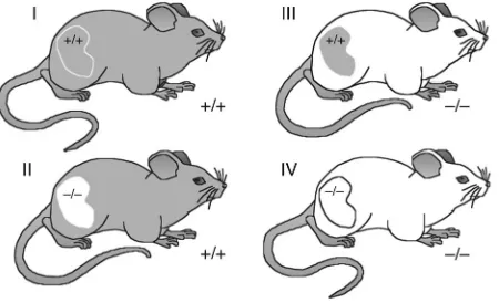
adrenal glands were retained. As shown in Figure 1, by varying the genotype of the transplant donors and recipients, we generated 4 groups of animals in which renal function was provided entirely by the single transplanted kidney. Group I animals were Agtr1a+/+
recipients (R+) that were transplanted with kidneys from
Agtr1a+/+ donors (D+) and thus had normal expression of
AT1A receptors in the donor renal and recipient nonrenal
tis-sues (D+R+). We will refer to group I as the D+R+ group, since
AT1A receptors were present in both kidney and somatic
tis-sues. Group II animals lacked expression of AT1A receptors
in the kidney, but had normal expression of receptors in systemic tissues (D–R+). In group III, AT
1A receptors were present in the
kid-ney but absent from all nonrenal tissues (D+R–). Finally, group IV
mice were completely deficient in AT1A receptors (D–R–).
Blood pressure measurements in Agtr1a+/+ and Agtr1a–/– mice. In order
to accurately measure blood pressures in conscious, unrestrained animals, we implanted radiotelemetry transmitters. Using this approach, we first measured blood pressures in otherwise unma-nipulated Agtr1a+/+ and Agtr1a–/– mice. Figure 2A shows that
blood pressures were significantly reduced in F1(C57BL/6 × 129)
Agtr1a–/– mice (89 ± 8 mmHg) compared with wild-type controls
(112 ± 3 mmHg; P = 0.03). The magnitude of the blood pressure reduction in Agtr1a–/– animals was very similar to that observed in
our previous experiments using tail cuff manometry (2). We also analyzed a series of 24-hour blood pressure recordings and found that the general nature of blood pressure fluctuations was similar in Agtr1a+/+ and Agtr1a–/– mice (Figure 2B), which indicated that
the absence of AT1A receptors does not affect qualitative patterns
of diurnal variation in blood pressure.
Distinct contributions of renal and systemic AT1 receptors to blood
pres-sure homeostasis. Among the transplanted animals, blood prespres-sures in group I (D+R+) were very similar to those of nontransplanted,
Agtr1a+/+ control mice (118 ± 5 vs. 112 ± 3 mmHg; P = 0.44), which
indicated that the transplant procedure does not significantly alter blood pressure. Figure 3 shows that the absence of AT1A receptors
in the kidney alone in group II (D–R+) animals was associated with
a significant reduction in blood pressure compared with the D+R+
group (99 ± 4 vs. 118 ± 5 mmHg; P = 0.006 vs. D+R+). This finding
is consistent with the prevailing view that regulation of sodium handling by the kidney is a major determinant of the chronic level of blood pressure and indicates that the absence of AT1A
recep-tor pathways in the kidney alone is sufficient to lower blood pres-sure. However, blood pressures were similarly reduced in group III (D+R–) mice, which lack AT
1A receptors in systemic tissues but
have normal renal expression of AT1A receptors (99 ± 2 vs. 118 ± 5
mmHg; P = 0.006). Thus, AT1A receptors in nonrenal tissues also
make a nonredundant contribution to the determination of the blood pressure level. Moreover, the presence of intact AT1A
recep-tor pathways in the kidneys of these animals does not compensate for the absence of receptors outside of the kidney. With respect to blood pressure homeostasis, the finding that blood pressures were virtually identical in the D–R+ and D+R– groups indicates that the
contributions of AT1A receptors in the kidney and systemic tissues
are very similar in magnitude.
Blood pressures were further reduced in group IV (D–R–)
[image:3.585.53.278.84.221.2]ani-mals, which completely lack AT1A receptors (86 ± 3 mmHg; P = 0.02
Figure 1
Kidney cross-transplantation groups. Wild-type (+/+) or AT1A receptor–
deficient (–/–) mice were transplanted with a wild-type or AT1A
recep-tor–deficient kidney. Nephrectomies were performed as described in Methods. Group I animals (D+R+) had a full complement of AT1A
recep-tors. Group II animals (D–R+) expressed AT1A receptors only outside the
kidney. Group III animals (D+R–) expressed AT1A receptors only within
the kidney. Group IV animals (D–R–) completely lacked AT1A receptors.
Recipients retained their own adrenal glands. n ≥ 6 per group.
Figure 2
Radiotelemetry blood pressure in untransplanted wild-type and AT1A receptor–deficient (Agtr1a–/–) mice. (A) Blood
pres-sure is reduced in Agtr1a–/– mice (black bar) vs. wild-type
con-trols (white bar); *P =0.03; n ≥ 4 per group. (B) Preservation of diurnal blood pressure variation in Agtr1a–/– mice. MAP, mean
[image:3.585.277.538.602.738.2]vs. D–R+; P = 0.002 vs. D+R–). This suggests that the mechanisms
for control of blood pressure by systemic and renal AT1A receptors
are nonoverlapping. Finally, blood pressures in D–R– animals were
very similar to those measured in intact, nontransplanted Agtr1a–/–
mice (89 ± 8 mmHg), which further illustrates the lack of any sig-nificant impact of the transplant procedure on blood pressure.
Effects of oral sodium loading on blood pressures. To assess the impact of altered sodium homeostasis on blood pressure regulation in the experimental groups, we examined the effects of high-salt feeding. After baseline blood pressures of mice on the normal (0.4% NaCl) diet were established, mice in each experimental group were fed a high-salt (6% NaCl) diet, and blood pressures were simultaneous-ly monitored. As shown in Figure 4, the high-salt diet caused sig-nificant increases in blood pressures only in the D–R+ (14.2% ± 5%
[from 99 ± 4 to 114 ± 9 mmHg]; P = 0.03 vs. normal salt) and the D–R– (20.1% ± 4.9% [from 85 ± 3 to 102 ± 5 mmHg]; P = 0.01 vs.
nor-mal salt) groups, lacking AT1A receptors in the kidney. By contrast,
in the D+R+ or D+R– groups with a normal complement of renal AT1A
receptors, high-salt feeding did not significantly affect blood pres-sures (118 ± 5 to 122 ± 7 mmHg for D+R+, 99 ± 2 to 105 ± 3 mmHg
for D+R–; P = not significant vs. normal salt in both groups). Thus,
the absence of AT1A receptors in the kidney was associated with
sodium sensitivity, whereas groups with normal renal AT1A receptor
expression were sodium resistant.
Extrarenal AT1A receptors, aldosterone, and blood pressure. As
dis-cussed above, there are a number of direct actions of AT1 receptors
that impact on blood pressure (19). In addition, AT1 receptors in
the zona glomerulosa of the adrenal gland indirectly affect blood pressure by stimulating aldosterone release and, in turn, promot-ing sodium reabsorption by the distal nephron (12, 13). We there-fore considered the possibility that impaired aldosterone release might explain the blood pressure reduction that we observed in D+R– mice, which lack AT
1A receptors in systemic tissues. To test
this, we collected urine from animals in each of the experimental groups and measured urinary aldosterone levels by ELISA.
As shown in Figure 5, aldosterone excretion was equivalent in the D+R+ and D–R+ groups (2,395 ± 260 vs. 3,177 ± 649 pg/24 h),
which is consistent with the fact that they have normal comple-ments of adrenal AT1A receptors. In contrast, aldosterone
excre-tion was reduced by approximately 50% in D+R– animals, lacking
AT1A receptors in nonrenal tissues including the adrenal glands
(1,158 ± 207 pg/24 h; P = 0.005 vs. D+R+; P = 0.01 vs. D–R+), which
suggests that the impaired aldosterone response in these animals is the cause of their reduced blood pressure. The aldosterone excretion in the D–R– mice, which completely lack AT
1A receptors,
was significantly higher than that in the D+R– group (2,643 ± 441
pg/24 h; P < 0.05). Since the adrenal glands in D–R– mice also lack
AT1A receptors, this augmented aldosterone excretion is likely
due to marked renin stimulation (see below) generating a high level of angiotensin II that stimulates the AT1B receptors in the
adrenal cortex. Other pathways for aldosterone stimulation may also play a role (20). Yet blood pressures are significantly lower in D–R– mice completely lacking AT
1A receptors compared with
those in the D+R– group, despite their higher aldosterone levels.
To determine whether lower aldosterone production in the D+R–
animals accounts for their lower blood pressures versus the D+R+
group, we performed an additional experiment using adrenalec-tomized D+R+ and D+R– animals in which aldosterone levels were
clamped by the infusion of equivalent amounts of aldosterone via an osmotic minipump. As shown in Figure 6A, infusing aldosterone at 1 μg/day provided supraphysiological aldosterone replacement, since urinary aldosterone excretion was increased almost 4-fold in the infused mice compared with mice with intact adrenal glands (P = 0.005). Nevertheless, this enhanced level of aldosterone had no effect on blood pressure in the D+R+ groups (118 ± 1 vs. 118 ± 5
mmHg; P = not significant). Moreover, urinary aldosterone excre-tion was not significantly different between the adrenalectomized D+R+ and adrenalectomized D+R– groups that received aldosterone
infusions (8,020 ± 2,627 vs. 8,828 ± 1,364 pg/24 h, respectively; P = not significant). However, despite supraphysiological and equivalent aldosterone levels, blood pressures were significantly reduced in the aldosterone-infused D+R– group compared with the
D+R+ group (106 ± 1 vs. 118 ± 1 mmHg; P < 0.0001) as shown in
Figure 6B. These data demonstrate that regulation of blood pres-sure by extrarenal AT1A receptors cannot be explained by changes
in aldosterone generation, which indicates that AT1 receptor
actions in other systemic tissues make nonredundant contribu-tions to blood pressure regulation.
Extrarenal AT1A receptors and renal sympathetic innervation. The
sym-pathetic nervous system, acting through renal nerves can modu-late sodium handling by the kidney (21). As AT1 receptors in the
CNS can stimulate sympathetic tone (22, 23), we considered the possibility that altered neural input to the kidney might represent a mechanism for the altered blood pressures in the D+R– group.
Furthermore, since the renal pedicle is transected in the course of harvesting the donor kidney, renal innervation may be impaired in this model. To examine these possibilities, we compared renal norepinephrine content in the D+R+ and D+R– groups with that of
[image:4.585.88.240.84.180.2]kidneys from Agtr1a+/+ and Agtr1a–/– mice. Norepinephrine content
Figure 3
Blood pressures in the cross-transplantation groups measured by radio-telemetry. *P =0.006 vs. group I; #P = 0.02 vs. group IV; §P =0.002 vs.
[image:4.585.333.506.587.681.2]group IV; ‡P =0.00007 vs. group I (n≥ 6 per group).
Figure 4
research article
The Journal of Clinical Investigation http://www.jci.org Volume 115 Number 4 April 2005 1095
is an accepted measure of renal sympathetic nerve activity (24). As shown in Figure 7, renal norepinephrine content was reduced significantly in both groups of transplanted kidneys compared with those in Agtr1a+/+ controls (P < 0.001), which suggested that
significant re-innervation of the transplanted kidneys had not occurred during the period of study. As there was no difference in norepinephrine levels between kidneys from the Agtr1a+/+ and
Agtr1a–/– mice (1,094 ± 132 vs. 1,286 ± 79 ng/g), the absence of AT1A
receptors does not substantially affect renal norepinephrine con-tent in the presence of intact renal nerves. Furthermore, norepi-nephrine levels were virtually identical in kidneys of the D+R+ and
D+R– mice (173 ± 53 vs. 163 ± 75 ng/g), which indicates that altered
sympathetic nerve input in the kidney cannot explain the reduced blood pressures in the D+R– animals.
Renal AT1A receptors and regulation of renin. Angiotensin II may
regulate its own synthesis by activation of AT1 receptors at the
juxtaglomerular apparatus (JGA), suppressing renin release (18). As this is one of several intrarenal actions of AT1A receptors that
could affect blood pressure, we examined the impact of renal and systemic AT1A receptors on renin mRNA expression in the kidney.
We found that renin mRNA levels in transplanted kidneys of D+R+ animals (29 ± 8 pg of renin mRNA/μg total mRNA) were
nearly identical to those of unmanipulated kidneys from wild-type mice (25 ± 4 pg of renin mRNA/μg total mRNA). In previous stud-ies, we and others have described markedly enhanced renin expres-sion and JGA hypertrophy in Agtr1a–/– mice with generalized
defi-ciency of AT1A receptors (25, 26). However, as shown in Figure 8,
this phenotype was not recapitulated in D–R+ animals, which lack
AT1A receptors only in the kidney. While kidney renin mRNA levels
tended to be higher in D–R+ mice (59 ± 19 pg of renin mRNA/μg
total mRNA) than in the D+R+ group, this difference was not
sta-tistically significant (P = 0.18). Thus, the absence of AT1A receptors
only in the kidney and the consequent lack of short-loop feedback did not markedly alter renin mRNA levels. Similarly, the absence of AT1A receptors only in nonrenal tissues of D+R– animals had
no effect on renin expression (22 ± 14 pg of renin mRNA/μg total mRNA) compared with the D+R+ group (Figure 8). By contrast, the
simultaneous absence of AT1A receptors in both renal and nonrenal
tissues of D–R– animals was associated with a marked upregulation
of renin expression to 708 ± 161 pg of renin mRNA/μg total mRNA (P < 0.003 vs. all other groups). Based on the findings from the D–R+
group, interruption of short-loop feedback in the kidney cannot explain the more than 10-fold increase in renin mRNA levels in the D–R– group. Accordingly, we postulate that this marked
augmen-tation in renin expression is due to the lower blood pressures in
this group. Moreover, since blood pressure reductions of almost 20 mmHg in the D–R+ and D+R– groups were not sufficient to trigger
this response, our data suggest that a threshold of blood pressure lowering must be achieved for activation of baroreceptor mecha-nisms that stimulate renin (27, 28).
We performed additional experiments to examine the contribu-tion of blood pressure to the stimulacontribu-tion of renin seen in the com-plete absence of AT1A receptors. To this end, we measured blood
pressure and renin mRNA expression in D–R– mice fed a high-salt
(6% NaCl) diet. As in our previous experiments on unmanipulated Agtr1a–/– mice (3), high-salt feeding caused a significant increase in
blood pressure in the D–R– mice (102 ± 5 mmHg) compared with
D–R– mice fed a normal (0.4% NaCl) diet (86 ± 3 mmHg; P = 0.01).
The blood pressures in the D–R– animals fed a high-salt diet increased
sufficiently, becoming not significantly different from those of the D–R+ and D+R– groups on a normal diet (99 ± 4 and 99 ± 2 mmHg
in D–R+ and D+R– groups, respectively). This normalized blood
pres-sure in the high-salt D–R– group was associated with a dramatic
suppression of renin mRNA levels (130 ± 58 pg of renin mRNA/μg total mRNA) compared with the D–R– animals on the normal diet
(708 ± 161 pg of renin mRNA/μg total mRNA; P = 0.003).
Discussion
The important role of the kidney in regulation of blood pressure has been long recognized (29), and the relationship between altera-tions in systemic blood pressure and changes in renal sodium excre-tion is well documented (30). For example, an elevaexcre-tion in perfusion pressure in the renal artery results in a rapid increase in sodium and water excretion by the kidney, so-called pressure natriuresis (30). Based on such observations, Guyton and coworkers suggested that whenever arterial pressure is elevated, activation of this pressure-natriuresis mechanism will cause sufficient excretion of sodium and water to return systemic pressures to normal (31). Because of its potent actions to modulate renal sodium excretion, angiotensin II has been implicated as a major determinant of these pressure-natriuresis relationships (19, 32). These actions may be mediated by direct effects of AT1 receptors on renal resistance vessels and
[image:5.585.77.250.83.174.2]epithe-lial cells or indirectly through stimulation of aldosterone produc-tion via AT1 receptors in the adrenal gland (32).
Figure 5
Urinary aldosterone excretion in the cross-transplantation groups.Urine samples were collected over 24 hours from mice in metabolic cages. Aldosterone was quantitated by ELISA (pg/24 h). *P =0.005 vs. group I;
[image:5.585.302.540.532.653.2]#P =0.01 vs. group II; ‡P <0.05 vs. group IV (n ≥ 6 per group).
Figure 6
Aldosterone clamp experiment. Adrenalectomies (ADX) were performed at the time of native nephrectomies. Osmotic minipumps containing aldosterone (ALDO) were implanted at the time of second nephrecto-my/adrenalectomy. (A) Urine samples were collected over 24 hours for aldosterone in ADX+ALDO groups (groups IA and IIIA) versus groups I and III from the initial experiment in Figure 5. §P≤ 0.005 vs. group I;
The importance of the RAS in clinical medicine is highlighted by the impressive cardiovascular efficacy of pharmacological agents that inhibit the synthesis or activity of angiotensin II. ACE inhibi-tors and ARBs are very effective and well tolerated antihyperten-sive agents (1). Along with their ability to lower blood pressure, these agents also ameliorate morbidity and mortality associated with cardiovascular diseases including congestive heart failure, coronary artery disease, and stroke (33, 34). The remarkably simi-lar profiles of ACE inhibitors and ARBs in these disorders suggest a common mechanism underlying their efficacy: reducing AT1
receptor signaling in critical target tissues. Along with their expres-sion in the kidney, AT1 receptors are present not only in the kidney
but in myriad sites that may influence blood pressure including the vasculature, adrenal glands, and brain. Yet the key sites that determine the level of blood pressure cannot be precisely localized using pharmacological antagonists that block AT1 receptors in all
tissues or in conventional gene targeting experiments. Accordingly, in the studies described here, we have used a renal cross-transplan-tation strategy to define the relative contributions of AT1A receptor
actions in the kidney and in extrarenal tissues.
We found that absence of AT1A receptors in the kidney in D–R+
mice is sufficient to reduce blood pressure by almost 20 mmHg (Figure 3), despite normal expression of receptors in all other tissues. These findings indicate that renal AT1A receptors have
unique and nonredundant actions in blood pressure homeostasis. As aldosterone levels are unaffected in these D–R+ animals, these
observations show that blood pressure is regulated by direct effects of AT1 receptors on kidney cells, independent of any impact of
min-eralocorticoids. We suggest that this reduction in blood pressure is a direct consequence of interruption of AT1 receptor actions in
the renal vasculature and/or renal epithelia that would otherwise reduce urinary sodium excretion. Our finding of sodium-sensitive blood pressure changes in animals lacking renal AT1A receptors
is consistent with this conclusion. While the baseline blood pres-sures were significantly lower in the D–R– compared with the D–R+
group, the absolute magnitude of the blood pressure increases with high-salt feeding were similar, 17 and 15 mmHg, respectively, representing the component of blood pressure reduction due to sodium and volume deficits. Furthermore, it is likely that inhibi-tion of these intrarenal pathways is an important mechanism for blood pressure lowering by ACE inhibitors and ARBs.
Although our studies support a vital role for the kidney in regu-lation of blood pressure, we find that AT1 receptors outside the
kid-ney also make a unique contribution to blood pressure homeosta-sis that is virtually equivalent to and independent of the intrarenal
actions of angiotensin II. Thus, we found that D+R– mice, which
lack AT1A receptors in extrarenal tissues but have the normal
complement of receptors in the kidney, also had blood pressure reductions of approximately 20 mmHg (Figure 3). We considered the possibility that alterations of aldosterone release due to the absence of AT1 receptors in the zona glomerulosa of the adrenal
gland might be responsible for low blood pressures in D+R– mice;
this would be consistent with the hypothesis that control of renal sodium handling is the final common pathway for chronic blood pressure homeostasis. To test this possibility, we measured aldosterone excretion in D+R– mice and found that absence of AT1A
receptors only in extrarenal tissues was associated with a reduc-tion in aldosterone excrereduc-tion of approximately 50% compared with controls (Figure 5). Preservation of a partial aldosterone response in these mice is probably due to the presence of AT1B receptors, the
minor murine AT1 receptor isoform. The adrenal cortex is one of
the few tissues with appreciable expression of AT1B receptors (10).
In order to determine whether the reduced aldosterone genera-tion in the D+R– mice was the cause of their low blood pressures, we
clamped aldosterone at supraphysiological levels by chronic infu-sion into adrenalectomized D+R– and D+R+ animals using osmotic
pumps. The aldosterone levels in the 2 groups were similar, yet the blood pressures remained lower in the D+R– group lacking
extrarenal AT1A receptors, compared with those in D+R+ controls
(Figure 6B). These findings corroborate other studies in which infusion of aldosterone alone failed to raise blood pressure (35) and suggest that the adrenal gland is not a critical site for blood pressure control by extrarenal AT1A receptors. We speculate instead
that AT1A receptors in the CNS and/or in the vasculature are more
likely to mediate the component of blood pressure regulation that is independent of the kidney. As our data on renal norepinephrine content indicate a persistent lack of sympathetic innervation in the transplanted kidneys (Figure 7), the contribution of extrarenal AT1 receptors to enhancing or modulating renal nerve activity and
thereby altering renal excretory functions may be underestimated in these experiments. Therefore, the major actions of extrarenal AT1 receptors to regulate blood pressure in this model are likely to
be due to effects on the vasculature. These could be direct actions of AT1A receptors on vascular smooth muscle cells (2) or indirect
effects of AT1A receptors in the CNS to modulate vascular resistance
(14). In this regard, recent studies have suggested that G-protein– coupled receptor actions in vascular smooth muscle make impor-tant contributions to chronic blood pressure homeostasis (36).
[image:6.585.78.248.83.172.2]The availability of renin affects the generation of angiotensin II by the RAS, and renin expression is tightly controlled at the JGA
Figure 7
Renal norepinephrine content. Norepinephrine levels were measured in kidneys harvested from unmanipulated Agtr1a+/+ and Agtr1a–/– mice
as well as in kidney grafts harvested from D+R+ and D+R– mice.
[image:6.585.324.510.598.690.2]*P < 0.001 vs. WT (n =5 per group).
Figure 8
research article
The Journal of Clinical Investigation http://www.jci.org Volume 115 Number 4 April 2005 1097
in the kidney (37). Angiotensin II is an important component in the regulatory pathways for renin and may control its own syn-thesis by activating AT1 receptors at the JGA, thereby suppressing
renin release (18, 25). The actions of ACE inhibitors and ARBs to increase renin mRNA expression and to cause JGA hypertrophy (38) have been cited as evidence supporting the existence of this so-called short-loop feedback mechanism. Although we find that renal AT1A receptors are key determinants of blood pressure, our
studies do not indicate a major role for these receptors in regulat-ing renin expression in the kidney.
In D–R+ animals, which lack AT1A receptors only in the kidney, we
found that renin mRNA levels are not significantly elevated despite the absence of AT1 receptors at the JGA and the significant
reduc-tion in blood pressure (Figure 8). Yet in the D–R– group completely
lacking AT1A receptors, renin mRNA expression is increased more
than 10-fold above baseline. Because blood pressures are reduced even further in D–R– compared with D–R+ animals, we posit that
renal baroreceptor mechanisms are primarily responsible for the marked stimulation of renin associated with interruption of AT1
receptor signaling (28). Moreover, since blood pressure reductions of almost 20 mmHg in D–R+ or D+R– groups were not sufficient to
trigger an increase in renin expression, our data also suggest that a threshold of blood pressure lowering must be achieved before the baroreceptor mechanisms are activated to stimulate renin produc-tion (27). The conclusion that blood pressure as such affects renin production is further supported by our observation that increasing blood pressure by dietary sodium loading significantly suppresses renin expression in D–R– mice. Taken together, our findings are
consistent with previous studies by Matsusaka and colleagues showing that JGA hypertrophy is correlated with reduced blood pressure rather than with the absence of AT1 receptors at the JGA
in chimeric AT1A-null mice (39).
In summary, AT1 receptors in the kidney have unique actions
to determine the normal level of blood pressure. These are direct effects of AT1 receptors on kidney cells that are independent of
aldosterone and likely target renal vasculature and/or epithelia to modulate sodium excretion. While the AT1A receptors in the kidney
are key determinants of blood pressure, they do not have a major role in regulating renin expression. Instead, our data indicate that baroreceptor mechanisms are dominant in the stimulation of renin production that is associated with absence of AT1 receptor
signal-ing. We have also demonstrated that AT1 receptors outside the
kid-ney make unequivocal and unique contributions to blood pressure homeostasis. These actions of extrarenal AT1A receptors cannot be
explained by altered aldosterone generation alone, and they affect the basal level of blood pressure despite the presence of intact AT1A
receptor pathways in the kidney. These studies illustrate the com-plexity of blood pressure regulation by the RAS and suggest that maximal efficacy of ACE inhibitors and ARBs requires complete blockade of renal and extrarenal pathways. Because these pharma-cological inhibitors only ameliorate but do not prevent progressive renal disease and other complications, a more precise understand-ing of their mechanisms of blood pressure lowerunderstand-ing and end-organ protection should lead to new opportunities for optimizing treat-ments for hypertension and its complications.
Methods
Animals. Mice lacking AT1A receptors for angiotensin II were generated
by homologous recombination in embryonic stem cells as previously described (2). Animals were bred and maintained in the animal facility of
the Durham Veterans Affairs Medical Center according to NIH guidelines with the approval of the Institutional Animal Care and Use Committees of the Duke University and Durham VA Medical Centers. Agtr1a genotypes, designated “+” for the wild-type allele and “–” for the targeted allele, were determined by PCR analysis of DNA isolated from tail biopsies using the following primers: Agtr1a+/+ upstream (5′
-GCATCATCTTTGTGGT-GGG-3′) with a sequence common to both alleles, Agtr1a+/+ downstream
(5′-ATCAGCACATCCAGGAATG-3′), and a sequence in the neomycin resistance cassette present only in the null allele (5′ -TGCCGAGAAAG-TATCCATCATGGCTGATGC-3′). F1(C57BL/6 × 129) Agtr1a+/+ and Agtr1a–/–
mice were generated from intercrosses of inbred 129/SvEv and C57BL/6
Agtr1a+/– mice in which the Agtr1a mutation had been back-crossed onto
each respective background for more than 12 generations. We studied male F1 mice that were 2–4 months old.
Mouse kidney transplantation. Transplantation of a single mouse kidney with bilateral native nephrectomy was performed as we have described previously (40). Briefly, animals were anesthetized with isoflurane, and the donor kidney, ureter, and bladder were harvested en bloc, including the renal artery with a small aortic cuff and the renal vein with a small caval cuff. These vascular cuffs were anastomosed to the recipient abdominal aorta and vena cava, respectively, below the level of the native renal vessels. Total ischemic time averaged 35–40 minutes. Donor and recipient bladders were attached dome to dome. The right native kidney was removed at the time of transplant, and the left native kidney was removed through a flank incision 1–3 days later; care was taken to preserve the recipient adrenal glands intact with their normal blood supply. Overall surgical mortality was approximately 5%. Each experimental group included at least 6 animals.
Data collection was initiated 2 weeks after transplantation to allow reso-lution of any ischemic injury associated with the transplant procedure. In addition, at the end of the experiments, sections of kidney grafts were examined to confirm normal renal structure, including the absence of isch-emic tubular damage. In previous studies, we have found that glomerular filtration rates in syngeneic mouse renal transplants are similar to those observed in uninephrectomized mice (40).
Radiotelemetry transmitter implantation. We used a radiotelemetry system (Transoma Medical) to monitor blood pressure in conscious mice, as described previously (41). Six to 8 days after the kidney transplantation procedure, mice were anesthetized with isofluorane, and a pressure-sensing catheter was implanted into the left carotid artery, as described previously (42). The transducer unit was then inserted into a subcutaneous pouch along the right flank generated by blunt dissection inferiorly from the orig-inal neck incision. Mice were allowed to recover for 7 days after surgery to regain their normal circadian rhythms before experiments were initiated. During the time that their blood pressures were being monitored, mice were housed in a monitoring room in the animal facility where quiet is maintained and no other activities are permitted. In separate experiments using identical procedures, catheters were also implanted into otherwise unmanipulated Agtr1a+/+ and Agtr1a–/– mice.
Blood pressure analysis. Arterial pressure recordings from unanesthetized, unrestrained animals were collected, stored, and analyzed using Dataquest A.R.T. software (version 2.2; Transoma Medical). The blood pressure data were collected continuously with sampling every 5 minutes for 10-second intervals (41) during the prescribed time periods. In some experiments, pres-sures were recorded continuously for at least 48 hours. Otherwise, in order to conserve battery function, we carried out blood pressure measurements between 10:00 am and 2:00 pm during the appointed days of the protocol.
pres-sure levels were established, mice from each of the experimental groups were placed on high-salt (6% NaCl) diet (Harlan Teklad) for 1 week while continuously monitoring blood pressures.After the blood pressure mea-surements were taken, the mice were placed in metabolic cages, and urine was collected for 24 hours. Urinary aldosterone concentrations were deter-mined by ELISA (Aldosterone Enzyme Immunoassay Kit) according to the manufacturer’s instructions (Cayman Chemical). At the end of the experi-ment, the animals were anesthetized, blood was collected via intracardiac puncture, and the transplanted kidney was harvested.
Aldosterone infusions.In separate experiments, we performed bilateral adre-nalectomy on selected groups of transplanted animals and infused them chronically with aldosterone in order to clamp aldosterone levels within a uniform range. Adrenalectomies were performed coincident with the native nephrectomies. At the time of the second nephrectomy and adrenal-ectomy, an osmotic minipump (ALZET model 2004; DURECT Corpora-tion) containing aldosterone (A12930; Pfaltz & Bauer) and dexamethasone (D-1756; Sigma-Aldrich) was placed in a subcutaneous pouch. The doses of aldosterone (1 μg/day) and dexamethasone (1.2 μg/100 g body wt/d) were chosen to provide near-physiological replacement based on published stud-ies (43, 44). The aldosterone and dexamethasone were dissolved in polyeth-ylene glycol 400 (Sigma-Aldrich) (45) and further diluted in normal saline. All other experimental procedures were carried out as described above.
Measurement of renal norepinephrine levels.Kidney specimens were placed in cold 0.1 N HCl and were homogenized (24). To remove lipids from the homogenate, 0.1% Triton X-100 (Sigma-Aldrich) was added. The samples were then incubated at 37°C for 30 minutes and centrifuged (model 5417C; Eppendorf North America) at 9,600 g for 5 minutes. Renal norepi-nephrine was extracted from the supernatants and quantitated by enzyme immunoassay (NorAdrenaline EIA) according to the manufacturer’s instructions (ALPCO Diagnostics).
Measurement of renin mRNA levels.Relative levels of mRNA for renin were determined by real time RT-PCR with the ABI Prism 7700 Sequence Detec-tion System (PE Biosystems Inc.) as described previously (46). Kidneys were harvested, and total RNA was isolated using an RNeasy Mini Kit per the manufacturer’s instructions (QIAGEN). The nucleotide sequences of the
PCR primers and their fluorogenic probes have been published previously (46). Real time RT-PCR amplifications were performed in a 96-well plate in the ABI Prism 7700 sequence detector (PE Biosystems Inc.) as previ-ously described (46). During the amplification, the fluorescence of FAM (or TET), TAMRA, and ROX (a passive reference dye) was measured by the 7700 sequence detector in each well of the 96-well plate. The numbers of copies of the PCR template in the starting sample were calculated using the Sequence Detector Software (SDS) included in the ABI Prism 7700 Sequence Detector System. A standard plasmid containing a DNA frag-ment for the renin gene was used as an external control, and amplification of the β-actin gene was used as an endogenous control. For each experi-mental sample, the amounts of the target and of the endogenous control were determined according to the appropriate standard curves.
Histopathology.To aid in the assessment of transplanted kidney viability, we fixed kidneys in formalin and sectioned and stained them with H&E. The histomorphology of these organs was then examined by light microscopy.
Statistical analysis. The values for each parameter within a group are expressed as the mean ± SEM. For comparisons between groups, statistical significance was assessed using ANOVA followed by unpaired Student’s t
test adjusted for multiple comparisons. A paired Student’s t test was used for comparisons within groups (normal salt vs. high salt).
Acknowledgments
The authors acknowledge outstanding administrative support from Norma Turner. This work was supported by NIH grants HL49277 and HL56122 and by funding from the Medical Research Service of the Veterans Health Administration.
Received for publication September 16, 2004, and accepted in revised form January 4, 2005.
Address correspondence to: Thomas M. Coffman, Building 6/ Nephrology (111I), VA Medical Center, 508 Fulton Street, Dur-ham, North Carolina 27705, USA. Phone: (919) 286-6947; Fax: (919) 286-6879; E-mail: tcoffman@acpub.duke.edu.
1. Husain, A., and Graham, R. 2000. Drugs, enzymes and receptors of the renin-angiotensin system: celebrating a century of discovery. Harwood Aca-demic. Amsterdam, The Netherlands. 3–66. 2. Ito, M., et al. 1995. Regulation of blood pressure by
the type 1A angiotensin II receptor gene. Proc. Natl. Acad. Sci. U. S. A.92:3521–3525.
3. Oliverio, M.I., Best, C.F., Smithies, O., and Coff-man, T.M. 2000. Regulation of sodium balance and blood pressure by the AT(1A) receptor for angiotensin II. Hypertension.35:550–554. 4. Guyton, A.C. 1991. Blood pressure
control--spe-cial role of the kidneys and body fluids. Science. 252:1813–1816.
5. Shanmugam, S., and Sandberg, K. 1996. Ontog-eny of angiotensin II receptors. Cell Biol. Int. 20:169–176.
6. Timmermans, P.B., et al. 1993. Angiotensin II receptors and angiotensin II receptor antagonists. Pharmacol. Rev.45:205–251.
7. Tharaux, P.L., and Coffman, T.M. 2001. Transgenic mice as a tool to study the renin-angiotensin sys-tem. Contrib. Nephrol.135:72–91.
8. Murphy, T.J., Alexander, R.W., Griendling, K.K., Runge, M.S., and Bernstein, K.E. 1991. Isolation of a cDNA encoding the vascular type-1 angiotensin II receptor. Nature.351:233–236.
9. Sasaki, K., et al. 1991. Cloning and expression of a complementary DNA encoding a bovine adrenal angiotensin II type-1 receptor. Nature.351:230–233. 10. Burson, J.M., Aguilera, G., Gross, K.W., and
Sig-mund, C.D. 1994. Differential expression of angiotensin receptor 1A and 1B in mouse. Am. J. Physiol.267:260–267.
11. Konishi, H., Kuroda, S., Inada, Y., and Fujisawa, Y. 1994. Novel subtype of human angiotensin II type 1 receptor: cDNA cloning and expression. Biochem. Biophys. Res. Commun.199:467–474.
12. Aguilera, G. 1992. Role of angiotensin II receptor subtypes on the regulation of aldosterone secre-tion in the adrenal glomerulosa zone in the rat. Mol. Cell. Endocrinol.90:53–60.
13. Masilamani, S., Kim, G.H., Mitchell, C., Wade, J.B., and Knepper, M.A. 1999. Aldosterone-medi-ated regulation of ENaC alpha, beta, and gamma subunit proteins in rat kidney. J. Clin. Invest. 104:R19–R23.
14. Davisson, R.L., Oliverio, M.I., Coffman, T.M., and Sigmund, C.D. 2000. Divergent functions of angiotensin II receptor isoforms in the brain. J. Clin. Invest.106:103–106.
15. Ichikawa, I., and Brenner, B.M. 1980. Importance of efferent arteriolar vascular tone in regulation of proximal tubule fluid reabsorption and glo-merulotubular balance in the rat. J. Clin. Invest. 65:1192–1201.
16. Navar, L.G., Carmines, P.K., Huang, W.C., and Mitchell, K.D. 1987. The tubular effects of angiotensin II. Kidney Int. Suppl.20:S81–S88. 17. Lorenz, J.N., et al. 1993. Effects of adenosine and
angiotensin on macula densa-stimulated renin secretion. Am. J. Physiol.265:F187–F194.
18. Shricker, K., Holmer, S., Kramer, B.K., Riegger, G.A., and Kurtz, A. 1997. The role of angiotensin II in the feedback control of renin gene expression. Pflugers Arch.434:166–172.
19. Hall, J.E. 1986. Control of sodium excretion by angiotensin II: intrarenal mechanisms and blood pressure regulation. Am. J. Physiol.250:R960–R972. 20. Okubo, S., et al. 1997. Angiotensin-independent
mechanism for aldosterone synthesis during chronic extracellular fluid volume depletion. J. Clin. Invest.99:855–860.
21. DiBona, G.F., and Sawin, L.L. 1983. Renal nerves in renal adaptation to dietary sodium restriction. Am. J. Physiol.245:F322–F328.
22. Muratani, H., Teruya, H., Sesoko, S., Takishita, S., and Fukiyama, K. 1996. Brain angiotensin and circulatory control. Clin. Exp. Pharmacol. Physiol. 23:458–464.
23. DiBona, G.F. 1999. Central sympathoexcitatory actions of angiotensin II: role of type 1 angiotensin II receptors. J. Am. Soc. Nephrol.10(Suppl.):S90–S94. 24. Mizelle, H.L., Hall, J.E., and Woods, L.L. 1988.
Interactions between angiotensin II and renal nerves during chronic sodium deprivation. Am. J. Physiol. Renal Physiol.255:F823–F827.
25. Oliverio, M.I., et al. 1998. Renal growth and development in mice lacking AT1A receptors for angiotensin II. Am. J. Physiol.274:F43–F50. 26. Sugaya, T., et al. 1995. Angiotensin II type 1a
research article
The Journal of Clinical Investigation http://www.jci.org Volume 115 Number 4 April 2005 1099 27. Kirchheim, H., Ehmke, H., and Persson, P. 1988.
Physiology of the renal baroreceptor mechanism of renin release and its role in congestive heart failure. Am. J. Cardiol.62:68E–71E.
28. Bock, H.A., Hermle, M., Brunner, F.P., and Thiel, G. 1992. Pressure dependent modulation of renin release in isolated perfused glomeruli. Kidney Int. 41:275–280.
29. Cowley, A.W., Jr., and Roman, R.J. 1996. The role of the kidney in hypertension. JAMA.275:1581–1589. 30. Aperia, A.C., Broberger, C.G., and Soderlund, S. 1971. Relationship between renal artery perfusion pressure and tubular sodium reabsorption. Am. J. Physiol.220:1205–1212.
31. Guyton, A.C., et al. 1972. Arterial pressure regula-tion. Overriding dominance of the kidneys in long-term regulation and in hypertension. Am. J. Med. 52:584–594.
32. Hall, J.E., Brands, M.W., and Henegar, J.R. 1999. Angiotensin II and long-term arterial pressure reg-ulation: the overriding dominance of the kidney. J. Am. Soc. Nephrol.10(Suppl. 12):S258–S265. 33. Anonymous. 1992. Effect of enalapril on mortality
and the development of heart failure in asymptom-atic patients with reduced left ventricular ejection fractions. The SOLVD Investigators. N. Engl. J. Med. 327:685–691.
34. Yusuf, S., et al. 2000. Effects of an angiotensin-con-verting-enzyme inhibitor, ramipril, on cardiovas-cular events in high-risk patients. The Heart Out-comes Prevention Evaluation Study Investigators. N. Engl. J. Med.342:145–153.
35. Lohmeier, T.E., Cowley, A.W., Jr., DeClue, J.W., and Guyton, A.C. 1978. Failure of chronic aldosterone infusion to increase arterial pressure in dogs with angiotensin-induced hypertension. Circ. Res. 43:381–390.
36. Tang, M.K., et al. 2003. Regulator of G-protein sig-naling-2 mediates vascular smooth muscle relax-ation and blood pressure. Nat. Med.9:1506–1512. 37. Tobian, L., Tomboulian, A., and Janecek, J. 1959.
The effect of high perfusion pressures on the granulation of juxtaglomerular cells in an isolated kidney. J. Clin. Invest.38:605–610.
38. Gomez, R.A., et al. 1990. Recruitment of renin gene-expressing cells in adult rat kidneys. Am. J. Physiol.259:F660–F665.
39. Matsusaka, T., et al. 1996. Chimeric mice carry-ing ‘regional’ targeted deletion of the angiotensin type 1A receptor gene. Evidence against the role for local angiotensin in the in vivo feedback regulation of renin synthesis in juxtaglomerular cells. J. Clin. Invest.98:1867–1877.
40. Coffman, T., et al. 1993. Improved renal function
in mouse kidney allografts lacking MHC class I antigens. J. Immunol.151:425–435.
41. Mills, P.A., et al. 2000. A new method for mea-surement of blood pressure, heart rate, and activ-ity in the mouse by radiotelemetry. J. Appl. Physiol. 88:1537–1544.
42. Butz, G.M., and Davisson, R.L. 2001. Long-term telemetric measurement of cardiovascular param-eters in awake mice: a physiological genomics tool. Physiol. Genomics.5:89–97.
43. Rubera, I., et al. 2003. Collecting duct-specific gene inactivation of alphaENaC in the mouse kidney does not impair sodium and potassium balance. J. Clin. Invest.112:554–565. doi:10.1172/ JCI200316956.
44. Kwon, T.H., et al. 2002. Regulation of collecting duct AQP3 expression: response to mineralocorti-coid. Am. J. Physiol. Renal Physiol.283:F1403–F1421. 45. Biller, K.J., Unwin, R.J., and Shirley, D.G. 2001. Dis-tal tubular electrolyte transport during inhibition of renal 11beta-hydroxysteroid dehydrogenase. Am. J. Physiol. Renal Physiol.280:F172–F179.