whole replacement off!
Ushma S. Neill
J Clin Invest.
2006;
116(9)
:2327-2329.
https://doi.org/10.1172/JCI29733
.
Estrogens and androgens play a key role in regulating bone mass. However, their clinical
use as bone anabolic agents is limited due to unwanted side effects, particularly in
reproductive organs. In 2002, the synthetic ligand estren was described to reproduce the
bone anabolic, nongenotropic effects of sex steroids while having no effect on the uterus or
seminal vesicles. But in the current issue of the
JCI
, Windahl et al. provide data showing
that estrens are
not
as suitable a replacement for estrogen as was initially reported (see the
related article beginning on page 2500). Though not catabolic, estrens triggered only minor,
nonsignificant increases in bone mass in gonadectomized mice, all the while inducing
hypertrophy of reproductive organs. Does this mean estrens should not be pursued as a
therapy for osteoporosis?
Commentary
Find the latest version:
Foxp3+ and suppressive but that express
no or low levels of CD25 (17). Addition-ally, one may consider whether Foxp3 in the human as opposed to the mouse is also expressed by activated T cells, independent-ly of any regulatory function.
The second concern, closely linked to the first, is the critical issue of the antigen spec-ificity of the Tregs studied, which is only very indirectly addressed (14). The results showing a biased T cell repertoire restricted to a given Vβ family (Vβ2 in this case) in a representative individual with persistent CMV infection are intriguing. However, the regulatory functional capacity of the CD4+CD25hiFoxp3+cell subset detected in
this Vβ2 anti-CMV population remains to be demonstrated.
To conclude, it appears plausible to extend to the human the dichotomy pro-posed in the mouse that distinguishes natural versus adaptive Tregs, which have distinct origins, namely, thymic-derived CD4+CD25+Foxp3+ cells in the case of
natural regulatory lymphocytes versus peripheral memory–type CD4+CD25–
pre-cursors in the case of adaptive Tregs. In this context, it will be important to further experimentally dissect the adaptive Treg subset to more directly address whether
or not the differences that have been pro-posed for each subset (e.g., Th2, Th3, Tr1, CD45RBlow T cells) in terms of phenotype
and putative cytokine dependency are indeed a reflection of their belonging to distinct cell lineages.
Address correspondence to: Jean-François Bach, INSERM U580, Hôpital Necker-Enfants Malades, 161 Rue de Sèvres, 75015 Paris, France. Phone: 33-144-49-53-73; Fax: 33-143-06-23-88; E-mail: bach@necker.fr.
1. Hori, S., Takahashi, T., and Sakaguchi, S. 2003. Control of autoimmunity by naturally arising reg-ulatory CD4+ T cells. Adv. Immunol. 81:331–371. 2. Bach, J.-F. 2003. Regulatory T cells under scrutiny.
Nat. Rev. Immunol. 3:189–198.
3. Chatenoud, L., Salomon, B., and Bluestone, J.A. 2001. Suppressor T cells–they’re back and critical for regulation of autoimmunity! Immunol. Rev. 182:149–163.
4. Bluestone, J.A., and Abbas, A.K. 2003. Natural ver-sus adaptive regulatory T cells. Nat. Rev. Immunol. 3:253–257.
5. Sakaguchi, S. 2005. Naturally arising Foxp3-expressing CD25+CD4+ regulatory T cells in immunological tolerance to self and non-self. Nat. Immunol. 6:345–352.
6. Waldmann, H., et al. 2006. Regulatory T cells in transplantation. Semin. Immunol. 18:111–119. 7. Coombes, J.L., Robinson, N.J., Maloy, K.J., Uhlig,
H.H., and Powrie, F. 2005. Regulatory T cells and intestinal homeostasis. Immunol. Rev. 204:184–194. 8. Groux, H., et al. 1997. A CD4+ T-cell subset inhibits
antigen-specific T-cell responses and prevents colitis. Nature. 389:737–742.
9. Roncarolo, M.G., and Levings, M.K. 2000. The role of different subsets of T regulatory cells in controlling autoimmunity. Curr. Opin. Immunol. 12:676–683. 10. Levings, M.K., et al. 2002. Human CD25+CD4+
T suppressor cell clones produce transforming growth factor beta, but not interleukin 10, and are distinct from type 1 T regulatory cells. J. Exp. Med. 196:1335–1346.
11. Weiner, H.L., et al. 1994. Oral tolerance: immuno-logic mechanisms and treatment of animal and human organ-specific autoimmune diseases by oral administration of autoantigens. Annu. Rev. Immunol. 12:809–837.
12. Ochi, H., et al. 2006. Oral CD3-specific antibody suppresses autoimmune encephalomyelitis by inducing CD4(+)CD25(-)LAP(+) T cells. Nat. Med. 12:627–635.
13. Tisch, R., Wang, B., Atkinson, M.A., Serreze, D.V., and Friedline, R. 2001. A glutamic acid decarbox-ylase 65-specific Th2 cell clone immunoregulates autoimmune diabetes in nonobese diabetic mice. J. Immunol. 166:6925–6936.
14. Vukmanovic-Stejic, M., et al. 2006. Human CD4+CD25hiFoxp3+ regulatory T cells are derived by rapid turnover of memory populations in vivo. J. Clin. Invest. 116:2423–2433. doi:10.1172/JCI28941. 15. Chen, W., et al. 2003. Conversion of peripheral
CD4+CD25- naive T cells to CD4+CD25+ regula-tory T cells by TGF-beta induction of transcription factor Foxp3. J. Exp. Med. 198:1875–1886.
16. Cobbold, S.P., et al. 2004. Induction of foxP3+ reg-ulatory T cells in the periphery of T cell receptor transgenic mice tolerized to transplants. J. Immunol. 172:6003–6010.
17. Liu, W., et al. 2006. CD127 expression inversely corre-lates with FoxP3 and suppressive function of human CD4(+) T reg cells. J. Exp. Med. 203:1701–1711.
You say estren, I say estrogen.
Let’s call the whole replacement off!
Ushma S. Neill
Journal of Clinical Investigation, Columbia University College of Physicians and Surgeons, New York, New York, USA.
Estrogens and androgens play a key role in regulating bone mass. However,
their clinical use as bone anabolic agents is limited due to unwanted side
effects, particularly in reproductive organs. In 2002, the synthetic ligand
estren was described to reproduce the bone anabolic, nongenotropic effects
of sex steroids while having no effect on the uterus or seminal vesicles. But in
the current issue of the
JCI
, Windahl et al. provide data showing that estrens
are
not
as suitable a replacement for estrogen as was initially reported (see
the related article beginning on page 2500). Though not catabolic, estrens
triggered only minor, nonsignificant increases in bone mass in
gonadecto-mized mice, all the while inducing hypertrophy of reproductive organs. Does
this mean estrens should not be pursued as a therapy for osteoporosis?
Estrogen and its receptors
The estrogen hormone family plays an essential role in the regulation of skeletal growth and homeostasis. While osteo-blasts, osteocytes, and osteoclasts can be indirect targets of hormone signaling, they are also direct targets of estrogen and
express functional estrogen and androgen receptors (ER and AR, respectively) (1). As estrogen or androgen deficiency can lead to rapid decreases in bone mass, therapies designed to return these sex hormones to their original levels would seem logi-cal. However, these strategies have been fraught with difficulty due to the complex nature of hormone signaling.
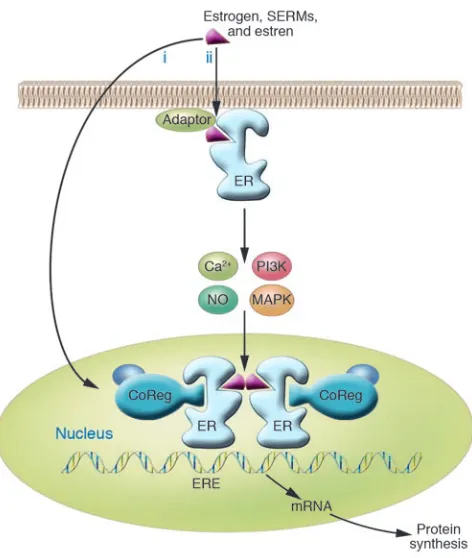
In the classical (genomic) model of estrogen signaling, estrogens bind to the ER in the nucleus (Figure 1). Over the course of several hours, the estrogen-ER complex then induces a direct response through estrogen response element sequences or an indirect response by trig-gering expression of other proteins such as transcription factors of the AP1 family, among others. This is viewed as the main
Nonstandard abbreviations used: AR, androgen receptor; ER, estrogen receptor; SERM, selective estro-gen receptor modulator.
Conflict of interest: The author has declared that no conflict of interest exists.
mode of action of estrogen. However, there is also evidence for a rapid, non-genomic response to estrogen. Signaling through the nongenomic pathway can lead to Ca2+
and NO release and activa- tion of various kinases. For a more in-depth review of estrogen signaling, see refs. 1 and 2.
Agonizing and antagonizing the genom- ic estrogen signaling pathway is compli-cated by the fact that ERs are expressed in multiple organs. The selective estrogen receptor modulator (SERM) tamoxifen, as an example, activates the ER in bone and
uterus but is an antagonist in the breast. Unfortunately, chronic administration of tamoxifen can lead to uterine cancer, so alternative SERMs and other methods for regulating the ER in specific organs have been sought. So far, all described SERMs have been shown to prevent bone loss, but their effects pale in comparison to the anabolic results seen with estrogen and androgen treatment.
Estren provides a solution
In 2002, Kousteni, Manolagas, and col-leagues described a synthetic ligand,
4-estren-3α,17β -diol (estren), that repro-duced the nongenomic effects of estrogen (3). In their hands, estren increased bone mass and strength in gonadectomized Swiss Webster mice but had no effects on uterine or seminal vesicle weight. Estrens also had no effect on the proliferation of MCF-7 human breast cancer cells. The effect of estren was attributed to activa-tion of transcription factors by several kinase cascades (4).
The potent effects of estrens on bone strength suggested that they could be used
as a bone anabolic agent in cases of estro-Figure 1
Pathways of estrogen, SERM, and estren signaling. In the genomic pathway of estro-gen action (i), estroestro-gen or SERMs bind to the ER, regulating transcription of target genes in the nucleus by binding to estrogen response element (ERE) regulatory sequences and by recruiting coregulatory proteins (CoRegs). Estrens were previously thought only to signal through the rapid, nongenomic pathway medi-ated by the ER locmedi-ated in or adjacent to the plasma membrane (ii), which may require the presence of adaptor proteins, which target the ER to the membrane. Activation of the mem-brane ER leads to a rapid change in cellular signaling molecules and stimulation of kinase activity, which in turn may affect transcription. Figure and legend adapted from ref. 2.
Table 1
Comparison of estren studies
Original estren study (3) Current study (7)
Animal model Ovariectomized and orchidectomized Ovariectomized and orchidectomized
Swiss Webster mice C57BL/6 and ovariectomized Swiss Webster mice Estren source Steraloids Inc., with slow-release pellets Steraloids Inc. and authors’ own synthesized estrens
from Innovative Research of America (98% identical) with slow-release pellets from Innovative Research of America
Estren dose 7.6 mg 7.6 mg
Age of mice in study 6 and 8 months old 3 months old
Estren effect on bone Statistically significant increases in BMD Preservation of BMD and strength but not anabolic and bone strength
Estren effect on uterus None Statistically significant increase in uterine weight
Estren effect on seminal vesicles None Statistically significant increase in seminal
vesicle weight Estren effect on breast cancer cell proliferation None (on day 2) Increased (on day 6)
[image:3.585.90.326.79.358.2]gen deficiency, such as menopause, given that their anabolic effects were restricted to bone. Estrens are currently in preclinical testing; however, the results of their use in humans have not yet been reported.
Upon closer inspection . . .
Since the original description of estren, a few reports have appeared that question whether estren acts in a nongenomic man-ner (5) and whether it really has no effects on the uterus (6). In the current issue of the JCI, Windahl, Baron, and colleagues report a systematic comparative analysis of estrens in gonadectomized mice to discern whether estrens are in fact nongenomic, bone anabolic compounds with no effects on reproductive organs (7).
The current study (7) reports that while estrens were able to prevent gonadectomy-induced bone loss, they showed no bone anabolic effects when given at the same doses and in the same manner as original-ly reported (Table 1). Furthermore, and in direct contrast to the original estren study (3), estren was shown to increase uterine and seminal vesicle weight and enhanced the proliferation of human breast cancer cells — the harmful effects estrens were designed to avoid.
Windahl et al. (7) show that estrens bind more strongly to the AR than the ER and suggest that they act more as androgens than estrogens. This was confirmed when the authors treated the gonadectomized animals with estrens and anti-androgens or anti-estrogens, as well as in ERα-KO mice: the addition of anti-androgen com-pletely blocked the response to estren, and removal of estrogen only partially blocked estren’s effects. In agreement with earlier
reports (5), estren was shown to have tran- scriptional activity — suggesting that est-rens could potentially exert their effects through the genomic pathway in addition to the nongenomic one.
So, who is right?
There are a few differences in the stud-ies that may lead us to believe that one or the other is more reliable (Table 1). First, the earlier estren study used Swiss Web-ster mice, while the current authors used C57BL/6 mice (but used Swiss Webster mice when comparing the effects of estrens on uterine weight). Could strain effects account for the differences seen? The real question is whether estrens are bone spe-cific in humans, which remains to be seen.
Also, the age of the animals studied was different — the current study used mice 3–5 months younger than those in the original study, but the authors note that Moverare et al. reported uterotrophic effects of estren in 11-month-old mice at the same doses used here (5). Despite the age difference, the fact that estrens could have an effect on reproductive organs at any stage of life raises serious concerns about their use as a SERM.
Data in the current article (7) show that when the dose of estren was reduced, the adverse effects on reproductive organs disappeared, but, unfortunately, so did the associated bone preservation capacity. Together, the data from the current study and others in the literature make a compel-ling case that estrens are not suitable for treatment of osteoporosis.
Time for a new SERM
Given that estren may not be the ideal SERM for treating osteoporosis, the search continues for what could become
a blockbuster drug. Some of the authors of the current study have also attempted to enter the fray by testing a new SERM, PSK3471 (7).
The miracle SERM for osteoporosis may be out there somewhere, but it has not been found yet. Perhaps it is PSK3471; perhaps it may still turn out to be estren — results from clinical testing in humans will pro-vide the definitive proof. But until then, the search must continue.
Acknowledgments
The author wishes to thank members of the bone research community for guidance in preparing this commentary.
Address correspondence to: Ushma S. Neill, Journal of Clinical Investigation, 630 West 168th Street, Box 57A, New York, New York 10032, USA. Phone: (212) 342-0497; Fax: (212) 342-0499; E-mail: editors@the-jci.org.
1. Weitzmann, M.N., and Pacifici, R. 2006. Estrogen deficiency and bone loss: an inflammatory tale. J. Clin. Invest. 116:1186–1194. doi:10.1172/JCI28550. 2. Deroo, B.J., and Korach, K.S. 2006. Estrogen recep-tors and human disease. J. Clin. Invest. 116:561–570. doi:10.1172/JCI27987.
3. Kousteni, S., et al. 2002. Reversal of bone loss in mice by nongenotropic signaling of sex steroids. Science. 298:843–846.
4. Kousteni, S., et al. 2003. Kinase-mediated regula-tion of common transcription factors accounts for the bone-protective effects of sex steroids. J. Clin. Invest. 111:1651–1664. doi:10.1172/JCI200317261. 5. Moverare, S., et al. 2003. Estren is a selective estrogen receptor modulator with transcriptional activity. Mol. Pharmacol. 64:1428–1433.
6. Hewitt, S.C., Collins, J., Grissom, S., Hamilton, K., and Korach, K.S. 2006. Estren behaves as a weak estrogen rather than a nongenomic selective activator in the mouse uterus. Endocrinology. 147:2203–2214. 7. Windahl, S.H., et al. 2006. Bone protection by
Deconstructing endothelial dysfunction:
soluble guanylyl cyclase oxidation
and the NO resistance syndrome
Mark T. Gladwin
Vascular Medicine Branch, National Heart, Lung, and Blood Institute, and Critical Care Medicine Department, Clinical Center, National Institutes of Health, Bethesda, Maryland, USA.
In this issue of the
JCI
, Stasch and colleagues suggest that a novel drug, BAY
58-2667, potently activates a pool of oxidized and heme-free soluble guanylyl
cyclase (sGC; see the related article beginning on page 2552). The increased
vasodilatory potency of BAY 58-2667 the authors found in a number of animal
models of endothelial dysfunction and in human blood vessels from patients
with diabetes suggests that there exists a subphenotype of endothelial
dys-function characterized by receptor-level NO resistance. Diseases associated
with NO resistance would appear to be ideally suited for therapies directed at
restoring redox homeostasis, sGC activity, and NO sensitivity.
Our molecular understanding of the pathogenesis of diabetes mellitus crystal-lized with the discovery of insulin and the catastrophic failure to produce insulin in type 1 disease. It would take decades to unravel the mechanisms underlying type 2 diabetes, a more common disease associ-ated with preserved insulin production but resistance to insulin at the receptor level. A similar march to discovery character- izes most endocrinopathies; e.g., the iden-tification of a failure to produce thyroid hormone in hypothyroidism and later the discovery of the more unusual generalized resistance to thyroid hormone. If we con- sider the diatomic free radical NO, a para-crine and endocrine signaling molecule (1), we should not be surprised by the ultimate discovery of “NO resistance syndromes.”
Endothelial NO is produced by the endothelial isoform of NOS, eNOS, via a 5-electron oxidation of l-arginine to
form l
-citrulline and NO. eNOS is acti-vated following stimulation with calcium ionophore, muscarinic receptor activation by acetylcholine, delivery of excess sub-strate arginine, and shear stress. NO then diffuses as a paracrine signaling molecule
to albuminal smooth muscle and binds to the hemes on the α/β heterodimer soluble guanylyl cyclase (sGC), which in turn converts GTP to cyclic GMP (cGMP) and activates cGMP-dependent protein kinases (Figure 1) (2–4). On the heels of the discovery of this pathway, it became clear that patients with coronary artery disease or its risk factors — diabetes, hypercho- lesterolemia, hypertension, atherosclero-sis, increasing age, and tobacco smoking — develop endothelial dysfunction. The observed impairments in stimulated and basal NO production are now classic: The expected blood flow responses to infusion with acetylcholine, an endothelium-depen- dent vasodilator, are reduced, and the nor- mal decrease in blood flow during NG-monomethyl l-arginine infusion, a direct
NOS inhibitor, are blunted (5). However, it is important to note that in all of these pathologies the vasodilatory responses to endothelium-independent exogenous NO, typically assessed by the infusion of sodium nitroprusside, are preserved.
This relatively simple signaling paradigm becomes increasingly complex as we begin to consider factors that modulate substrate transport and availability for NOS, phos- phorylation, and posttranslational modi-fications of NOS; oxidative uncoupling of the enzyme; and downstream stability of the secondary messenger cGMP. Indeed, the enzymes arginase I and II can degrade arginine, and oxidative stress can uncou-ple eNOS, leading to a state of l-arginine
resistance (6–8). Once again, we must note
that in all of these pathologies the vasodi- latory responses to authentic NO are gen-erally preserved.
Is there a subphenotype of endothelial dysfunction characterized by NO resistance?
In this issue of the JCI, Stasch and col-leagues present provocative findings that clinical states of endothelial dysfunction can be associated with the accumulation of oxidized and heme-free sGC that cannot be activated by NO (9). Indeed, the oxidation of purified sGC enzyme, endothelial cells, platelets, or aortic ring bioassay prepara- tions with 1H-[1,2,4]oxadiazolo [3,4-a]quin- oxalin-1-one (ODQ) or peroxynitrite pro-duces a state of NO resistance in which both NO-dependent cGMP accumulation and vasodilation are impaired. Remark-ably, the authors provide extensive and compelling experimental evidence that the NO- and heme-independent activa-tor of sGC, 4-[((4-carboxybutyl){2-[(4-phenethylbenzyl)oxy] phenethyl}amino) methyl [benzoic]acid (BAY 58-2667), can potently bind to and activate these oxi-dized and/or heme-free sGCs, producing selective sGC activation and vasodilation of diseased blood vessels (Figure 1). This binding also appears to inhibit ubiquitin-dependent sGC protein degradation. The thesis that oxidized and heme-free sGC contributes to endothelial dysfunction in clinical conditions is further supported by in vivo and vascular ring experiments in the spontaneously hypertensive rat, Watanabe hyperlipidemic rabbits, and ApoE–/– mice
on a high-fat diet as well as in isolated human mesocolon arteries from patients with type 2 diabetes.
While these studies suggest that ath-erosclerosis and its risk factors would be associated with impaired sGC function and resistance to exogenous NO, we know that patients with endothelial dysfunction have preserved responses to nitroprusside
Nonstandard abbreviations used: BAY 58-2667, 4-[((4-carboxybutyl){2-[(4-phenethylbenzyl)oxy] phenethyl}amino) methyl [benzoic]acid; cGMP, cyclic GMP; DEA/NO, donor 2-(N,N -diethylamino)-diazeno-late-2-oxide; sGC, soluble guanylyl cyclase.
Conflict of interest: The author has declared that no conflict of interest exists.