Cytoprotective effects of nitrite during in vivo
ischemia-reperfusion of the heart and liver
Mark R. Duranski, … , Mark T. Gladwin, David J. Lefer
J Clin Invest. 2005;
115(5)
:1232-1240.
https://doi.org/10.1172/JCI22493
.
Nitrite represents a circulating and tissue storage form of NO whose bioactivation is
mediated by the enzymatic action of xanthine oxidoreductase, nonenzymatic
disproportionation, and reduction by deoxyhemoglobin, myoglobin, and tissue heme
proteins. Because the rate of NO generation from nitrite is linearly dependent on reductions
in oxygen and pH levels, we hypothesized that nitrite would be reduced to NO in ischemic
tissue and exert NO-dependent protective effects. Solutions of sodium nitrite were
administered in the setting of hepatic and cardiac ischemia-reperfusion (I/R) injury in mice.
In hepatic I/R, nitrite exerted profound dose-dependent protective effects on cellular
necrosis and apoptosis, with highly significant protective effects observed at
near-physiological nitrite concentrations. In myocardial I/R injury, nitrite reduced cardiac infarct
size by 67%. Consistent with hypoxia-dependent nitrite bioactivation, nitrite was reduced to
NO, S-nitrosothiols, N-nitros-amines, and iron-nitrosylated heme proteins within 1–30
minutes of reperfusion. Nitrite-mediated protection of both the liver and the heart was
dependent on NO generation and independent of eNOS and heme oxygenase-1 enzyme
activities. These results suggest that nitrite is a biological storage reserve of NO subserving
a critical function in tissue protection from ischemic injury. These studies reveal an
unexpected and novel therapy for diseases such as myocardial infarction, organ
preservation and transplantation, and shock states.
Article
Cardiology
Find the latest version:
Cytoprotective effects of nitrite during in vivo
ischemia-reperfusion of the heart and liver
Mark R. Duranski,1 James J.M. Greer,1 Andre Dejam,2 Sathya Jaganmohan,1 Neil Hogg,3
William Langston,4 Rakesh P. Patel,5 Shaw-Fang Yet,6 Xunde Wang,7,8 Christopher G. Kevil,4
Mark T. Gladwin,7,8 and David J. Lefer1
1Departments of Physiology and Cardiology, Louisiana State University Health Sciences Center, Shreveport, Louisiana, USA. 2Laboratory of Chemical Biology,
National Institute of Diabetes and Digestive and Kidney Diseases, NIH, Bethesda, Maryland, USA. 3Department of Biophysics
and Free Radical Research Center, Medical College of Wisconsin, Milwaukee, Wisconsin, USA. 4Department of Pathology, Louisiana State University
Health Sciences Center, Shreveport, Louisiana, USA. 5Department of Pathology and Center for Free Radical Biology, University of Alabama at Birmingham,
Birmingham, Alabama, USA. 6Pulmonary and Critical Care Division, Brigham and Women’s Hospital, Boston, Massachusetts, USA. 7Vascular Therapeutics Section, Cardiovascular Branch, National Heart, Lung and Blood Institute, NIH, Bethesda, Maryland, USA.
8Critical Care Medicine Department, Clinical Center, NIH, Bethesda, Maryland, USA.
Nitrite represents a circulating and tissue storage form of NO whose bioactivation is mediated by the
enzymat-ic action of xanthine oxidoreductase, nonenzymatenzymat-ic disproportionation, and reduction by deoxyhemoglobin,
myoglobin, and tissue heme proteins. Because the rate of NO generation from nitrite is linearly dependent on
reductions in oxygen and pH levels, we hypothesized that nitrite would be reduced to NO in ischemic tissue and
exert NO-dependent protective effects. Solutions of sodium nitrite were administered in the setting of hepatic
and cardiac ischemia-reperfusion (I/R) injury in mice. In hepatic I/R, nitrite exerted profound
dose-depen-dent protective effects on cellular necrosis and apoptosis, with highly significant protective effects observed at
near-physiological nitrite concentrations. In myocardial I/R injury, nitrite reduced cardiac infarct size by 67%.
Consistent with hypoxia-dependent nitrite bioactivation, nitrite was reduced to NO, S-nitrosothiols,
N-nitros-amines, and iron-nitrosylated heme proteins within 1–30 minutes of reperfusion. Nitrite-mediated protection of
both the liver and the heart was dependent on NO generation and independent of eNOS and heme oxygenase-1
enzyme activities. These results suggest that nitrite is a biological storage reserve of NO subserving a critical
function in tissue protection from ischemic injury. These studies reveal an unexpected and novel therapy for
diseases such as myocardial infarction, organ preservation and transplantation, and shock states.
Introduction
The anion nitrite (NO2–) forms as a consequence of NO
oxida-tion and is present at concentraoxida-tions of 0.3–1.0 μM in plasma and 1–20 μM in tissue (1–5). This nitrite may be reduced to NO dur-ing hypoxia and acidosis (1, 4–13). At very low tissue pH and oxy-gen tension, nitrite may be reduced to NO by disproportionation (acidic reduction) (13) or by the enzymatic action of xanthine oxidoreductase (6, 7, 12, 13). However, within physiological rang-es of pH and oxygen tension, nitrite has been considered an inert metabolic end product of NO oxidation with limited intrinsic biological activity (14). Challenging this dogma, we have recently demonstrated that near-physiological levels of nitrite are reduced to NO by reaction with deoxyhemoglobin along the physiological oxygen gradient, a chemical reaction whose rate is oxygen and pH dependent and that potentially contributes to hypoxic vasodila-tion (8). We therefore hypothesized that hypoxia-dependent NO production from nitrite in ischemic tissue might limit ischemia-reperfusion (I/R) injury.
Although reperfusion of ischemic tissues provides oxygen and metabolic substrates necessary for the recovery and survival of reversibly injured cells, reperfusion itself actually results in the acceleration of cellular necrosis (15). I/R is characterized by the formation of oxygen radicals upon reintroduction of molecular oxygen to ischemic tissues, resulting in widespread lipid and pro-tein oxidative modifications, mitochondrial injury, and tissue apoptosis and necrosis (16). In addition, after reperfusion of isch-emic tissues, blood flow may not return uniformly to all portions of the ischemic tissues, a phenomenon that has been termed the “no-reflow” phenomenon (17). Reductions in blood flow after reperfusion are thought to contribute to cellular injury and necro-sis (17). The sudden re-introduction of blood into ischemic tissue also results in massive tissue disruption, enzyme release, reduc-tions in high energy phosphate stores, mitochondrial injury, and necrosis (18, 19). Furthermore, previous studies have also indicat-ed that the I/R injury is characterizindicat-ed by an inappropriate inflam-matory response in the microcirculation, resulting in leukocyte– endothelial cell interactions that are mediated by the upregulation of both leukocyte and endothelial cell adhesion molecules (20, 21). Intensive research efforts have been focused on the amelioration of various pathophysiological components of I/R injury to limit the extent of tissue injury and necrosis.
Nitric oxide, NO donors, and NO synthase activation or trans-genic overexpression have been shown to exert protective effects on this process in a number of models (22–27) but in other mod-els appear harmful (28–31). Evaluation of those studies suggests a critical effect of dose and duration of NO exposure, resulting in a Nonstandard abbreviations used: AAR, area at risk; ALT, alanine aminotransferase;
AST, aspartate aminotransferase; CT, comparative threshold; HO-1, heme oxygenase-1; I/R, ischemia-reperfusion; ODQ, [1H-[1,2,4]Oxadiazole[4,3-a]quinoxalin-1-one]; PTIO, 2-phenyl-4,4,5,5-tetramethylimidazoline-1-oxyl-3-oxide; RSNO, mercury-labile NO-modified protein; RxNO, mercury-stable NO-modified protein; sGC, soluble guanylate cyclase; ZnDPBG, zinc (II) deuteroporphyrin IX 2,4-bis-ethylene glycol.
Conflict of interest: The NIH has submitted a provisional patent on the use of nitrite in ischemia-reperfusion. D.J. Lefer and M.T. Gladwin are listed as co-inventors.
research article
The Journal of Clinical Investigation http://www.jci.org Volume 115 Number 5 May 2005 1233
narrow therapeutic safety window for NO in I/R pathophysiology (32, 33). An additional limitation is that NO formation from NO synthase requires oxygen as substrate, a molecule whose availability becomes limited during ischemia. We therefore considered the use of nitrite in this context for the following reasons: (a) it is a natu-rally occurring substance with no potentially toxic “leaving group”; (b) it is selectively reduced to NO in tissues with low oxygen tension and low pH (4, 6–13, 34, 35); and (c) NO is known to maintain heme proteins in a reduced and liganded state (36–38), to limit free iron– and heme-mediated oxidative chemistry (39–41), to tran-siently inhibit cytochrome c oxidase and mitochondrial respiration (42–45), and to modulate apoptotic effectors (46), all mechanisms that might participate in cytotoxicity after severe ischemia.
To test this hypothesis, we evaluated the effects of nitrite therapy compared with those of control therapy with vehicle or nitrate in well characterized in vivo murine models of hepatic and
myocar-dial I/R injury. We provide strong evidence for a profound pro-tective effect of low concentrations of nitrite on cellular necrosis and apoptosis, mediated by a hypoxia-dependent bioconversion of nitrite to NO and nitrosated or nitrosylated proteins.
Results
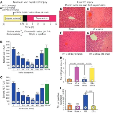
[image:3.585.110.484.81.451.2]Intraperitoneal nitrite limits hepatic I/R injury. Intraperitoneal deliv-ery of 1.2–960 nmol sodium nitrite during hepatic ischemia limited serum elevations of the liver transaminases aspartate aminotransferase (AST) and alanine aminotransferase (ALT) in a dose-dependent way (Figure 1, A–C), with a peak effect occurring at a dose of 48 nmol nitrite. In sharp contrast, treatment with 0.9% saline or sodium nitrate (48 nmol) did not exert any protec-tive effects in the setting of hepatic I/R injury. Consistent with consumption of the intraperitoneal nitrite by the liver, plasma nitrite levels were not significantly elevated (baseline nitrite levels
Figure 1
of 594 ± 83 nM, which increased to 727 ± 40 nM; n = 3; P = 0.16) in mice treated with 48 nmol nitrite, the most effective dose.
Additional studies were performed for evaluation of the effects of nitrite treatment on hepatocellular injury in mice after in vivo hepatic ischemia (45 minutes) and more prolonged reperfusion (24 hours; Figure 1, D–I). The administration of 48 nmol nitrite significantly reduced hepatocellular injury at 24 hours of reperfusion compared with the administration of saline or nitrate. In addition, nitrite therapy significantly attenuated the extent of hepatocellular apoptosis after 45 minutes of hepatic ischemia and 24 hours of reperfusion (P < 0.001; Figure 1I). The extent of hepatic cell apoptosis in nitrite-treated animals subjected to I/R was similar to that observed in sham-operated control animals (P = NS). Consistent with this treatment having a U-shaped effi-cacy profile, administration of 960 nmol nitrite to mice subjected to hepatic I/R injury failed to exert a significant protective effect (also evidenced by the transaminase profile in Figure 1, B and C).
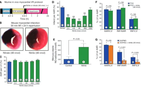
Intraventricular nitrite limits myocardial I/R injury. To determine whether the potent cytoprotective effects of nitrite on liver I/R injury could be generalized to other organ systems, we next per-formed studies to evaluate the potential cardioprotective effects of acute nitrite therapy in the setting of coronary artery occlu-sion and reperfuocclu-sion. The experimental protocol for the myo-cardial I/R studies is depicted in Figure 2A. The administration of nitrite (48 nmol) into the left ventricular cavity at 5 minutes prior to reperfusion significantly limited myocardial infarct size (P < 0.001; Figure 2B) compared with the control treatment of 48 nmol nitrate. Despite the results showing similar myocardial area at risk (AAR) (P = NS between groups; Figure 2C), nitrate treat-ment decreased myocardial infarct size relative to the AAR and the left ventricle by 67% compared with nitrate-treated controls.
[image:4.585.47.539.81.385.2]In additional groups of animals, we investigated the effects of various dosages of nitrite on myocardial infarct size after ischemia and reperfusion (Figure 2, C and D). Dosages of nitrite from 2.4
Figure 2
research article
The Journal of Clinical Investigation http://www.jci.org Volume 115 Number 5 May 2005 1235
to 1,920 nmol were investigated. Data for myocardial AAR per left ventricle are presented in Figure 2C and clearly indicate that the AAR per left ventricle was similar in all study groups (P = NS between groups). We observed a dose-dependent decrease in myo-cardial infarct size with a maximal protective effect at 48 nmol (Figure 2D). However, administration of 1,920 nmol of nitrite failed to exert any significant cardioprotective effect.
To establish the plasma concentrations of nitrite that were car-dioprotective, in additional experiments we injected increasing amounts of nitrite into the left ventricular chamber and sacrificed the animals and measured the concentration of nitrite in blood 5 minutes later. The plasma nitrite concentration correlated with the amount of nitrite injected (μM nitrite concentration in plas-ma, 0.965 + 0.1971 ×X + 0.0006815 ×X2, where X = the amount
of nitrite injected in nanomoles; n = 16; r = 0.96; P < 0.001). For the cardiac I/R experiments, the amounts injected and the mea-sured concentrations in plasma were 0 nmol and 0.965 μM (basal plasma concentration of nitrite), 1.2 nmol and 1.2 μM, 2.4 nmol and 1.4 μM, 4.8 nmol and 1.9 μM, 48 nmol and 11.9 μM, 480 nmol and 252.6 μM, and 960 nmol and 818.3 μM. Consistent with the intravascular conversion of nitrite to NO (8), after the injection of 48 nmol nitrite, a significant increase in iron-nitrosylated hemo-globin was observed (Figure 2E).
We performed additional studies of myocardial I/R injury to determine possible mechanisms of nitrite-mediated cardiopro-tection (Figure 2, F and G). Pretreatment with the NO scavenger 2-phenyl-4,4,5,5-tetramethylimidazoline-1-oxyl-3-oxide (PTIO) completely abolished the protective effects of nitrite therapy (Figure 2F). In contrast, treatment with the heme oxygenase-1 (HO-1) inhibitor zinc (II) deuteroporphyrin IX 2,4-bis-ethylene glycol (ZnDPBG) did not attenuate the cardioprotective actions of nitrite therapy in the ischemic-reperfused myocardium (Figure 2G).
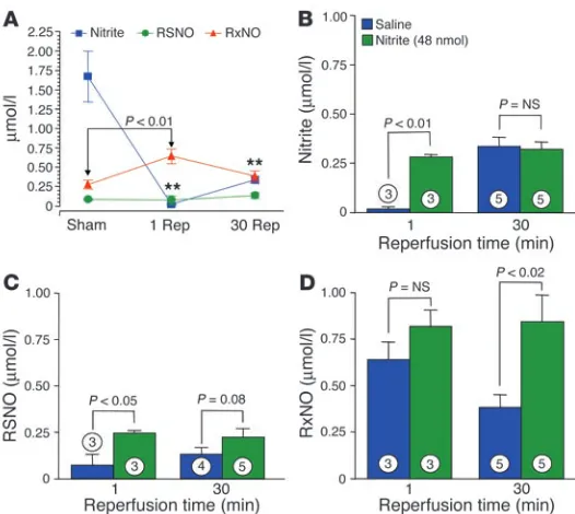
Nitrite-mediated cytoprotection is associated with the reduction of nitrite to NO and formation of S-, N-, and heme-nitrosated or nitrosylated hepatic proteins. Consistent with previously described cellular reduction of nitrite to NO and S-nitrosothiols/N-nitrosamines (4, 8, 9, 34), 1 minute after reperfusion the levels of nitrite in the livers of
saline-treated control mice subjected to ischemia decreased from 1.75 μM to undetectable (P < 0.001 versus sham group) and levels of mercury-stable NO-modified proteins (representing N-nitrosamines and iron-nitrosyl proteins and collectively referred to here as RxNOs) increased to approximately 750 nM (P < 0.001; Figure 3A). Interestingly, for nitrite treat-ment compared with saline treattreat-ment, there was a signifi-cant increase in liver levels of nitrite (P < 0.01 versus saline; Figure 3B), S-nitrosothiols (Figure 3C), and RxNOs (Figure 3D) after reperfusion in the nitrite-treated mice.
These data suggest that nitrite is bioactivated during hypoxic stress and are consistent with our recent studies showing deoxy-hemoglobin-dependent reduction of nitrite to NO (8), as well as recent studies by Bryan and colleagues demonstrating an acute conversion of tissue nitrite to mercury-labile NO-modified pro-teins (RSNOs) and RxNOs after a systemic anoxic insult (4). The low levels of nitrite that are cytoprotective (1.2 nmol at the lowest dose; Figure 1, B and C) and the observed reductive decomposition of “native” liver nitrite in the saline-treated control animals (Fig-ure 3A) suggest that this may be an innate mechanism for hypoxic NO production and cytoprotection.
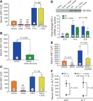
Cytoprotective effects of nitrite are NO dependent but independent of NO synthase and HO-1 enzymatic activities. Further supporting the idea of a mechanism involving the hypoxic reduction of nitrite to NO, the NO inhibitor PTIO completely inhibited protective effects of nitrite in both heart (Figure 2F) and liver (Figure 4A). In contrast, nitrite remained cytoprotection in eNOS-deficient mice (P < 0.001; Figure 4B), suggesting that NO production from nitrite during I/R is eNOS independent. We investigated the role of soluble guanylate cyclase (sGC) in nitrite-mediated protection against hepatic I/R injury (Figure 4C). Pretreatment with the sGC inhibitor [1H-[1,2,4]Oxa-diazole[4,3-a]quinoxalin-1-one] (ODQ) completely inhibited the protective effects of nitrite in the setting of hepatic I/R injury.
Although HO-1 mRNA and protein are both significantly induced after hepatic I/R in this model, we did not observe any effects of nitrite treatment (48 nmol) on HO-1 mRNA or protein levels during I/R (Figure 4D). Furthermore, in mice pretreated with ZnDPBG, a specific and potent HO-1 inhibitor, nitrite significantly limited liver tissue injury, suggesting an HO-1–independent effect (P < 0.05; Figure 4E), similar to experimental results during car-diac I/R (Figure 2G). Additional studies were performed utilizing HO-1–deficient (HO-1–/–) mice that were treated with nitrite and
subjected to hepatic I/R injury for further evaluation of the role of HO-1 in nitrite-mediated cytoprotection. We observed robust
pro-Figure 3
[image:5.585.42.305.80.315.2]tection against hepatic I/R injury in HO-1–/– mice treated with 48
nmol nitrite (P < 0.01 versus the untreated HO-1–/– group; Figure
4F), which further suggests that nitrite-induced protection does not involve HO-1 activation or signaling.
Discussion
In the present studies, nitrite treatment significantly increased the levels of liver nitrite and nitrosated or nitrosylated species (iron-nitrosyl proteins, RSNO, and RXNO), compared with saline and nitrate control treatment, and conferred a substantial and unex-pected dose-dependent cytoprotective effect, limiting necrosis and apoptosis and preserving organ function. Remarkably, a protective effect was observed with doses as low as 1.2 nmol nitrite, suggest-ing that this may represent an endogenous protective mechanism that buffers severe metabolic or pathophysiological stress.
Recent data suggest that nitrite concentrations vary between blood and different organs and are typically in the high nanomo-lar to low micromonanomo-lar range. However, until recently, the high con-centrations required to vasodilate aortic ring preparations led to its dismissal as an important biologically active molecule. Indeed, Furchgott et al. demonstrated in 1953 that 100 μM nitrite stimu-lated vasodilation of aortic ring preparations, a process later shown to be mediated by activation of sGC (47–50). From a physiological standpoint, the in vivo conversion of nitrite to NO was thought to be limited to the stomach and severely ischemic heart, where acidic reduction or disproportionation at very low pH produces gastric mucosal vasodilation (5, 11, 51) and apparent cardiac tissue injury and heme iron-nitrosylation (at high nitrite concentrations
in ischemic ex vivoheart preparations) (10), respectively. Although xanthine oxidoreductase–dependent nitrite reduction can occur at very low oxygen tensions and has recently been suggested to mediate nitrite-dependent cytoprotection in a Langendorff heart I/R model (52), NO production from this system is detectable only in the pres-ence of high concentrations of superoxide dismutase (53, 54).
We recently reported that infusions of sodium nitrite into the human circulation produced significant vasodilation at both phar-macological and near-physiological concentrations (8). The bioac-tivation of nitrite appeared to be mediated by a nitrite reductase activity of deoxygenated hemoglobin, ultimately forming NO and iron-nitrosylated hemoglobin and, to a lesser extent, S-nitrosated protein species. Based on these data, a role for circulating nitrite in mediating hypoxic vasodilation was proposed, with the oxygen sensor in this case being hemoglobin (8). We hypothesize that a similar nitrite reductase activity of deoxyhemoglobin, deoxymyo-globin, and/or other deoxygenated heme proteins may account for the formation of nitrosated or nitrosylated proteins and apparent NO-dependent cytoprotection observed during liver and cardiac ischemia in our study here. The contribution of this mechanism versus pathways involving xanthine oxido-reductase and acidic reduction will require further study.
[image:6.585.238.539.412.739.2]The precise mechanism of how nitrite confers tissue protec-tion remains unclear, although a critical and direct role for NO is implied from data shown in our work here. Previous studies of NO and I/R have yielded conflicting reports regarding the effects of NO on the severity of I/R injury, with some studies suggesting that NO actually contributed to reperfusion injury (30, 33). Our
Figure 4
research article
The Journal of Clinical Investigation http://www.jci.org Volume 115 Number 5 May 2005 1237
laboratory has previously demonstrated that NO donors as well as the NO precursor L-arginine protect against myocardial I/R injury
(22, 24, 55). More recently, we demonstrated that the severity of myocardial I/R injury is markedly exacerbated in mice completely deficient in eNOS (eNOS–/– mice) (55), whereas mice with eNOS
overexpression are protected against myocardial infarction and subsequent congestive heart failure (25, 26, 56). Conflicting data on the effects of NO on I/R injury are probably related to the dose of NO and the conditions during ischemia and reperfusion (32). It is now well appreciated that very high, nonphysiological levels of NO (i.e., high micromolar and millimolar) actually promote cellu-lar necrosis and apoptosis (57), while the demonstrated cytoprotec-tive effects of NO typically involve nanomolar or low micromolar concentrations of NO (22, 23, 32). Additionally, studies investigat-ing NO and NO-releasinvestigat-ing agents under in vitro conditions of I/R have consistently reported deleterious effects of NO (32), in con-trast to in vivo studies of I/R that have reported beneficial effects of NO therapy (22, 23). How NO mediates protection is also not clear, with multiple mechanisms being reported, including sGC activation, inhibition of cytochrome C oxidase, and inhibition of deleterious mitochondrial calcium uptake (42–45).
Although these data suggest that the effects of nitrite occur secondary to NO formation, the ultimate mechanism of nitrite-dependent cytoprotection is currently unknown. An intriguing possibility is the intermediate formation of S-nitrosothiols and N-nitrosamines, known to form via reactions of nitrite with deoxy-hemoglobin and possibly tissue heme proteins (4, 8, 9, 35, 38, 58, 59). Consistent with hypoxia-dependent formation of S-nitroso-thiols in red blood cells and tissues from nitrite, hepatic levels of these species were significantly higher after reperfusion (1–30 min-utes) in livers exposed to ischemia and nitrite. Within the relative reductive environment intracellularly, S-nitrosothiols formed via nitrite will be readily reduced to NO and activate sGC. Alternatively, S-nitrosation and subsequent effects on activity of critical proteins important in I/R-induced injury and apoptotic cell death may lead to protection (46). In addition, our data reveal a dynamic regula-tion of hepatic RxNOs, a pool of mercury-stable NO-modified proteins that include N-nitrosamines and iron-nitrosyl proteins (4, 50, 60), during I/R. In saline-treated groups, RxNO levels increased at 1 minute of reperfusion and then decreased after 30 minutes of reperfusion, whereas a sustained elevation in RxNO levels was observed in nitrite-treated mice, suggesting that the maintenance of RxNOs could be important in protecting tissues from I/R injury.
Finally, it is interesting that similar mechanisms might be at play in vivo and in isolated muscle during reductive meat-curing pro-cedures. Nitrite has been utilized for curing meat for more than a century. During meat curing, the formation of iron-nitrosylated heme limits release of iron from the porphyrin molecule, prevents iron porphyrin– and metal ion–catalyzed lipid oxidation, Fenton chemistry, and ferryl heme formation (39–41, 61). Nitrite and NO produced from nitrite under hypoxic conditions can reduce fer-ryl hemoglobin (36, 37) and catalyze ferriheme protein reductive nitrosylation (38), and nitrite can react with deoxyhemoglobin to form NO, iron-nitrosyl-heme, and S- and N-nitrosated intermedi-ates (4, 8, 9, 34, 62). While it is clear that the formation of these reaction products in the mouse liver after I/R are associated with cytoprotection, more work will be needed to determine the exact mechanisms of nitrite-induced cytoprotection.
In conclusion, these data demonstrate a remarkable function for the relatively simple inorganic anion nitrite as a potent inhibitor
of liver and cardiac I/R injury and infarction in the mouse. The effects of nitrite appear to be NO dependent, with a rapid con-version of nitrite to NO and nitrosated or nitrosylated proteins after reperfusion. Given the known safety of nitrite as a naturally occurring anion and as an FDA-approved therapeutic for cyanide poisoning, these data evince a novel, safe, and inexpensive therapy for I/R injury. Such a therapy could be used to prevent or modu-late organ dysfunction after coronary and peripheral vasculature reperfusion, high-risk abdominal surgery (such as aortic aneurism repair that leads to renal acute tubular necrosis), cardiopulmonary resuscitation, and, perhaps most importantly, solid organ trans-plantation. Further studies are required to establish the efficacy of nitrite in various organs and disease states.
Methods
Chemicals and reagents. Sodium nitrite (S-2252) and sodium nitrate (S-8170) were obtained from Sigma-Aldrich. Sodium nitrite and sodium nitrate were dissolved in phosphate-buffered saline and the pH was adjusted to 7.4. In all experiments, a final volume of 50 μl containing 1.2–1,920 nmol sodium nitrite or sodium nitrate was administered to the mice. Carboxy-PTIO [2-(4-carboxyphenyl)-4,4,5,5-tetramethylimidazoline-1-oxyl-3-oxide potassium salt], a direct intravascular NO scavenger, was utilized to inhibit NO-depen-dent effects after both hepatic and myocardial I/R injury. Carboxy-PTIO (Alexis Biochemicals) was dissolved in phosphate-buffered saline and was administered intravenously at a dose of 1 mg/kg in a volume of 50 μl 30 minutes prior to either hepatic or myocardial ischemia. ZnDPBG (Alexis Biochemicals), an HO-1 inhibitor, was injected i.p. at a dose of 10 mg/kg in a volume of 50 μl 30 minutes prior to the induction of either hepatic or myocardial ischemia. ZnDBG has been shown previously to be a highly specific and potent inhibitor of HO-1 (63).The sGC inhibitor ODQ (Alexis Biochemicals) was utilized in experiments of hepatic I/R injury at a dose of 20 mg/kg in a volume of 50 μl 30 minutes prior to hepatic ischemia.
Animals. All of the mice utilized in the present studies were C57BL6/J at 8–10 weeks of age obtained from The Jackson Laboratory. In additional experiments of hepatic I/R injury we utilized eNOS–/– mice. The eNOS–/–
mice were originally generously donated by Paul Huang (Massachusetts General Hospital, Boston, Massachusetts, USA) and were generated in our breeding colony at Louisiana State University Health Sciences Center. The eNOS–/– mice were utilized at 8–10 weeks of age. HO-1–/– mice were obtained
from Shaw-Fang Yet (Harvard Medical School, Boston, Massachusetts, USA) and were utilized at 8–10 weeks of age.
removed. The duration of hepatic reperfusion was 5 hours in the studies of serum liver transaminase levels (i.e., AST or ALT) and 24 hours for the studies of liver histopathology (i.e., hepatocellular infarction).
Liver enzyme determinations. Serum samples were analyzed for AST and ALT using a spectrophotometric method (Sigma-Aldrich) (66). These enzymes are liver specific and are released from the liver during injury (64, 65).
Liver histopathology studies. Histopathology of liver tissue was performed as reported previously (64). Liver tissue was fixed in 10% buffered formalin for 24 hours and was embedded in paraffin, and 10-μm sections were stained with hematoxylin and eosin. Histopathology scoring was performed on randomly selected high-power fields by investigators blinded to sample identity using the following criteria: 0, no hepatocellular damage; 1, mild injury characterized by cytoplasmic vacuolization and focal nuclear pykno-sis; 2, moderate injury with dilated sinusoids, cytosolic vacuolization, and blurring of intercellular borders; 3, moderate to severe injury with coagula-tive necrosis, abundant sinusoidal dilation, red blood cell extravasation into hepatic chords, and hypereosinophilia and margination of neutrophils; 4, severe necrosis with loss of hepatic architecture, disintegration of hepatic chords, hemorrhage, and neutrophil infiltration.
Hepatocellular apoptosis was determined using a TUNEL staining kit from Roche Diagnostics Corp. according to the manufacturer’s rec-ommendations. Briefly, liver tissue from various treatments was fixed in buffered formalin and 10-μm sections were prepared. Sections were permeablilzed on ice for 2 minutes and were incubated in 50 μl TUNEL solution for 30 minutes at 37°C. Sections were then treated with 50 μl substrate solution for 10 minutes and were mounted under glass cover slips. The number of apoptotic nuclei was determined from 5 randomly selected 40× fields per specimen. A total of 6 specimens per treatment group (16 slides per group) were analyzed and compared using 1-way ANOVA with Bonferroni’s post-testing.
Myocardial I/R protocol.Surgicalligation of the left main coronary artery was performed similar to methods described previously (25). Briefly, mice were anesthetized by intraperitoneal injection of ketamine (50 mg/kg) and pentobarbital sodium (50 mg/kg). The animals were then attached to a surgical board with their ventral side up. The mice were orally intubated with PE-90 polyethylene tubing connected to PE-240 tubing and then were connected to a Model 683 rodent ventilator (Harvard Apparatus). The tidal volume was set at 2.2 milliliters and the respiratory rate was set at 122 breaths per minute. The mice were supplemented with 100% oxygen via the ventilator side port. A median sternotomy was performed using an electric cautery and the proximal left main coronary artery was visualized and completely ligated with 7-0 silk suture mounted on a tapered needle (BV-1; Ethicon). In the initial experiments of myocardial infarct size, coro-nary occlusion was maintained for 30 minutes, followed by removal of the suture and reperfusion for 24 hours.
Myocardial infarct size determination. At 24 hours of reperfusion, the mice were anesthetized as described previously, intubated, and connected to a rodent ventilator. A catheter (PE-10 tubing) was placed in the com-mon carotid artery to allow for Evans blue dye injection. A median ster-notomy was performed and the left main coronary artery was re-ligated in the same location as before. Evans blue dye (1.2 ml of a 2.0% solution; Sigma-Aldrich) was injected into the carotid artery catheter into the heart for delineation of the ischemic zone from the nonischemic zone. The heart was rapidly excised and serially sectioned along the short axis in 5 1-mm-thick sections that were then incubated in 1.0% 2,3,5-triphenyltet-razolium chloride (Sigma-Aldrich) for 5 minutes at 37°C for demarcation of the viable and nonviable myocardium within the risk zone. Each of the 5 1-mm-thick myocardial slices were weighed and the areas of infarction, AAR, and nonischemic left ventricle were assessed with computer-assisted planimetry (NIH Image 1.57) by an observer blinded to sample identity. All
of the procedures for the left ventricular AAR and infarct size determina-tion have been described previously (25).
HO-1 Western blot analysis.Liver tissue collected at 2,4, and 6 hours after I/R injury was homogenized in radioimmunoprecipitation assay buffer with protease and phosphatase inhibitors and was centrifuged 3 times for 30 minutes at 4°C; the supernatant was transferred to a fresh tube after each centrifugation. Protein concentrations were determined using Bio-Rad reagents (BioBio-Rad Laboratories), and 50 μg total protein from each sample was loaded. Western blots were performed using mouse mAb to HO-1 (Stressgen) at a 1:3,000 dilution and goat anti-mouse secondary HRP conjugate at a 1:2,000 dilution (Amersham Biosciences).
Blood and tissue nitrite and nitrosated or nitrosylated protein and hemoglobin determination. The concentration of nitrite in plasma and the levels of iron-nitrosylated hemoglobin were measured using tri-idodide–based reductive chemiluminescence as described and validated previously (67, 68). Liver tissue was homogenized using an amended protocol published by Bryan and col-leagues (4). Harvested liver tissue was blotted dry on filter paper, weighed, and homogenized immediately in ice-cold buffer containing N-ethylmaleimide (10 mmol/l)/diethylenetriaminepenta-acetic acid (2 mmol/l) (3:1 dilution, wt/vol). The buffer/tissue mixture was then homogenized with a Wheaton glass-on-glass homogenizer. Tissue homogenates were kept on ice and were analyzed within 5 minutes. The homogenate was subsequently injected directly into tri-iodine for measurement of the sum of nitrite, RxNOs, and RSNOs. For determining the levels of specific NO adducts (RxNOs and RSNOs), the sample was treated with and without 5 mM mercuric chloride (RSNO becomes nitrite in presence of mercuric chloride and RxNO is stable) and subsequently treated with acid sulfanilamide (0.5%) to eliminate nitrite.
Real-time PCR protocol. Total RNA from liver tissue was isolated using the RNeasy kit (Qiagen Inc.) and the isolated RNA concentration was determined using a nanodrop ND-1000 spectrophotometer (Nanodrop Technologies). The quality and integrity of total RNA was assessed on an Agilent 2100 bioanalyzer. Quantitative real-time PCR assays for HO-1 and GAPDH transcripts were carried out using gene-specific double-fluores-cence–labeled probes in a 7900 Sequence Detector (PE Applied Biosystems). Probes and primers for HO-1 and GAPDH were obtained from Applied Biosystems as Assays On Demand gene expression products.
In brief, first-strand cDNA was synthesized using 1 μg of total RNA in a 20 μl reverse transcriptase reaction mixture with Invitrogen Corp.’s Superscript cDNA synthesis kit, following the manufacturer’s directions. PCR amplification was performed in a 384-well plate with a 20-μl reaction mixture containing 300 nm of forward and reverse primers, 200 nm probe, 200 nm dNTP in 1× real-time PCR buffer, and passive reference (ROX) fluorochrome. The thermal cycling conditions were 2 minutes at 50°C and 10 minutes at 95°C, followed by 40 cycles of 15 seconds of denaturation at 95°C and 1 minute of annealing and extension at 60°C. The comparative threshold (CT) PCR cycle detection method (∆∆ CT method) that compares the differences in CT values of control and treated groups was used to cal-culate the relative fold change in gene expression between groups.
Statistical analyses. Data were analyzed by 2-way ANOVA with post-hoc Bonferroni analysis using StatView software version 5.0 (SAS Institute). Data are reported as mean ± SEM. P values less than 0.05 were consid-ered significant.
Acknowledgments
research article
The Journal of Clinical Investigation http://www.jci.org Volume 115 Number 5 May 2005 1239
Received for publication June 22, 2004, and accepted in revised form March 8, 2005.
Address correspondence to: David J. Lefer, Louisiana State Uni-versity Health Sciences Center, 1501 Kings Highway, Shreveport, Louisiana 71130, USA. Phone: (318) 6974; Fax: (318)
675-4217; E-mail: dlefer@lsuhsc.edu. Or to: Mark T. Gladwin, Building 10-CRC, Room 5-5140, 10 Center Drive; MS-1454, NIH, Bethesda, Maryland 20892-1454, USA. Phone: (301) 435-2310; Fax: (301) 402-1213; E-mail: mgladwin@nih.gov.
David J. Lefer and Mark T. Gladwin are co-senior authors.
1. Gladwin, M.T., et al. 2000. Role of circulating nitrite and S-nitrosohemoglobin in the regulation of regional blood flow in humans. Proc. Natl. Acad. Sci. U. S. A.97:11482–11487.
2. Rodriguez, J., Maloney, R.E., Rassaf, T., Bryan, N.S., and Feelisch, M. 2003. Chemical nature of nitric oxide storage forms in rat vascular tissue. Proc. Natl. Acad. Sci. U. S. A.100:336–341.
3. Rassaf, T., et al. 2003. NO adducts in mammalian red blood cells: too much or too little? Nat. Med.
9:481–483.
4. Bryan, N.S., et al. 2004. Cellular targets and mecha-nisms of nitros(yl)ation: an insight into their nature and kinetics in vivo. Proc. Natl. Acad. Sci. U. S. A.
101:4308–4313.
5. Gladwin, M.T. 2004. Haldane, hot dogs, halito-sis, and hypoxic vasodilation: the emerging biol-ogy of the nitrite anion. J. Clin. Invest.113:19–21. doi:10.1172/JCI200420664.
6. Godber, B.L., et al. 2000. Reduction of nitrite to nitric oxide catalyzed by xanthine oxidoreductase.
J. Biol. Chem.275:7757–7763.
7. Li, H., Samouilov, A., Liu, X., and Zweier, J.L. 2001. Characterization of the magnitude and kinetics of xanthine oxidase-catalyzed nitrite reduction. Evaluation of its role in nitric oxide generation in anoxic tissues. J. Biol. Chem.276:24482–24489. 8. Cosby, K., et al. 2003. Nitrite reduction to nitric
oxide by deoxyhemoglobin vasodilates the human circulation. Nat. Med.9:1498–1505.
9. Nagababu, E., Ramasamy, S., Abernethy, D.R., and Rifkind, J.M. 2003. Active nitric oxide produced in the red cell under hypoxic conditions by deoxyhe-moglobin-mediated nitrite reduction. J. Biol. Chem.
278:46349–46356.
10. Tiravanti, E., Samouilov, A., and Zweier, J.L. 2004. Nitrosyl-heme complexes are formed in the isch-emic heart: evidence of nitrite-derived nitric oxide formation, storage, and signaling in post-ischemic tissues. J. Biol. Chem.279:11065–11073.
11. Zweier, J.L., Wang, P., Samouilov, A., and Kup-pusamy, P. 1995. Enzyme-independent forma-tion of nitric oxide in biological tissues. Nat. Med.
1:804–809.
12. Millar, T.M., et al. 1998. Xanthine oxidoreductase catalyses the reduction of nitrates and nitrite to nitric oxide under hypoxic conditions. FEBS Lett.
427:225–228.
13. Zhang, Z., et al. 1997. Human xanthine oxidase converts nitrite ions into nitric oxide (NO). Biochem. Soc. Trans.25:524S.
14. Lauer, T., et al. 2001. Plasma nitrite rather than nitrate reflects regional endothelial nitric oxide synthase activity but lacks intrinsic vasodilator action. Proc. Natl. Acad. Sci. U. S. A.98:12814–12819. 15. Braunwald, E., and Kloner, R.A. 1985. Myocardial
reperfusion: A double-edged sword? J. Clin. Invest.
76:1713–1719.
16. McCord, J.M., Roy, R.S., and Schaffer, S.W. 1985. Free radicals and myocardial ischemia. The role of xanthine oxidase. Adv. Myocardiol.5:183–189. 17. Kloner, R.A., Ganote, C.E., and Jennings, R.B.
1974. The “no-reflow” phenomenon after tempo-rary coronary occlusion in the dog. J. Clin. Invest.
54:1496–1508.
18. Nayler, W.G. 1981. The role of calcium in the isch-emic myocardium. Am. J. Pathol.102:262–270. 19. Shen, A.C., and Jennings, R.B. 1972. Myocardial
calcium and magnesium in acute ischemic injury.
Am. J. Pathol.67:417–440.
20. Lefer, A.M., and Lefer, D.J. 1996. The role of nitric oxide and cell adhesion molecules on the microcirculation in ischaemia-reperfusion. Cardio-vasc. Res.32:743–751.
21. Entman, M.L., et al. 1991. Inflammation in the course of early myocardial ischemia. FASEB J.
5:2529–2537.
22. Lefer, D.J. 1995. Myocardial protective actions of nitric oxide donors after myocardial ischemia and reperfusion. New Horiz.3:105–112.
23. Lefer, D.J., Nakanishi, K., Johnston, W.E., and Vint-en-Johansen, J. 1993. Antineutrophil and myocar-dial protecting actions of a novel nitric oxide donor after acute myocardial ischemia and reperfusion of dogs. Circulation.88:2337–2350.
24. Nakanishi, K., et al. 1992. Intracoronary L-argi-nine during reperfusion improves endothelial function and reduces infarct size. Am. J. Physiol.
263:H1650–H1658.
25. Jones, S.P., et al. 2004. Endothelial nitric oxide synthase overexpression attenuates myocardial reperfusion injury. Am. J. Physiol. Heart Circ. Physiol.
286:H276–H282.
26. Jones, S.P., et al. 2003. Endothelial nitric oxide synthase overexpression attenuates congestive heart failure in mice. Proc. Natl. Acad. Sci. U. S. A.
100:4891–4896.
27. Kanno, S., et al. 2000. Attenuation of myocardial ischemia/reperfusion injury by superinduction of inducible nitric oxide synthase. Circulation.
101:2742–2748.
28. Flogel, U., Decking, U.K., Godecke, A., and Schrad-er, J. 1999. Contribution of NO to ischemia-reperfusion injury in the saline-perfused heart: a study in endothelial NO synthase knockout mice.
J. Mol. Cell. Cardiol.31:827–836.
29. Menezes, J., et al. 1999. A novel nitric oxide scaven-ger decreases liver injury and improves survival after hemorrhagic shock. Am. J. Physiol.277:G144–G151. 30. Woolfson, R.G., Patel, V.C., Neild, G.H., and Yel-lon, D.M. 1995. Inhibition of nitric oxide synthesis reduces infarct size by an adenosine-dependent mechanism. Circulation.91:1545–1551.
31. Schulz, R., and Wambolt, R. 1995. Inhibition of nitric oxide synthesis protects the isolated work-ing rabbit heart from ischaemia-reperfusion injury.
Cardiovasc. Res.30:432–439.
32. Bolli, R. 2001. Cardioprotective function of induc-ible nitric oxide synthase and role of nitric oxide in myocardial ischemia and preconditioning: an overview of a decade of research. J. Mol. Cell Cardiol.
33:1897–1918.
33. Wink, D.A., et al. 2003. Orthogonal properties of the redox siblings nitroxyl and nitric oxide in the cardiovascular system: a novel redox paradigm. Am. J. Physiol. Heart Circ. Physiol.285:H2264–H2276. 34. Doyle, M.P., Pickering, R.A., DeWeert, T.M.,
Hoek-stra, J.W., and Pater, D. 1981. Kinetics and mecha-nism of the oxidation of human deoxyhemoglobin by nitrites. J. Biol. Chem.256:12393–12398. 35. Luchsinger, B.P., et al. 2003. Routes to
S-nitroso-hemoglobin formation with heme redox and pref-erential reactivity in the beta subunits. Proc. Natl. Acad. Sci. U. S. A.100:461–466.
36. Herold, S., and Rehmann, F.J. 2003. Kinetics of the reactions of nitrogen monoxide and nitrite with fer-ryl hemoglobin. Free Radic. Biol. Med.34:531–545. 37. Herold, S., and Rehmann, F.J. 2001. Kinetic and
mechanistic studies of the reactions of nitrogen monoxide and nitrite with ferryl myoglobin. J. Biol. Inorg. Chem.6:543–555.
38. Fernandez, B.O., Lorkovic, I.M., and Ford, P.C. 2003. Nitrite catalyzes reductive nitrosylation of the water-soluble ferri-heme model FeIII(TPPS) to FeII(TPPS)(NO). Inorg. Chem.42:2–4.
39. Kanner, J., and Harel, S. 1985. Initiation of mem-branal lipid peroxidation by activated metmyo-globin and methemometmyo-globin. Arch. Biochem. Biophys.
237:314–321.
40. Kanner, J., and Harel, S. 1985. Lipid peroxidation and oxidation of several compounds by H2O2 acti-vated metmyoglobin. Lipids.20:625–628. 41. Kanner, J., Harel, S., and Granit, R. 1992. Nitric
oxide, an inhibitor of lipid oxidation by lipoxy-genase, cyclooxygenase and hemoglobin. Lipids.
27:46–49.
42. Torres, J., Sharpe, M.A., Rosquist, A., Cooper, C.E., and Wilson, M.T. 2000. Cytochrome c oxidase rap-idly metabolises nitric oxide to nitrite. FEBS Lett.
475:263–266.
43. Brown, G.C., and Cooper, C.E. 1994. Nanomolar concentrations of nitric oxide reversibly inhibit synaptosomal respiration by competing with oxy-gen at cytochrome oxidase. FEBS Lett.356:295–298. 44. Cleeter, M.W., Cooper, J.M., Darley-Usmar, V.M.,
Moncada, S., and Schapira, A.H. 1994. Reversible inhibition of cytochrome c oxidase, the terminal enzyme of the mitochondrial respiratory chain, by nitric oxide. Implications for neurodegenerative diseases. FEBS Lett.345:50–54.
45. Rakhit, R.D., Mojet, M.H., Marber, M.S., and Duchen, M.R. 2001. Mitochondria as targets for nitric oxide-induced protection during simulated ischemia and reoxygenation in isolated neonatal cardiomyocytes. Circulation.103:2617–2623. 46. Mannick, J.B., et al. 1999. Fas-induced caspase
denitrosylation. Science.284:651–654.
47. Kimura, H., Mittal, C.K., and Murad, F. 1975. Activa-tion of guanylate cyclase from rat liver and other tis-sues by sodium azide. J. Biol. Chem.250:8016–8022. 48. Mittal, C.K., Arnold, W.P., and Murad, F. 1978.
Characterization of protein inhibitors of guanylate cyclase activation from rat heart and bovine lung.
J. Biol. Chem.253:1266–1271.
49. Ignarro, L.J., and Gruetter, C.A. 1980. Require-ment of thiols for activation of coronary arterial guanylate cyclase by glyceryl trinitrate and sodium nitrite: possible involvement of S-nitrosothiols.
Biochim. Biophys. Acta.631:221–231.
50. Ignarro, L.J., et al. 1981. Mechanism of vascular smooth muscle relaxation by organic nitrates, nitrites, nitroprusside and nitric oxide: evidence for the involvement of S-nitrosothiols as active inter-mediates. J. Pharmacol. Exp. Ther.218:739–749. 51. Bjorne, H.H., et al. 2004. Nitrite in saliva increases
gastric mucosal blood flow and mucus thick-ness. J. Clin. Invest.113:106–114. doi:10.1172/ JCI200419019.
52. Webb, A., et al. 2004. Reduction of nitrite to nitric oxide during ischemia protects against myocardial ischemia-reperfusion damage. Proc. Natl. Acad. Sci. U. S. A.101:13683–13688.
53. Li, H., Samouilov, A., Liu, X., and Zweier, J.L. 2004. Characterization of the effects of oxygen on xanthine oxidase-mediated nitric oxide formation.
J. Biol. Chem.279:16939–16946.
Characterization of the magnitude and kinetics of xanthine oxidase-catalyzed nitrate reduction: eval-uation of its role in nitrite and nitric oxide genera-tion in anoxic tissues. Biochemistry.42:1150–1159. 55. Pabla, R., Buda, A.J., Flynn, D.M., Salzberg, D.B.,
and Lefer, D.J. 1995. Intracoronary nitric oxide improves postischemic coronary blood flow and myocardial contractile function. Am. J. Physiol.
269:H1113–H1121.
56. Jones, S.P., et al. 1999. Myocardial ischemia-reperfusion injury is exacerbated in absence of endothelial cell nitric oxide synthase. Am. J. Physiol.
276:H1567–H1573.
57. Dimmeler, S., and Zeiher, A.M. 1997. Nitric oxide and apoptosis: another paradigm for the double-edged role of nitric oxide. Nitric Oxide.4:275–281. 58. Han, T.H., Hyduke, D.R., Vaughn, M.W., Fukuto,
J.M., and Liao, J.C. 2002. Nitric oxide reaction with red blood cells and hemoglobin under het-erogeneous conditions. Proc. Natl. Acad. Sci. U. S. A.
99:7763–7768.
59. Crawford, J.H., White, C.R., and Patel, R.P. 2003. Vasoactivity of S-nitrosohemoglobin: role of oxygen, heme, and NO oxidation states. Blood.
101:4408–4415.
60. Rassaf, T., Bryan, N.S., Kelm, M., and Feelisch, M. 2002. Concomitant presence of N-nitroso and S-nitroso proteins in human plasma. Free Radic. Biol. Med.33:1590–1596.
61. Pegg, R.B., and Shahidi, F. 2000. Nitrite Curing of Meat. Food & Nutrition Press. Trumball, Con-necticut, USA.
62. Herold, S. 2004. Nitrotyrosine, dityrosine, and nitrotryptophan formation from metmyoglobin, hydrogen peroxide, and nitrite. Free Radic. Biol. Med.
36:565–579.
63. Chernick, R.J., Martasek, P., Levere, R.D., Marg-reiter, R., and Abraham, N.G. 1989. Sensitivity of human tissue heme oxygenase to a new synthetic metalloporphyrin. Hepatology.10:365–369.
64. Hines, I.N., et al. 2001. Enhanced post-ischemic liver injury in iNOS-deficient mice: a cautionary note. Biochem. Biophys. Res. Commun.284:972–976. 65. Hines, I.N., et al. 2003. Regulation of
postisch-emic liver injury following different durations of ischemia. Am. J. Physiol. Gastrointest. Liver Physiol.
284:G536–G545.
66. Harada, H., et al. 2003. Sexual dimorphism in reduced-size liver ischemia and reperfusion inju-ry in mice: role of endothelial cell nitric oxide synthase. Proc. Natl. Acad. Sci. U. S. A.100:739–744. 67. Gladwin, M.T., et al. 2002. S-nitrosohemoglobin
is unstable in the reductive red cell environment and lacks O2/NO-linked allosteric function. J. Biol. Chem.277:27818–27828.