Helicobacter pylori strain-specific differences in
genetic content, identified by microarray,
influence host inflammatory responses
Dawn A. Israel, … , Stanley Falkow, Richard M. Peek Jr.
J Clin Invest.
2001;
107(5)
:611-620.
https://doi.org/10.1172/JCI11450
.
Helicobacter pylori
enhances the risk for ulcer disease and gastric cancer, yet only a
minority of
H. pylori
–colonized individuals develop disease. We examined the ability of two
H. pylori
isolates to induce differential host responses in vivo or in vitro, and then used an
H.
pylori
whole genome microarray to identify bacterial determinants related to pathogenesis.
Gastric ulcer strain B128 induced more severe gastritis, proliferation, and apoptosis in gerbil
mucosa than did duodenal ulcer strain G1.1, and gastric ulceration and atrophy occurred
only in B128
+gerbils. In vitro, gerbil-passaged B128 derivatives significantly increased IL-8
secretion and apoptosis compared with G1.1 strains. DNA hybridization to the microarray
identified several strain-specific differences in gene composition including a large deletion
of the
cag
pathogenicity island in strain G1.1. Partial and complete disruption of the
cag
island in strain B128 attenuated induction of IL-8 in vitro and significantly decreased gastric
inflammation in vivo. These results indicate that the ability of
H. pylori
to regulate epithelial
cell responses related to inflammation depends on the presence of an intact
cag
pathogenicity island. Use of an
H
.
pylori
whole genome microarray is an effective method to
identify differences in gene content between
H. pylori
strains that induce distinct
pathological outcomes in a rodent model of
H. pylori
infection.
Article
Introduction
Chronic gastritis induced by Helicobacter pylori
enhances the risk for a diverse spectrum of diseases including duodenal and gastric ulceration and non-cardia gastric cancer (1–3). However, the majority of H. pylori–colonized individuals remain asymptomatic; propensity to develop disease may depend on host characteristics, particular bacterial factors, or to the specific interactions between host and microbe. One histological pattern induced by H. pyloriis pangastritis, which may subsequently progress to gastric ulceration (GU), atrophy, and intestinal metaplasia and lower the threshold for distal gastric cancer (4, 5). In contrast, patients with antral-predominant gastritis are more likely to develop duodenal ulcer (DU) disease, yet less likely to develop distal gastric cancer, underscoring the dichotomy of these two histological pathways (5).
Given that H. pyloriis the most common etiologic agent of chronic gastritis, it is likely that this organism plays a causative role early in the progression to
neo-plasia. Therefore, attention has focused on mecha-nisms by which H. pylorimay alter cellular processes with premalignant potential, such as apoptosis and proliferation (6, 7). H. pyloriinfection of Mongolian gerbils induces gastritis (8, 9), which then may lead to distal gastric adenocarcinoma (10), and experimental
H. pyloriinfection alters gastric epithelial cell apoptosis and proliferation in this model of carcinogenesis (9). In addition to differences in host response, bacterial determinants also contribute to disease outcome. One strain-specific H. pylorigene is cagA, a component of the
cag pathogenicity island; several genes within this island encode products that are homologs of proteins of the type IV bacterial secretion pathway (11, 12). In vivo, cagA+strains augment the risk for peptic ulcer dis-ease, atrophic gastritis, and distal gastric cancer com-pared with cagA–strains (13–16). A second polymorphic
H. pylorilocus is vacA, which encodes the vacuolating cytotoxin. H. pyloristrains of the s1a vacAsubtype are usually strongly toxigenic and cagA+(17) and also have
Helicobacter pylori strain-specific differences
in genetic content, identified by microarray,
influence host inflammatory responses
Dawn A. Israel,
1Nina Salama,
2Carrie N. Arnold,
2Steven F. Moss,
3Takafumi Ando,
4Hans-Peter Wirth,
4Kyi T. Tham,
5,6Margorita Camorlinga,
4Martin J. Blaser,
4,6Stanley Falkow,
2and Richard M. Peek, Jr.
1,61Division of Gastroenterology, Vanderbilt University School of Medicine, Nashville, Tennessee, USA
2Department of Microbiology and Immunology, Stanford University School of Medicine, Stanford, California, USA 3Division of Gastroenterology, Rhode Island Hospital, Brown University School of Medicine, Providence, Rhode Island, USA 4Division of Infectious Diseases, and
5Department of Pathology, Vanderbilt University School of Medicine, Nashville, Tennessee, USA 6Department of Veterans Affairs Medical Center, Nashville, Tennessee, USA
Address correspondence to: Richard M. Peek, Jr., Division of Gastroenterology, Vanderbilt University School of Medicine, C-2104 Medical Center North, Nashville, Tennessee 37232-2279, USA.
Phone: (615) 322-5200; Fax: (615) 343-6229; E-mail: [email protected]. Received for publication September 29, 2000, and accepted in revised form January 29, 2001.
Helicobacter pylorienhances the risk for ulcer disease and gastric cancer, yet only a minority of
H. pylori–colonized individuals develop disease. We examined the ability of two H. pyloriisolates to induce differential host responses in vivo or in vitro, and then used an H. pyloriwhole genome microarray to identify bacterial determinants related to pathogenesis. Gastric ulcer strain B128 induced more severe gastritis, proliferation, and apoptosis in gerbil mucosa than did duodenal ulcer strain G1.1, and gastric ulceration and atrophy occurred only in B128+gerbils. In vitro,
gerbil-pas-saged B128 derivatives significantly increased IL-8 secretion and apoptosis compared with G1.1 strains. DNA hybridization to the microarray identified several strain-specific differences in gene composition including a large deletion of the cagpathogenicity island in strain G1.1. Partial and com-plete disruption of the cagisland in strain B128 attenuated induction of IL-8 in vitro and significantly decreased gastric inflammation in vivo. These results indicate that the ability of H. pylorito regulate epithelial cell responses related to inflammation depends on the presence of an intact cag patho-genicity island. Use of an H. pyloriwhole genome microarray is an effective method to identify dif-ferences in gene content between H. pyloristrains that induce distinct pathological outcomes in a rodent model of H. pyloriinfection.
been associated with more severe clinical outcomes (17). Recently, another H. pylorigene, iceA, has been identified that is induced after contact with epithelial cells (18). The iceAgene exists as two distinct genotypes,
iceA1and iceA2, and carriage of iceA1strains has been associated with peptic ulceration in some (18, 19), but not all (20), studies.
Although H. pylori cagA+vacAs1a iceA1strains are more frequently associated with disease, important geographic differences in susceptibility to disease exist, as clear-cut markers for H. pyloristrains that affect Western populations have little or no predictive power in East Asian populations (20, 21). H. pylori iso-lates exhibit extensive genetic diversity involving point mutations and intragenic variation of larger segments (22, 23), which hinders identification of strain-specific, pathogenesis-related genes using molecular fingerprinting techniques such as RFLPs or random arbitrarily primed PCR. A more comprehen-sive approach to understanding the different patho-genic effects of these strains requires that each gene within the bacterial genome be interrogated simulta-neously. The availability of complete genome sequences for two different H. pyloristrains (22, 23) and development of microarrays containing repre-sentations of each of these genes (24) provide both of the prerequisites for this approach. Therefore, we sought to determine whether H. pyloriisolates from patients with divergent cancer risks (i.e., GU versus DU) differentially affected epithelial cellular respons-es in vivo and in vitro and to identify and characterize genetic differences responsible for these events.
Methods
Animals, housing, and H. pylori challenge. Mongolian ger-bils 4–8 weeks of age were purchased either from Har-lan Sprague Dawley Inc. (Hsd:MON; Indianapolis, Indiana, USA) or Charles River Laboratories (Crl:[MON]BR[outbred]; Wilmington, Massachusetts, USA). All procedures were approved by the Institu-tional Animal Care Committee of Vanderbilt Univer-sity. Gerbil-passaged cagA+vacAs1a iceA1strains B128 (18) and G1.1 (8) were originally isolated from patients with GU and those with DUs, respectively. Isogenic
picB–mutants used for inoculation were generated within strain B128 as described later. Gerbils were oro-gastrically challenged with sterile Brucella broth or H. pyloriand sacrificed between 1 and 52 weeks after inoc-ulation, as described previously (8). One half of the glandular stomach was fixed in 10% neutral buffered formalin for histological examination, and the other half was homogenized in sterile PBS (pH 7.4); plated on selective Trypticase soy agar plates containing van-comycin (20 µg/ml), nalidixic acid (10 µg/ml), baci-tracin (30 µg/ml), and amphotericin B (2 µg/ml); and grown for 4–5 days, as described previously (8). Colonies were identified as H. pyloribased on their characteristic morphology, and by urease and oxidase activities; colony counts were expressed as log CFU per
stomach (8). B128 picB–mutant strains were also test-ed for kanamycin resistance, as describtest-ed elsewhere (8).
Histological examination and serum antibodies to H. pylori. Hematoxylin and eosin sections were examined by a single experienced pathologist (KT). For semiquantita-tive estimates, the following parameters were graded from 0 to 3: acute and chronic inflammation, epithe-lial cell degeneration, and erosions (9). To quantitate parietal cell mass, total thickness of the gastric corpus glandular layer, which is composed predominantly of parietal cells, was measured in all gerbils (9). Gerbils were tail bled before sacrifice and by cardiac puncture at sacrifice, and serum Immunoglobulin G (IgG) anti-body levels were measured by ELISA (8).
Immunohistochemistry. Proliferation and apoptosis were quantitated in situ using immunohistochemical stains for the proliferating cell nuclear antigen (PCNA) or terminal uridine deoxynucleotidyl nick end-label-ing (TUNEL), respectively, as described (9). At least 20 well-oriented gastric glands were scored, and results are expressed as the mean number of PCNA-positive or TUNEL-positive cells per gland. Apoptosis and pro-liferation scores were determined on antral sections from 97 animals (77 H. pylori–infected and 20 con-trols), and corpus sections from 41 animals (36 infect-ed and five controls).
H. pylori strains examined in vitro. Experiments were performed with H. pylori strains B128 and G1.1 already described here, or with their respective gerbil-passaged derivatives. Isogenic picB/cagE (HP0544) null mutants were constructed within GU strain B128 by insertional mutagenesis, using aphA (con-ferring kanamycin resistance) as described previous-ly (25). The cagisland deletion mutants were gener-ated within strain B128 by allelic exchange, using the chloramphenicol acetyl transferase (cat) gene (25). Briefly, the 5′and 3′flanking regions of the cagisland were amplified from strain 26695 cag–(26) in which the cagisland has been replaced with cat(25) using the primers 5′CCAATTTCACTCGCTATGACGGCATG3′ and 5′AAGCTTTGTCTATTCTAAAATGCAAC3′, and H. pyloriB128 ∆cagmutants were generated by natural transformation and allelic exchange using this puri-fied PCR product, as described elsewhere (26). Iso-genic picBand ∆cag mutants were selected with either kanamycin (25 µg/ml) or chloramphenicol (10
µg/ml), respectively.
Assessment of AGS cell viability, apoptosis, and IL-8 pro-duction. AGS human gastric epithelial cells (ATCC CRL 1739) were grown in RPMI 1640 (Life Technologies Inc., Rockville, Maryland, USA) supplemented with 10% FBS (Sigma Chemical Co., St. Louis, Missouri, USA) and 20 µg/ml gentamicin in an atmosphere of 5% CO2at 37°C (25). For coculture experiments, H. pylori
1,000:1, respectively, based on reports that H. pylori
reproducibly induce apoptosis and IL-8 secretion in AGS cells at these ratios (12, 25). For viability, AGS cells were seeded to a density of 5 ×10⁴cells per well in 24-well plates, incubated overnight, and inoculated with H. pylori(5 ×10⁶CFU per well) or media alone for up to 48 hours, as described elsewhere (25). Viability was determined by trypan blue exclusion using phase-contrast microscopy. DNA fragmentation, reflecting apoptosis, was quantified using a commercially avail-able ELISA (Roche Molecular Biochemicals, Indi-anapolis, Indiana, USA); AGS cells (5 ×10³per well) in 96-well plates were incubated in triplicate with H. pylori
or media alone for 24–48 hours and lysed, and after centrifugation supernatants were used for ELISA (25). For quantitation of IL-8, AGS cell monolayers in six-well plates were cocultured with or without H. pylorifor 24 hours in triplicate, and supernatants removed from the wells were centrifuged at 15,000 gbefore freezing at –70°C as described previously (12). IL-8 protein was measured by ELISA (R&D Systems Inc., Minneapolis, Minnesota, USA) on supernatants (12).
Microarray analysis of strains B128 and G1.1. The genom-ic content of GU strain B128 and DU strain G1.1 was determined using an H. pyloriwhole genome microar-ray containing 1,660 unique H. pylorigenes (24). Briefly, Cy5-labeled probes were prepared for each strain from 2 µg of genomic DNA using Klenow fragment, and a Cy3-labeled reference probe was prepared from a mix-ture of 1 µg of DNA each from strains 26695 and J99, the strains used for construction of the microarray (24). Cy5 and Cy3 probes were mixed, hybridized to the microarrays, and washed (27), and arrays were scanned using an Axon scanner and further processed using Genepix 3.0 software (Axon Instruments Inc., Foster City, California, USA). Data for each channel were nor-malized using the Stanford Microarray Database (28). The geometric mean of the normalized red/green (R/G) ratio was computed using data from two arrays yield-ing one to four readyield-ings per gene (each array contains two spots per gene). Spots were excluded due to slide abnormalities or low signal (<100 arbitrary units in the green channel). The cutoff for absence of a gene was a
normalized R/G ratio < 0.5 based on test hybridizations (24). Data were simplified into a binary score (gene present in a given strain = 1, absent = 0) and analyzed by average hierarchical clustering using Cluster soft-ware and displayed using TreeView (29, 30).
Statistical analysis. The Mann-Whitney Utest for non-parametric data was used to compare scores between samples, whereas scores for inflammation, apoptosis, and proliferation within the same animals were com-pared by linear regression analysis. Apoptosis, prolifer-ation, inflammprolifer-ation, and IL-8 data are presented as mean ± SD. Significance was defined as P ≤0.05.
Results
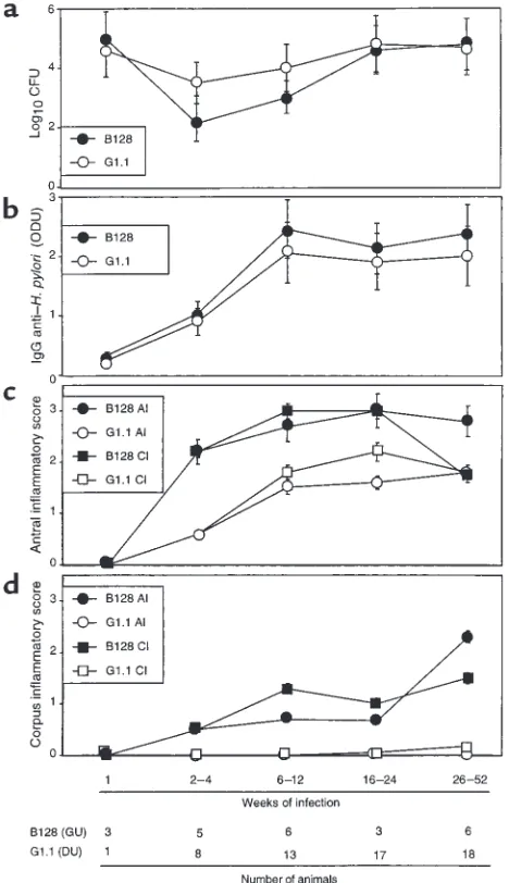
[image:4.612.307.541.324.730.2]H. pylori strains induce distinct patterns of inflammation and injury in Mongolian gerbils. In total, 104 gerbils were chal-lenged either with H. pyloriDU strain G1.1 (n = 60), GU strain B128 (n = 24), or Brucella broth alone (n = 20). Of 84 gerbils challenged with H. pylori, 80 (57 G1.1, 23 B128) were successfully infected, and there were no sig-nificant differences in colonization efficiency (P = 0.4).
Figure 1
We next asked whether colonization density, serum antibody response, or gastric inflammation and injury differed between the gerbil populations. There were no differences in colonization density (Figure 1a) or anti–H. pylori IgG antibody responses (Figure 1b) between B128- and G1.1-infected animals. All gerbils infected for more than 2–4 weeks developed acute and chronic antral gastritis, and scores increased until 16–24 weeks (Figure 1c). Similar to the antrum, 14 (93%) of 15 gerbils infected with GU strain B128 for more than 2–4 weeks developed corpus gastritis, and scores steadily increased throughout the observation period (Figure 1d). In contrast, only 3 (5%) of 57 gerbils infected with DU strain G1.1 had any corpus inflammation (P ≤0.001 compared with B128). Acute and chronic inflammato-ry scores (Figure 1, c and d), and the degree of epithelial cell degeneration (data not shown) were significantly (P ≤0.03) higher at all gastric sites in animals infected with B128 compared with G1.1. Thus, GU strain B128 induced a more severe and global inflammatory response than did DU strain G1.1.
Because gastric ulcer disease is associated with an increased risk for gastric adenocarcinoma (5), we next sought to determine whether GU strain B128 enhanced the development of premalignant mucosal lesions. Antral erosions and glandular displacement each were significantly (P ≤0.03) more frequent among gerbils infected with GU strain B128 (19/23 and 10/23 animals, respectively) compared with DU strain G1.1 (3/57 and 0/57 animals, respectively). Gastric ulcers developed in 4 (17%) of 23 and 0 (0%) of 57 animals infected with GU strain B128 and DU strain G1.1, respectively (P < 0.001), and all ulcers were located within the gastric antrum. Given that parietal cell loss can lead to aberrant epithe-lial cellular differentiation (31), we determined whether the respective H. pyloristrains variably altered parietal cell mass. Infection with GU strain B128 for more than
26 weeks led to parietal cell loss and glandular atrophy in 5 (83%) of 6 gerbils, but these changes did not occur at any time point in the G1.1-infected animals.
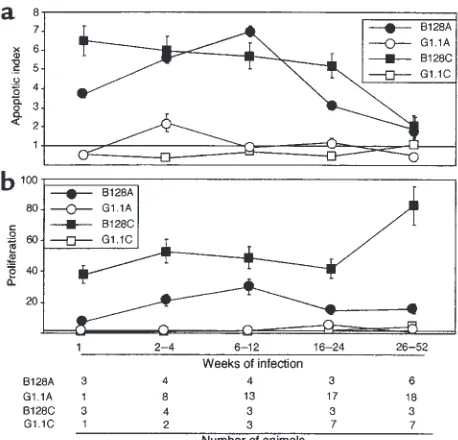
H. pylori GU strain B128 induces more severe mucosal cel-lular imbalances. Because the severity and pattern of gastritis differed among B128- and G1.1-infected ani-mals and because mucosal inflammation may con-tribute to cell turnover (7, 9), we next addressed whether apoptosis or proliferation was differentially affected by these H. pylori strains. Gerbils infected with GU strain B128 had significantly (P ≤0.05) higher antral and corpus apoptotic indices 1–24 weeks after inoculation than did broth- or G1.1-challenged ani-mals (Figure 2a). In contrast, antral apoptosis scores in G1.1-infected animals were significantly increased only at 2–4 weeks, and corpus apoptosis scores were no different from controls (Figure 2a). We have previous-ly shown that H. pylori–infected Mongolian gerbils develop hyperproliferation, although with different timing and morphological location than apoptosis (9). After challenge with GU strain B128, mucosal prolif-eration indices increased rapidly and were significant-ly higher at all time points than either G1.1-infected or control animals (Figure 2b; P < 0.001 for each). Thus, GU strain B128 induces a more intense tissue response than G1.1 as reflected by enhanced mucosal inflam-mation, cell proliferation, and cell death.
[image:5.612.305.534.56.276.2]H. pylori GU strain B128 enhances cytokine production and apoptosis in vitro. Given that GU strain B128 altered inflammation and cellular turnover more profoundly in vivo, we examined whether similar effects could be observed in vitro. Levels of IL-8, a proinflammatory cytokine that has potent chemoattractive and neu-trophil-activating properties, are significantly related to severity of gastric inflammation among H. pylori–positive persons (32, 33). Comparing IL-8 pro-duction in AGS cells after coculture with our prototype Figure 2
H. pyloristrains, GU strain B128 stimulated signifi-cantly (P = 0.003 versus G1.1) higher levels of IL-8 (Fig-ure 3a), results that parallel the in vivo response to this strain. AGS cells cocultured with either H. pyloristrain had significantly (P < 0.05) reduced viability compared with controls (Figure 3a); however, GU strain B128 sig-nificantly (P < 0.001) decreased viability compared with DU strain G1.1 (Figure 3a). To determine whether the reduction in viability reflected apoptosis, we quanti-tated DNA fragmentation in AGS cells after bacterial coculture. H. pylorisignificantly increased cytoplasmic oligonucleosomal fragments compared with AGS cells alone (P < 0.001 for each time point; Figure 3a), and again, GU strain B128 significantly enhanced this effect (P < 0.001; Figure 3a).
The ability of strains B128 and G1.1 to differential-ly alter IL-8 secretion and apoptosis in AGS cells reflected the responses to these same strains in vivo. Therefore, we next asked whether the bacterial prop-erties responsible for these changes had been affected by prolonged in vivo growth. Similar to the results obtained with the parental strains (Figure 3a), all B128 strains after prolonged gerbil colonization induced significantly higher levels of apoptosis and IL-8 pro-duction than G1.1 derivatives (P ≤0.01; Figure 3b), although no consistent intrafamilial strain differences were identified. These data indicate that the properties
necessary for induction of epithelial cell responses remain relatively stable during persistent H. pylori col-onization of the gastric niche.
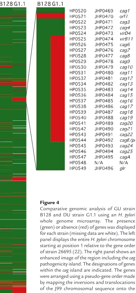
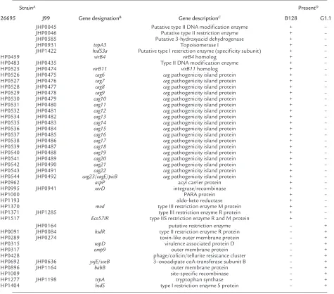
H. pylori whole genome microarray reveals a partial cag island deletion in strain G1.1. Having established that strains B128 and G1.1 have different phenotypes, we next used an H. pyloriwhole genome microarray to examine differences in gene content between these prototype strains. A total of 153 strain-specific genes (98 B128-specific and 55 G1.1-specific; Figure 4) were identified in the two isolates, and 44 (29%) of these (Table 1) were predicted to encode gene products with known, diverse functions such as recombination (xerD) and basic cellular metabolism (yxjE, trpA,
topA3). A substantial proportion of these ORFs had significant homology to genes encoding putative restriction-modification (R-M) enzymes (Table 1), findings that substantiate recent reports (34–36) demonstrating the presence of unique complements of R-M systems within individual H. pyloristrains. The microarray also identified major differences in gene content that localize to regions of linked genes, one of which includes a portion of the cag pathogenicity island (Figure 4). GU strain B128 contains 26 of 27 cag
island genes, whereas DU strain G1.1 possesses only seven of the 27 caggenes, indicating a strain-specific deletion of this region (Figure 4; Table 1). This
dele-Figure 3
[image:6.612.259.536.388.738.2]tion consisted of contiguous genes that were located in the midregion of the island and included picB/cagE
(HP0544, JHP0492), but not cagA(HP0547, JHP0495) (22, 23) (Figure 4; Table 1).
Inactivation of components of the cag island attenuates IL-8 induction and development of gastritis. Because previous studies have demonstrated that genes within the cag
island contribute to induction of IL-8 and apoptosis in vitro (11, 12, 25), we next disrupted picB/cagEand also deleted the entire cagisland in GU strain B128 to deter-mine whether these components were required for the phenotypes observed in the current study. Compared to parental strain B128, loss of picB/cagEand/or the entire
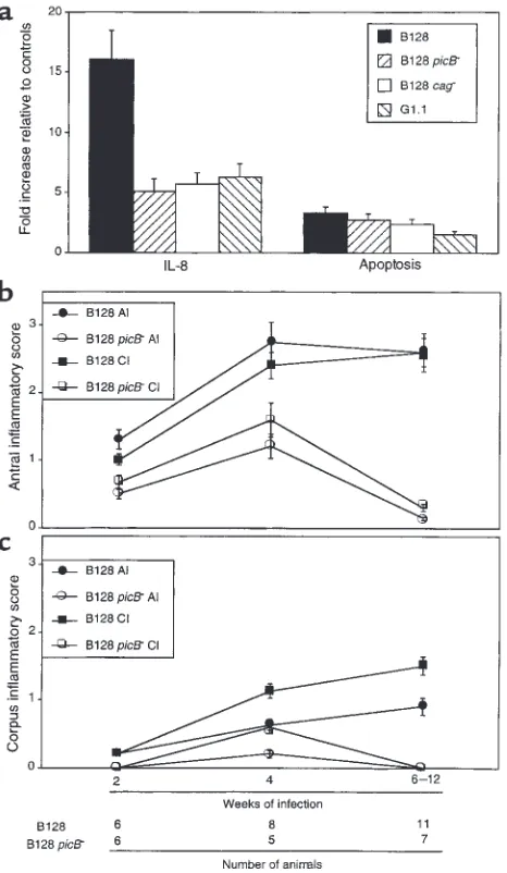
cagisland significantly reduced IL-8 secretion (P ≤0.001 versus B128) to levels that were similar to those induced by DU strain G1.1 (Figure 5a). Disruption of picB/cagE
alone was sufficient to produce the phenotype obtained by deletion of the entire cagisland. In contrast, the abil-ity of these same mutant strains to induce apoptosis was not significantly different from the wild-type strain (P = 0.32 and 0.20 versus B128, respectively; Figure 5a). We next sought to corroborate these in vitro findings by infecting gerbils with either wild-type B128 or its isogenic picB–derivative. Of 44 gerbils challenged with either B128 (n = 26) or B128 picB–(n = 18), 43 (25 B128 and 18 picB–) were successfully colonized. All strains recovered from mutant-challenged animals were kanamycin-resistant and contained the aphAcassette as determined by PCR using primers that flanked the insertion site within picB, indicating genomic stability of the antibiotic resistance cassette during in vivo col-onization. There were no differences in colonization levels between the populations (data not shown). How-ever, compared with the parental strain B128, infection with the picB–mutant significantly attenuated devel-opment of antral inflammation (P ≤0.03 versus B128; Figure 5b) as early as 2 weeks after challenge, and cor-pus inflammation by 4 weeks after infection (P ≤0.04 versus B128; Figure 5c). Within the antra of picB– -chal-lenged gerbils, neutrophilic infiltration was reduced to a greater degree compared with mononuclear cell infil-tration (Figure 5b). In addition to inflammation, the severity of epithelial cell degeneration and erosion was significantly (P ≤0.04 for each) decreased in gerbils infected with the picB–mutant compared with the wild-type strain (data not shown). These findings confirm our in vitro results (Figure 5a) and support the utility of whole genome microarray to identify strain-specific
H. pyloricomponents relevant to pathogenesis in vivo.
Discussion
Although certain H. pyloristrains are associated with pathological outcomes, the specific mechanisms that lead to these relationships have not been fully delin-eated. One strain-specific H. pyloriconstituent associ-ated with an augmented risk for ulcer disease and dis-tal gastric cancer is the cag pathogenicity island (13–16). After H. pyloriadherence to host cells, CagA is transported into the epithelial cell, where it becomes phosphorylated (37–39), and these events trigger changes in cellular morphology similar to those induced by hepatocyte growth factor (40). A CagA-independent consequence of cagisland-mediated H. pylori–epithelial cell contact is activation of intracellu-lar signaling pathways including NF-κB, which may contribute to IL-8 secretion (41). Thus, cagA+strains appear to be disproportionately represented among persons who develop serious sequelae of H. pylori car-riage, and cagisland genes are necessary for induction of epithelial cell responses relevant to pathogenesis.
[image:7.612.67.294.53.542.2]In the current study, we examined two strains that possessed similar virulence genotypic profiles (cagA+ Figure 4
Comparative genomic analysis of GU strain B128 and DU strain G1.1 using an H. pylori
whole genome microarray. The presence (green) or absence (red) of genes was displayed for each strain (missing data are white). The left panel displays the entire H. pylorichromosome starting at position 1 relative to the gene order of strain 26695 (22). The right panel shows an enhanced image of the region including the cag
vacAs1a iceA1), but which induced distinct phenotyp-ic responses. GU strain B128 induced more severe inflammation within gerbil mucosa and significantly higher levels of IL-8 in vitro.Using an H. pyloriwhole genome microarray, we found that although both pro-totype strains possessed the cagAgene and hence were
cagA+, their gene content within the cagisland differed substantially. GU strain B128 contains a near com-plete copy of the island (26 of 27 genes), whereas in DU strain G1.1, a large deletion is present. These results prompted us to re-examine epithelial cell responses without the influence of caggene products, and we confirmed the findings of previous investiga-tors (11, 12) that inactivation of picB/cagEor deletion of the entire cagisland significantly attenuated the ability of GU strain B128 to induce cytokine release in vitro. Infection of gerbils with an isogenic picB–
[image:8.612.57.532.287.705.2]deriv-ative recapitulated these events in vivo as mucosal inflammation, but not colonization, was significantly decreased in animals challenged with mutant strains compared with those infected with parental B128. The inflammatory component most profoundly affected was polymorphonuclear cell infiltration, which is con-sistent with the demonstration that loss of picBis asso-ciated with decreased production of IL-8, a cytokine with potent chemotactic and activating properties for neutrophils. Our previous studies have shown that neither CagA nor VacA is required for induction of inflammation in this model of H. pylori–induced gas-tritis (9), and genomic profiling of strains B128 and G1.1 support these observations, as both strains con-tained cagAand vacA. These findings underscore and confirm the importance of the entire cagisland as a bacterial locus related to high-grade host responses
Table 1
Strain-specific B128 and G1.1 genes with putative functions
StrainA PresentD
26695 J99 Gene designationB Gene descriptionC B128 G1.1
JHP0045 Putative type II DNA modification enzyme + –
JHP0046 Putative type II restriction enzyme + –
JHP0585 Putative 3-hydroxyacid dehydrogenase + –
JHP0931 topA3 Topoisomerase I + –
JHP1422 hsdS3a Putative type I restriction enzyme (specificity subunit) + –
HP0459 virB4 virB4homolog + –
HP0483 JHP0435 Type II DNA modification enzyme + –
HP0525 JHP0474 virB11 virB11homolog + –
HP0526 JHP0475 cag6 cagpathogenicity island protein + –
HP0527 JHP0476 cag7 cagpathogenicity island protein + –
HP0528 JHP0477 cag8 cagpathogenicity island protein + –
HP0529 JHP0478 cag9 cagpathogenicity island protein + –
HP0530 JHP0479 cag10 cagpathogenicity island protein + –
HP0531 JHP0480 cag11 cagpathogenicity island protein + –
HP0532 JHP0481 cag12 cagpathogenicity island protein + –
HP0534 JHP0482 cag13 cagpathogenicity island protein + –
HP0535 JHP0483 cag14 cagpathogenicity island protein + –
HP0536 JHP0484 cag15 cagpathogenicity island protein + –
HP0537 JHP0485 cag16 cagpathogenicity island protein + –
HP0538 JHP0486 cag17 cagpathogenicity island protein + –
HP0539 JHP0487 cag18 cagpathogenicity island protein + –
HP0540 JHP0488 cag19 cagpathogenicity island protein + –
HP0541 JHP0489 cag20 cagpathogenicity island protein + –
HP0542 JHP0490 cag21 cagpathogenicity island protein + –
HP0543 JHP0491 cag22 cagpathogenicity island protein + –
HP0544 JHP0492 cag23/cagE/picB cagpathogenicity island protein + –
HP0962 acpP acyl carrier protein + –
HP0995 JHP0941 xerD integrase/recombinase + –
HP1000 PARA protein + –
HP1193 aldo-keto reductase + –
HP1370 mod type III restriction enzyme M protein + –
HP1371 JHP1285 type III restriction enzyme R protein + –
HP1517 Eco57IR type IIS restriction enzyme R and M protein + –
JHP0164 putative restriction enzyme – +
HP0091 JHP0084 hsdR type II restriction enzyme R protein – +
HP0289 JHP0274 toxin-like outer membrane protein – +
HP0315 vapD virulence associated protein D – +
HP0317 omp9 outer membrane protein – +
HP0428 phage/colicin/tellurite resistance cluster – +
HP0692 JHP0636 yxjE/scoB 3–oxoadipate coA-transferase subunit B – +
HP0896 JHP1164 babB outer membrane protein – +
HP1009 site-specific recombinase – +
HP1277 JHP1198 trpA tryptophan synthase – +
HP1404 hsdS type I restriction enzyme S protein – +
AUnique identifier from H. pylorigenes in annotated sequences of 26695 and J99. BPutative gene name if available. CDescription of putative gene function.
DPresence (+) as determined by an average fluorescence ratio of greater or equal to 0.5 from two independent hybridizations. Absence (–) was defined as an
and provide further evidence that strain-specific bac-terial components identified by microarray are rele-vant to pathology within the gastric niche.
Many of the genetic differences identified between the two studied strains involved the cagisland; howev-er, deletion of this locus did not significantly affect the ability of GU strain B128 to induce apoptosis in vitro. Other H. pyloricomponents that may induce apoptosis include urease, a highly conserved H. pyloriconstituent,
which can induce apoptosis in vitro by binding to MHC class II molecules expressed on the surfaces of gastric epithelial cells (42). However, although H. pylori
can directly induce apoptosis in isolated in vitro sys-tems, apoptosis in vivo is likely to be regulated by host mediators present within inflamed mucosa. The immune response elicited by H. pyloriin humans and by Helicobacter felisin mice is Th1-predominant (43, 44), and IFN-γ, a Th1 lymphocyte–derived cytokine that is increased within colonized mucosa, is synergistic with
H. pyloriin inducing Fas-Fas ligand–regulated (FasL-regulated) apoptosis of gastric epithelial cells in vitro (45, 46). H. pylori infection of IFN-γ–deficient mice leads to decreased levels of gastric inflammation com-pared with wild-type mice (47, 48), and in vivo neutral-ization of IFN-γin mice infected with H. felissimilarly reduces the severity of gastritis (43). Adoptive transfer of Th1 cells from H. pylori–infected mice into infected recipients increases the severity of gastritis (49), and concurrent helminth infection modulates a proin-flammatory Th1-mediated murine gastritis and atro-phy response to H. felistoward a less injurious Th2-type response (50). H. felisinfection of Fas-deficient mice is also associated with reduced levels of inflammation compared with infected wild-type mice, and mucosal apoptosis scores are decreased in parallel (51).
One hypothesis suggested by the previous findings described here is that Th1-derived cytokines (i.e., IFN-γ) within inflamed mucosa may increase apop-tosis by regulating Fas-FasL interactions between inflammatory cells and gastric epithelial cells. How-ever, additional host genetic factors also may con-tribute to the ability of H. pylorito alter apoptosis in vivo. Mice lacking secretory phospholipase A2that are infected by H. felisdevelop increased epithelial cell apoptosis (52). Apoptosis in response to H. pylorialso may reflect heterogeneity of class II MHC host geno-types, as binding of H. pylorito these IFN-γ–inducible molecules can directly stimulate apoptosis (42). El-Omar et al. have recently identified particular poly-morphisms of the human IL-1βgene promoter that are genetic risk factors for both precursor lesions of gastric cancer (atrophic gastritis) as well as gastric adenocarcinoma per se among H. pylori–infected per-sons (53). IL-1βcan stimulate multiple intracellular signaling pathways involved in apoptosis, including Fas and NF-κB (41, 45, 46), and thus differing levels of IL-1βexpression within H. pylori–colonized gastric mucosa may regulate levels of apoptosis in vivo.
[image:9.612.59.292.53.454.2]IL-1βis also a potent inhibitor of gastric acid secre-tion, and polymorphisms within its promoter region may differentially influence additional physiological events that dictate disease outcomes among H. pylori–colonized populations. Two recent investigations have revealed that acid outputs in gerbils chronically infected with H. pyloriare either no different (54) or reduced (55) compared with uninfected controls. Fur-ther, mucosal IL-1βmRNA levels reciprocally increase as acidity decreases and administration of recombinant Figure 5
IL-1 receptor antagonist normalizes acid outputs among gerbils carrying H. pylori(55). Collectively, these data indicate that the host inflammatory response is an important determinant of specific pathological out-comes among H. pylori–infected populations.
Understanding H. pyloridiversification and adapta-tion during long-term colonizaadapta-tion is crucial toward delineating mechanisms by which these organisms may lead to disease. As evidenced by our results, the use of microarray-based comparative genomics pro-vides a powerful tool for identifying strain-specific factors that may contribute to phenotypic variability among H. pylori isolates assumed to have similar genetic composition. One limitation of this tech-nique, however, is that it cannot detect small dele-tions, point mutadele-tions, deletions within intergenic regions, rearrangements, ORFs that are not present in either strain 26695 or J99, or deletions of homologous repetitive elements. However, as microarrays of greater resolution are developed and new strain-spe-cific ORFs are identified, it should soon be possible to determine the relative frequency and importance of these additional sources of genetic variability. Geno-typic markers could then be developed to not only to identify individuals at risk for specific clinical seque-lae of infection, but also to permit selective targeting of therapy for disease prevention.
In conclusion, we have shown that H. pyloristrains with similar virulence markers can induce distinct patterns of gastric inflammation and injury in an ani-mal model of H. pylori–induced carcinogenesis and that these findings can be corroborated in vitro and in vivo. DNA hybridization to an H. pylori whole genome microarray revealed that differences in the ability of H. pyloristrains to induce epithelial cell responses related to inflammation are dependent on the presence of an intact cag pathogenicity island, although the specific function of numerous addi-tional strain-specific loci and their significance in pathogenesis remain to be determined.
Acknowledgments
Supported in part by the NIH (KO8 DK02381, R29 CA77955, R01 DK50837, and RO1 AI38459), and by the Medical Research Service of the Department of Vet-erans Affairs. We thank Tyler Richmond and Uma Krishna for excellent technical assistance.
1. Peterson, W.L. 1991. Helicobacter pyloriand peptic ulcer disease. N. Engl. J. Med. 324:1043–1048.
2. Nomura, A., et al. 1991. Helicobacter pyloriinfection and gastric carci-noma among Japanese Americans in Hawaii. N. Engl. J. Med.
325:1132–1136.
3. Parsonnet, J., et al. 1991. Helicobacter pyloriinfection and the risk of gas-tric carcinoma. N. Engl. J. Med. 325:1127–1131.
4. Correa, P., et al. 1990. Gastric precancerous process in a high risk pop-ulation: cohort follow-up. Cancer Res. 50:4737–4740.
5. Hansson, L.E., et al. 1996. The risk of stomach cancer in patients with gastric or duodenal ulcer disease. N. Engl. J. Med. 335:242–249. 6. Moss, S.F., Calam, J., Agarwal, B., Wang, S., and Holt, P.R. 1996.
Induc-tion of gastric epithelial apoptosis by Helicobacter pylori. Gut.
38:498–501.
7. Peek, R.M., Jr., et al. 1997. Helicobacter pylori cagA+ strains and
dissoci-ation of gastric epithelial cell proliferdissoci-ation from apoptosis. J. Natl. Can-cer Inst. 89:863–868.
8. Wirth, H.P., Beins, M.H., Yang, M., Tham, K.T., and Blaser, M.J. 1998. Experimental infection of Mongolian gerbils with wild-type and mutant Helicobacter pyloristrains. Infect. Immun. 66:4856–4866. 9. Peek, R.M., et al. 2000. Helicobacter pylorialters gastric epithelial cell
cycle events and gastrin secretion in Mongolian gerbils. Gastroenterolo-gy. 118:48–59.
10. Watanabe, T., Tada, M., Nagai, H., Sasaki, S., and Nakao, M. 1998. Heli-cobacter pyloriinfection induces gastric cancer in Mongolian gerbils. Gastroenterology. 115:642–648.
11. Censini, S., et al. 1996. cag, a pathogenicity island of Helicobacter pylori, encodes type I-specific and disease-associated virulence factors. Proc. Natl. Acad. Sci. USA. 93:14648–14653.
12. Tummuru, M.K., Sharma, S.A., and Blaser, M.J. 1995. Helicobacter pylori picB, a homologue of the Bordetella pertussistoxin secretion protein, is required for induction of IL-8 in gastric epithelial cells. Mol. Microbiol.
18:867–876.
13. Crabtree, J.E., et al. 1991. Mucosal IgA recognition of Helicobacter pylori 120 kDa protein, peptic ulceration, and gastric pathology. Lancet.
338:332–335.
14. Kuipers, E.J., Perez-Perez, G.I., Meuwissen, S.G., and Blaser, M.J. 1995. Helicobacter pyloriand atrophic gastritis: importance of the cagAstatus. J. Natl. Cancer Inst. 87:1777–1780.
15. Crabtree, J.E., et al. 1993. Systemic and mucosal humoral responses to Helicobacter pyloriin gastric cancer. Gut. 34:1339–1343.
16. Blaser, M.J., et al. 1995. Infection with Helicobacter pyloristrains pos-sessing cagAis associated with an increased risk of developing adeno-carcinoma of the stomach. Cancer Res. 55:2111–2115.
17. Atherton, J.C., et al. 1995. Mosaicism in vacuolating cytotoxin alleles of Helicobacter pylori: Association of specific vacAtypes with cytotoxin production and peptic ulceration. J. Biol. Chem. 270:17771–17777. 18. Peek, R.M., Jr., et al. 1998. Adherence to gastric epithelial cells induces
expression of a Helicobacter pylorigene, iceA, that is associated with clin-ical outcome. Proc. Assoc. Am. Physicians. 110:531–544.
19. van Doorn, L.J., et al. 1998. Clinical relevance of the cagA, vacA, and iceA status of Helicobacter pylori. Gastroenterology. 115:58–66.
20. Ito, Y., et al. 2000. Sequence analysis and clinical significance of the iceA gene from Helicobacter pylori strains in Japan. J. Clin. Microbiol.
38:483–488.
21. Maeda, S., et al. 1997. High seropositivity of anti-CagA antibody in Helicobacter pylori-infected patients irrelevant to peptic ulcers and nor-mal mucosa in Japan. Dig. Dis. Sci. 42:1841–1847.
22. Tomb, J.F., et al. 1997. The complete genome sequence of the gastric pathogen Helicobacter pylori. Nature. 388:539–547.
23. Alm, R.A., et al. 1999. Genomic-sequence comparison of two unrelat-ed isolates of the human gastric pathogen Helicobacter pylori. Nature.
397:176–180.
24. Salama, N., et al. 2000. A whole genome microarray reveals genetic diversity among Helicobacter pyloristrains. Proc. Natl. Acad. Sci. USA.
97:14668–14673.
25. Peek, R.M., Jr., et al. 1999. Helicobacter pyloristrain-specific genotypes and modulation of the gastric epithelial cell cycle. Cancer Res.
59:6124–6131.
26. Akopyants, N.S., et al. 1998. Analyses of the cagpathogenicity island of Helicobacter pylori. Mol. Microbiol. 28:37–53.
27. Eisen, M.B., and Brown, P.O. 1999. DNA arrays for analysis of gene expression. Methods Enzymol. 303:179–205.
28. Stanford Microarray Database. http://genome-www4.stanford.edu/ MicroArray/help/results_normalization.html.
29. http://www.microarrays.org/software.html. Accessed August, 2000. 30. Eisen, M.B., Spellman, P.T., Brown, P.O., and Botstein, D. 1998. Clus-ter analysis and display of genome-wide expression patClus-terns. Proc. Natl. Acad. Sci. USA. 95:14863–14868.
31. Guruge, J.L., et al. 1998. Epithelial attachment alters the outcome of Helicobacter pyloriinfection. Proc. Natl. Acad. Sci. USA. 95:3925–3930. 32. Peek, R.M., Jr., et al. 1995. Heightened inflammatory response and cytokine expression in vivoto cagA+ Helicobacter pyloristrains. Lab. Invest. 73:760–770.
33. Crabtree, J.E., Peichl, P., Wyatt, J.I., Stachl, U., and Lindley, I.J. 1993. Gastric interleukin-8 and IgA IL-8 autoantibodies in Helicobacter pylori infection. Scand. J. Immunol. 37:65–70.
34. Akopyants, N.S., et al. 1998. PCR-based subtractive hybridization and differences in gene content among strains of Helicobacter pylori. Proc. Natl. Acad. Sci. USA. 95:13108–13113.
35. Xu, Q., Morgan, R.D., Roberts, R.J., and Blaser, M.J. 2000. Identifica-tion of type II restricIdentifica-tion and modificaIdentifica-tion systems in Helicobacter pylori reveals their substantial diversity among strains. Proc. Natl. Acad. Sci. USA. 97:9671–9676.
37. Odenbreit, S., et al. 2000. Translocation of Helicobacter pyloriCagA into gastric epithelial cells by type IV secretion. Science. 287:1497–1500. 38. Stein, M., Rappuoli, R., and Covacci, A. 2000. Tyrosine phosphorylation
of the Helicobacter pyloriCagA antigen after cag-driven host cell translo-cation. Proc. Natl. Acad. Sci. USA. 97:1263–1268.
39. Asahi, M., et al. 2000. Helicobacter pyloriCagA protein can be tyrosine phosphorylated in gastric epithelial cells. J. Exp. Med. 191:593–602. 40. Segal, E.D., Cha, J., Lo, J., Falkow, S., and Tompkins, L. 1999. Altered
states: involvement of phosphorylated CagA in the induction of host cel-lular growth changes by Helicobacter pylori. Proc. Natl. Acad. Sci. USA.
96:14559–14564.
41. Keates, S., Hitti, Y.S., Upton, M., and Kelly, C.P. 1997. Helicobacter pylori infection activates NF-kappa B in gastric epithelial cells. Gastroenterolo-gy. 113:1099–1109.
42. Fan, X., et al. 2000. Helicobacter pyloriurease binds to class II MHC on gas-tric epithelial cells and induces their apoptosis. J. Immunol.
165:1918–1924.
43. Mohammadi, M., Czinn, S., Redline, R., and Nedrud, J. 1996. Helicobac-ter-specific cell-mediated immune responses display a predominant Th1 phenotype and promote a delayed-type hypersensitive response in the stomachs of mice. J. Immunol.156:4729–4738.
44. Bamford, K.B., et al. 1998. Lymphocytes in the human gastric mucosa during Helicobacter pylorihave a T helper cell 1 phenotype. Gastroenterol-ogy. 114:482–492.
45. Wagner, S., et al. 1997. Regulation of gastric epithelial cell growth by Heli-cobacter pylori: evidence for a major role of apoptosis. Gastroenterology.
113:1836–1847.
46. Wang, J., et al. 2000. Helicobacter pylorimodulates lymphoepithelial cell interactions leading to epithelial cell damage through Fas/Fas ligand
interactions. Infect. Immun.68:4303–4311.
47. Sawai, N., et al. 1999. Role of gamma interferon in Helicobacter pylori -induced gastric inflammatory responses in a mouse model. Infect. Immun.67:279–285.
48. Smythies, L.E., et al. 2000. Helicobacter pylori-induced mucosal inflam-mation is Th1 mediated and exacerbated in IL-4, but not IFN-γ, gene deficient mice. J. Immunol.165:1022–1029.
49. Mohammadi, M., Nedrud, J., Redline, R., Lycke, N., and Czinn, S.J. 1997. Murine CD4 T-cell responses to Helicobacterinfection: TH1 cells enhance gastritis and TH2 cells reduce bacterial load. Gastroenterology. 113:1848–1857.
50. Fox, J.G., et al. 2000. Concurrent enteric helminth infection modulates inflammation and gastric immune responses and reduces Helicobacter -induced gastric atrophy. Nat. Med.6:536–542.
51. Houghton, J., Bloch, L.M., Goldstein, M., von Hagen, S., and Korah, R.M. 2000. In vivo disruption of the Fas pathway abrogates gastric growth alterations secondary to Helicobacter infection. J. Infect. Dis.182:856–864. 52. Wang, T.C., et al. 1998. Mice lacking secretory phospholipase A2 show altered apoptosis and differentiation with Helicobacter felisinfection. Gas-troenterology. 114:675–689.
53. El-Omar, E.M., et al. 2000. Interleukin-1 polymorphisms associated with increased risk of gastric cancer. Nature. 404:398–402.
54. Keto, Y., Takahashi, S., and Okabe, S. 1999. Healing of Helicobacter pylori -induced gastric ulcers in Mongolian gerbils: combined treatment with omeprazole and clarithromycin. Dig. Dis. Sci.44:257–265.