http://dx.doi.org/10.4236/ojanes.2015.59037
How to cite this paper: Baral, B.K., Ide, S., Uzawa, M., Kiyosawa, K., Sasaki, H. and Kawamata, M. (2015) Conscious Sedation and Awake Fiberoptic Intubation in a Patient with Difficult Mask Ventilation—A Case Report. Open Journal of Anesthesiol-ogy, 5, 206-210. http://dx.doi.org/10.4236/ojanes.2015.59037
Conscious Sedation and Awake Fiberoptic
Intubation in a Patient with Difficult Mask
Ventilation—A Case Report
Bidur Kumar Baral
1,2, Susumu Ide
1*, Masahiro Uzawa
1, Kenkichi Kiyosawa
1,
Hiroyuki Sasaki
3, Mikito Kawamata
11Department of Anesthesiology and Resuscitology, Shinshu University School of Medicine, Matsumoto, Japan 2Department of Anesthesiology and Critical Care, Norvic International Hospital, Kathmandu, Nepal
3Department of Radiology, Shinshu University Hospital, Matsumoto, Japan
Email: *ide@shinshu-u.ac.jp
Received 28 August 2015; accepted 27 September 2015; published 30 September 2015
Copyright © 2015 by authors and Scientific Research Publishing Inc.
This work is licensed under the Creative Commons Attribution International License (CC BY).
http://creativecommons.org/licenses/by/4.0/
Abstract
Since maxillofacial malignancy is a common cause of facial defects and disfigurement of the face that may make fitting of a mask difficult and cause air leakage from the side, thus making mask ventilation difficult. In addition, distorted anatomy of the airway and base of the skull in such pa-tients may cause difficult intubation (DI). We experienced a case with a huge facial defect due to maxillary carcinoma, in which difficult mask ventilation (DMV) and DI were predicted. After eval-uation by three-dimensional airway computed tomography, the airway was secured with con-scious sedation using dexmedetomidine, and awake fiberoptic intubation was safely performed. Three-dimensional airway computed tomography seems to be a good tool for successful intuba-tion when DMV and DI are predicted.
Keywords
Conscious Sedation, Difficult Mask Ventilation, Awake Fiberoptic Intubation
1. Introduction
Difficult airway is still a challenge for anesthesiologists. It includes difficult intubation (DI), difficult mask ven-tilation (DMV) or both. Since mask venven-tilation is a basic fundamental skill in airway management, every anes-thesiologist should acquire skills for mask ventilation and should be knowledgeable about the causes of DMV
and consider alternative options including oropharyngeal airway, nasopharyngeal airway, laryngeal mask airway, transtracheal jet ventilation, cricothyrotomy, and awake fiberoptic intubation (AFOI) when the mask ventilation is difficult [1]. Mask ventilation is the first step of airway management before endotracheal intubation or inser-tion of any airway devices. It is a rescue technique when endotracheal intubainser-tion has failed or has become diffi-cult. Therefore mask ventilation became a major step in any difficult airway algorithm. Every Anesthesiologist should also concentrate on prediction of DI. Prediction of DMV and concomitant DI may be the most difficult situation for anesthesiologists. Here we present a case of a huge maxillofacial defect due to squamous cell car-cinoma of the maxilla. After evaluation by three-dimensional airway computed tomography, we managed the airway with conscious sedation using dexmedetomidine and AFOI.
2. Case Summary
A 58-year-oldmale, height 168 cm, weight 45 kg, and American Society of Anesthesiologists physical status 1, was presented with a history of a large wound on the left side of his face and swelling. He was diagnosed with squamous cell carcinoma of the maxilla 4 years ago and had been treated with chemotherapy and radiotherapy. He underwent extended total maxillectomy 3 years ago. His wound laterbecame infected, and chronic osteomye-litis of the mandible developed. He was diagnosed as having chronic osteomyeosteomye-litis of the left mandible with large facial defect on left side of face. He was planned for debridement, sequestrectomy and reconstruction of the defect with anterolateral free flap of the thigh. On examination, his vital parameters were within normal ranges. His general physical examination and systemic examination were unremarkable. Results of laboratory tests were within normal ranges.
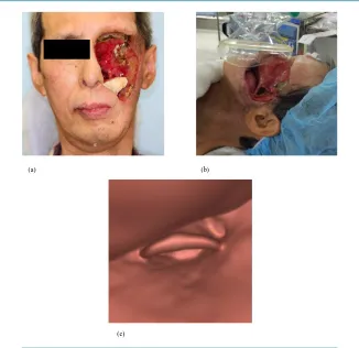
Airway assessment showed that mouth opening was restricted (less than 1 finger). He had a large ulcerated wound on the left side of his face that extended from the left supra orbit to lower jaw and involved the eye. Large amounts of tissue and muscle were lost on the left cheek (Figure 1(a)). A mandibular protrusion test could not be performed due to pain. Neck movement was normal and thyromental distance was 6.5 cm.
The surgery was plannedunder general anesthesia. Considering the DMV (Figure 1(b)), we had planned to secure the airway with AFOI under conscious sedation. DI was also predicted because of distorted anatomy of the airway and base of the skull. Three-dimensional computed tomography (3D-CT) of the airway was obtained before surgery, and access to the trachea was pre-evaluated for tracheal intubation (Figure 1(c)). Finally, we planned nasal intubation at the right side using AFOI. The procedure for AFOI was explained in detail to the pa-tient and papa-tient’s family and informed consent was obtained. The papa-tient was kept nil per oral for 8 hours prior to surgery.
On the day of surgery, the patient was transferred to the operation theatre, monitors (i.e. electrocardiogram, non-invasive blood pressure, pulse oximeter, temperature probe) were connected. An intravenousline was se-cured with an 18 G intravenous cannula. Arterial cannulation was done on his left hand for invasive blood pres-sure monitoring. The patient was catheterized with a Foley catheter. The availability and working condition of all components of the difficult airway trolley were checked. Arescue ventilation device was prepared, and standby arrangement for emergency tracheostomy was also prepared. For nasal decongestion and anesthesia, the right nasal cavity was packed with ribbon gauze soaked in 1% lidocaine and epinephrine solution (1:200,000) and was leftin place for 10 minutes. For conscious sedation, injection dexmedetomidine at 6 mcg/kg/hr was started and sedation was assessed with the Richmond agitation-sedation scale (RASS) [2]. After 10 minutes of dexmedetomidine infusion, the RASS score was 0 to −1, and the dose of dexmedetomidine was decreased to 0.4 mcg/kg/hr. There was no hypotension or bradycardiaduring the infusion of dexmedetomidine.
(a) (b)
[image:3.595.149.475.77.393.2](c)
Figure 1. Large ulcerated wound on the left side of the face (a), difficult mask ventilation (b), and three-dimensional computed tomography (3D-CT) of the airway (c).
entry and end-tidal CO2 tracing. The ET tube was then fixed. Sixty mg of propofol was injected intravenously,
and eyelash reflex was noted. Thirty mg of rocuronium was injected intravenously after the disappearance of eyelash reflex. Then dexmedetomidine infusion was stopped and the patient was kept on a mechanical ventilator, allowing the surgeon to start the surgery. The patient was maintained intraoperatively with oxygen, remifentanil, rocuronium and sevoflurane. The patienttolerated the procedure well, and he had no recall of fiberoptic intuba-tion.
Surgery lasted for 13 hours, and the intraoperative period was uneventful. Total blood loss was 300 ml. After completion of surgery, the patient was transferred to the intensive care unit and kept on mechanical ventilation with propofol sedation. We did not extubated the patient because of anticipation of glottic edema, anticipated DI and long surgery. The patient was successfully extubated on the next day. The postoperative period was un-eventful, and the patient was discharged after two weeks.
3. Discussion
Mask ventilation is the initial and mostessential step in airway management. Every anesthesiologist should ac-quire the skills for mask ventilation [1]. DMV is a situation in which it is not possible for the anesthesiologist to provide adequate face mask ventilation due to an inadequate mask seal, excessive gas leakage or excessive re-sistance to ingress or egress of gas [3]. There is a wide variation in the incidence of DMV. The incidence of DMV was reported to be 5% by Langeron et al. [4] and 1.4% by Asai et al. [5]. However, DMV is underesti-mated by most anesthesiologists. DMV may occur before intubation or after failure of endotracheal intubation
[6]. All anesthesiologists should therefore have knowledge of DMV, causes of DMV and alternative techniques when mask ventilation has become difficult or has failed [7].
side of his cheek, perioral edema and chronic osteomyelitis of the mandible. These conditions made proper fit-ting of the mask difficult and would cause air leakage from the side(Figure 1(a) and Figure 1(b)), and there would also be a risk of spread of infection to the brain. The mouth opening was <1 finger, and the use of an oral alternative airway device for intubation was limited. The patient had also received radiation and had distorted airway anatomy, suggesting that DI was predicted. Thus, the airway was pre-evaluated by using 3D-CT of the airway (Figure 1(c)), indicating thatthe orolaryngeal space was narrow but that the epiglottis was placed in the middle. AFOI under conscious sedation was considered to be safe and the only option to avoid complications. According to Benumof et al. [8], AOFI is also the safest approach of the airway management in the patients with predicted DMV. Inadequateventilation occurs in up to 38% of patients with facial injuries reported by Caplan et al. [9]. Benumof et al. [8] estimated that up to 30% of deathsare due to inability of successful airway manage-ment.
Major challenges during AFOI are providing adequate sedation, maintaining a patent airway and ensuring adequate spontaneous ventilation. Several classes of drugs including benzodiazepines, opioids, alpha2 agonists,
propofol, and ketamine have been reported for conscious sedation during AFOI [2]. Among them, dexmedeto-midine is a highly selective, potent alpha2 adrenergic receptor agonist. It has the ability to produce profound
se-dation without causing respiratory depression. In addition, dexmedetomidine decreases salivary secretion through sympatholytic and vagomimetic effects, which is advantageous for fiberoptic intubation [10] [11]. Furthermore, dexmedetomidine provided an optimal intubating condition, less hemodynamic instability and better patient to-lerance [12] [13]. Dexmedetomidine thus has many properties that make it suitable for AFOI, and it has been highly recommended for AFOI. In addition to successful use of dexmedetomidine for AFOI in patients with se-vere submandibular abscess, dexmedetomidine also provided an optimal intubating condition in our patient.
dos Reis Falcao et al. [14] reported a similar type of case with huge facial defect having DMV. They managed the case with transorbital intubation using propofol and fentanyl under spontaneous ventilation. In our case, transorbital intubation was not possible and nasal intubation was required because the plan of surgery was re-construction of the facial defect. Pre-evaluation of the airway by using 3D-CT provided useful information for planning of airway management.
4. Conclusion
In conclusion, for patients who are predicted to have DMV, conscious sedation using dexmedetomidine and AFOI is a good alternative.
References
[1] El-Orbany, M. and Woehlck, H.J. (2009) Difficult Mask Ventilation. Anesthesia and Analgesia, 109, 1870-1880. http://dx.doi.org/10.1213/ANE.0b013e3181b5881c
[2] Sessler, C.N., Gosnell, M.S., Grap, M.J., Brophy, G.M., O’Neal, P.V., Keane, K.A., Tesoro, E.P. and Elswick, R.K. (2002) The Richmond Agitation-Sedation Scale: Validity and Reliability in Adult Intensive Care Unit Patients. Ameri-can Journal of Respiratory and Critical Care Medicine, 166, 1338-1344. http://dx.doi.org/10.1164/rccm.2107138 [3] American Society of Anesthesiologists Task Force on Management of the Difficult Airway (2003) Practice Guidelines
for Management of the Difficult Airway: An Updated Report by the American Society of Anesthesiologists Task Force on Management of the Difficult Airway. Anesthesiology, 98, 1269-1277.
http://dx.doi.org/10.1097/00000542-200305000-00032
[4] Langeron, O., Masso, E., Huraux, C., Guggiari, M., Bianchi, A., Coriat, P. and Riou, B. (2000) Prediction of Difficult Mask Ventilation. Anesthesiology, 92, 1229-1236. http://dx.doi.org/10.1097/00000542-200005000-00009
[5] Asai, T., Koga, K. and Vaughan, R.S. (1998) Respiratory Complications Associated with Tracheal Intubation and Ex-tubation. British Journal of Anaesthesia, 80, 767-775. http://dx.doi.org/10.1093/bja/80.6.767
[6] Johnston, K.D. and Rai, M.R. (2013) Conscious Sedation for Awake Fibreoptic Intubation: A Review of the Literature.
Canadian Journal of Anaesthesia, 60, 584-599. http://dx.doi.org/10.1007/s12630-013-9915-9
[7] Kheterpal, S., Han, R., Tremper, K.K., Shanks, A., Tait, A.R., O’Reilly, M. and Ludwig, T.A. (2006) Incidence and Predictors of Difficult and Impossible Mask Ventilation. Anesthesiology, 105, 885-891.
http://dx.doi.org/10.1097/00000542-200611000-00007
[9] Caplan, R.A., Posner, K.L., Ward, R.J. and Cheney, F.W. (1990) Adverse Respiratory Events in Anesthesia: A Closed Claims Analysis. Anesthesiology, 72, 828-833. http://dx.doi.org/10.1097/00000542-199005000-00010
[10] Belleville, J.P., Ward, D.S., Bloor, B.C. and Maze, M. (1992) Effects of Intravenous Dexmedetomidine in Humans. I. Sedation, Ventilation, and Metabolic Rate. Anesthesiology, 77, 1125-1133.
http://dx.doi.org/10.1097/00000542-199212000-00013
[11] Hall, J.E., Uhrich, T.D., Barney, J.A., Arain, S.R. and Ebert, T.J. (2000) Sedative, Amnestic, and Analgesic Properties of Small-Dose Dexmedetomidine Infusions. Anesthesia and Analgesia, 90, 699-705.
http://dx.doi.org/10.1097/00000539-200003000-00035
[12] Chu, K.S., Wang, F.Y., Hsu, H.T., Lu, I.C., Wang, H.M. and Tsai, C.J. (2010) The Effectiveness of Dexmedetomidine Infusion for Sedating Oral Cancer Patients Undergoing Awake Fibreoptic Nasal Intubation. European Journal of Anaesthesiology, 27, 36-40. http://dx.doi.org/10.1097/EJA.0b013e32832e0d2b
[13] Bergese, S.D., Khabiri, B., Roberts, W.D., Howie, M.B., McSweeney, T.D. and Gerhardt, M.A. (2007) Dexmedetomi-dine for Conscious Sedation in Difficult Awake Fiberoptic Intubation Cases. Journal of Clinical Anesthesia, 19, 141- 144. http://dx.doi.org/10.1016/j.jclinane.2006.07.005