http://dx.doi.org/10.4236/ss.2014.56043
How to cite this paper: Mandhan, P., Ismail, F.A.H., Ali, M.J. and Soofi, M.E. (2014) Appendicular Neuroendocrine Tumors in Children. Surgical Science, 5, 246-251. http://dx.doi.org/10.4236/ss.2014.56043
Appendicular Neuroendocrine Tumors
in Children
Parkash Mandhan
1*, Falah Ali Hasan Ismail
1, Mansour J. Ali
1, Madiha Emran Soofi
21Department of Pediatric Surgery, Hamad General Hospital, Hamad Medical Corporation, Doha, Qatar 2Department of Anatomical Pathology, Hamad General Hospital, Hamad Medical Corporation, Doha, Qatar
Email: *kidscisurg@icloud.com, fismail76@yahoo.com, maliI31@hmc.org.qa, emran_amir@hotmail.com
Received 3 May 2014; revised 1 June 2014; accepted 9 June 2014
Copyright © 2014 by authors and Scientific Research Publishing Inc.
This work is licensed under the Creative Commons Attribution International License (CC BY). http://creativecommons.org/licenses/by/4.0/
Abstract
Appendicular Neuroendocrine Tumors (ANETs) in pediatric age group are infrequent. Though children may present like symptoms of acute appendicitis, these tumors are incidentally picked up during routine histological examination of resected appendix. We report our experience with ANETs in children from a tertiary care hospital in Arabian Peninsula. During 6-year period, there were 700 appendectomies performed in children (≤14 years) and we collected only 3 cases of ANETs diagnosed from histological examination of resected appendix. Appendectomy alone has been sufficient in all cases and they are disease free at follow-up till 3 years after surgery. We have reviewed the clinical presentation, diagnosis and management of these cases. With no typical clin-ical picture, ANET is usually an incidental finding hence we propose that the review of the histolo-gy of resected appendix is mandatory to identify the prevalence of ANETs in pediatric population. For most patients, appendectomy is the appropriate treatment and the outcome is excellent after appendectomy.
Keywords
Children, Appendicitis, Carcinoid Tumor, Neuroendocrine Tumor, Appendix
1. Background
In children, Appendicular Neuroendocrine Tumors (carcinoid tumors of the appendix) ANETs are rare and an incidence of about one case per thousand of evaluated patients has been proposed but variations of this rate have also been reported in the literature [1] [2]. Neuroendocrine Tumors are known to occur throughout the gastroin-testinal tract, but most common sites are appendix, small bowel and rectum [3] [4]. The clinical presentation of
*
ANETs may be similar to that of acute appendicitis, but in majority these tumors are either identified on histo-logical examination of resected appendix or incidentally found during abdominal surgery performed for another pathology [5]. Untreated ANETs may cause recurrent episodes of abdominal pain due to incomplete or complete obstruction of the appendiceal lumen. ANETs are usually benign neoplasm and the uncommon occurrence of metastasis to liver and retroperitoneum has been reported [6].
We report our experience with ANETs in children from a tertiary care hospital, which covers pediatric surgic-al service for a country with 2 million populations. During 6-year period, there were 700 appendectomies per-formed in children (≤14 years) and we collected only 3 cases of ANETs diagnosed incidentally from histological examination of resected appendix specimens. We have reviewed the clinical presentation, diagnosis and man-agement of these cases.
2. Case Reports
2.1. Case 1
A 14-year-old boy was admitted through emergency room in January 2013 with acute onset of right lower qua-drant pain associated with nausea for 1 day. He had similar attack of pain 5 years back, which resolved sponta-neously in a day. His physical examination in emergency room revealed mild tenderness in right lower quadrant. His Full blood work up showed WBC 18,600/μl, CRP 13 mg/L. Ultrasound abdomen showed a blind end struc-ture in the right iliac fossa measuring 10 mm in maximum transverse diameter with intraluminal foci or possibly a fecolith and also there was free fluid in the pelvis. Assessment after 12-h revealed localized tenderness in right iliac fossa. He was taken to operating theater and laparoscopy revealed sub hepatic inflamed appendix, which was removed. His postoperative course was uneventful.
Histopathology report of the resected appendix showed a 0.8 × 0.5 cm well differentiated neuroendocrine tu-mor in the middle of the appendix, which was extending through muscularis mucosa into the surrounding fat (Figure 1(a)) reaching up to subseroa with lymphovascular invasion (Figure 1(b)). Immunohistochemistry with chromogranin (Figure 2(a)) and synaptophysin (Figure 2(b)) were positive in tumor cells. These findings were consistent with ANET. Further evaluation with CT scan of abdomen and 5-HIAA (5-hydroxyindoleacetic acid) were performed and the results were unremarkable. Patient has been in follow up for last ten months and is dis-ease-free.
2.2. Case 2
A nine-year-old boy was admitted via emergency room in August 2012 with 1-day history of right lower qua-drant pain associated with nausea and low-grade fever. Past history was insignificant. On examination he had tenderness in right lower quadrant. His laboratory work showed WBC 11,200/μl, CRP 18 mg/L and ultrasound abdomen revealed inflamed appendix and phlegmon formation. After intravenous fluids and antibiotics, he had laparoscopy that revealed an inflamed appendix and free fluid in the pelvis. He has uncomplicated appendecto-my and an uneventful postoperative course. His histopathology report showed a 0.4 × 0.4 cm nodule in the
[image:2.595.136.492.540.687.2]
(a) (b)
(a)
[image:3.595.133.494.87.224.2](b)
Figure 2. Immunohistochemical stains for chromogranin (a) and synaptophysin (b) staining the tumor cells brown. Note the normal mucosal glands are not taking the stain (arrows). Magnifica-tion (a) & (b) = 40X.
midappendix and solid nests and trabeculae of neuroendocrine cells with round uniform nuclei infiltrating to subserosal fat with no lymphovascular. The proximal margins were tumor free and had positive chromograni-nand synaptophysin in tumor cells. The findings were suggestive of ANET (Figure 3). His CT scan of abdomen and 5-HIAA were unremarkable. He has been in regular follow up for 15 months and is disease free.
2.3. Case 3
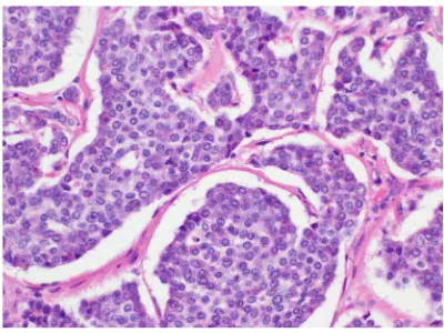
A thirteen-year-old male was seen in emergency in July 2007 with periumbilical pain and vomiting for 10-days. In last 24-h his pain shifted to supra pubic area and he was noted to have fever. Patients’ past history was insig-nificant. His examination revealed right lower quadrant tenderness and temperature of 39.1˚C. His WBC was 17,600/μl and ultrasound abdomen was suggestive of acute appendicitis. He was started on intravenous fluids and antibiotics. After stabilization, he was taken to operation room where laparoscopic examination showed se-verely inflamed appendix. He had laparoscopic appendectomy and in postoperative period he developed port site infection, which was treated with wound cleaning and antibiotics. His histopathology showed 1 × 0.8 cm neuro-endocrine (carcinoid) tumor at the tip of appendix, extending to serosa with perineural and lymphovascular in-vasion (Figure 4) but the margins were tumor free. He was further evaluated with CT scan of abdomen and 24-h urine 5-HIAA, which were normal. He remained in follow up till 3 years after surgery and was disease free. Later he has lost follow up as family has moved out from country.
3. Discussion
Neuroendocrine tumors are considered to be of neuroectodermal origin and classified as part of the other amine precursor uptake and decarboxylation neoplasms [3] [4]. The reported incidence of ANETs in children (≤14 years) range from 0.08% to 0.7% in surgical specimen [5] [7] and prevalence in our study was 0.03 in 700 sur-gical resected specimen of pediatric appendectomies carried out at our institute in last 6 years.
Presentation of ANETs in children is variable. Most common presentation is like acute appendicitis, whereas in others ANET may be an incidental finding during surgery. Some children may present with recurrent abdo-minal pain due to partial obstruction of the appendiceal lumen or rarely children with ANETs have presented with clinical signs of peritonitis [8]. Symptoms of carcinoid syndrome such as flushing and diarrhea due to in-creased urine excretion of 5-HIAA have also been reported with liver or retroperitoneal metastases [7]. However, in majority of children aneuroendocrine tumor is usually an incidental finding during histological examination of the resected appendix [1] [2] [7] as happened in all cases in this case series. In all our patients ANETs were discovered during routine histological examination of resected appendix followed by specific Immuno-histo- chemical staining (Figure 1 and Figure 2) and none of these children presented with any carcinoid syndrome symptoms. Post-operative imaging with CT abdomen and 5-HIAA excretions were also unremarkable suggest-ing that ANETs are usually non-aggressive in paediatric age group.
Figure 3. H & E staining of resected appendix show-ing normal mucosa on the left and neuroendocrine tumor on the right side invading deep in the underly-ing layer (Magnification 10X).
Figure 4. High power magnification of H & E image of a resected appendix specimen showing neuroendo-crine tumor and features include granular chromatin pattern, low NC ratio and relatively uniform calls in the nesting pattern (Magnification 40X).
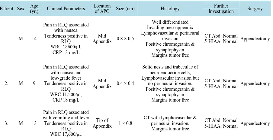
the mid appendix (2 cases) with a diameter of 1 cm or less (Table 1).
The management of ANETs depends much on the size, site and depth of the primary tumor. It is generally agreed that appendectomy is sufficient in children with incidental small (<2 cm) ANET localized at tip or in mid appendix and no further surgical intervention is indicated if follow up CT scan of abdomen and 24-h urine 5-HIAA are normal [2] [10]. This practice was followed up in our case series since the diagnosis was made from the appendectomy specimens and additional post-operative evaluation with CT abdomen and urinary excretion of 5-HIAA in all cases were not indicative of any further intervention.
The management of large (>2 cm) ANETs located at base of appendix and extending into the mesoappendix has been a controversial subject. A more extensive surgical procedure such as right hemicolectomy, ileocecal resection and caecotomy has been recommended in adults [2]. However, in paediatric age group with large ANETs a more limited surgical approach has been advocated [8]. Till to date, there is only one documented case of metastatic ANET [6]; this child had a large (5 cm) ANET and had right hemicolectomy, which contained one lymph node with a metastatic deposit. At 3-year follow-up this patient remained clinically and radiologically disease-free.
[image:4.595.213.413.288.438.2]Table 1. Characteristics of 3 patients with appendiceal neuroendocrine tumors.
Patient Sex Age
(yr.) Clinical Parameters
Location
of APC Size (cm) Histology
Further
Investigation Surgery
1. M 14
Pain in RLQ associated with nausea Tenderness positive in
RLQ WBC 18600/μl,
CRP 13 mg/L
Mid
Appendix 0.8 × 0.5
Well differentiated Invading mesoappendix Lymphovascular & perineural
invasion Positive chromogranin &
synaptophysin Margins tumor free
CT Abd: Normal
5-HIAA: Normal Appendectomy
2. M 9
Pain in RLQ associated with nausea and low-grade fever Tenderness positive in
RLQ WBC 11,200/μl,
CRP 18 mg/L
Mid
Appendix 0.4 × 0.4
Solid nests and trabeculae of neuroendocrine cells, Lymphovascular invasion but
no perineural invasion, Positive chromogranin &
synaptophysin Margins tumor free
CT Abd: Normal
5-HIAA: Normal Appendectomy
3. M 13
Pain in RLQ associated with vomiting and fever Tenderness positive in
RLQ WBC 17,600/μl,
Tip of
Appendix 1 × 0.8
CT with lymphovascular & perineural invasion, Margins tumor free
CT Abd: Normal
5-HIAA: Normal Appendectomy
M = Male; RLQ = Right Lower Quadrant; WBC = White blood cells; CRP = C-reactive protein; CT Abd = Computerized Tomography of abdomen; 5-HIAA = 5-hydroxyindoleacetic acid.
first-line screening tools [8] [9] [12]. Since our case series is too small to draw a recommendation, we still agree with Kulkarni et al. that children with small (<2 cm) ANETs and with no associated risk factors should have a regular annual clinical follow up to monitor the outcome and prognosis.
4. Conclusion
In summary, ANETs in pediatric age group usually present like acute appendicitis and are discovered inciden-tally during histological examination of the resected appendix. ANETs in children are less aggressive and the prognosis is excellent after appendectomy. We advocate obligatory review of histopathology of resected speci-men to docuspeci-ment prevalence of these tumors in children.
Acknowledgements
We gratefully acknowledge the Medical Research Centre, Hamad Medical Corporation for their support in pub-lishing this article.
Conflict of Interest Statement
The authors declare that there are no conflicts of interest.
References
[1] Emmanouil, H., Paraskevi, P., Vasiliki, S.-F., et al. (2010) Carcinoid Tumors of the Appendix in Children: Experience from a Tertiary Center in Northern Greece.Journal of Pediatric Gastroenterology & Nutrition, 51, 622-625.
http://dx.doi.org/10.1097/MPG.0b013e3181e05358
[2] Scott, A. and Upadhyay, V. (2011) Carcinoid Tumours of the Appendix in Children in Auckland, New Zealand: 1965- 2008.The New Zealand Medical Journal, 124, 56-60.
[3] Allan, B., Davis, J., Perez, E., Lew, J. and Sola, J. (2013) Malignant Neuroendocrine Tumors: Incidence and Outcomes in Pediatric Patients.European Journal of Pediatric Surgery, 23, 394-399. http://dx.doi.org/10.1055/s-0033-1333643
[4] Schmittenbecher, P.P. (2001) Carcinoid Tumours of the Appendix in Children—Epidemiology, Clinical Aspects and Procedures.European Journal of Pediatric Surgery, 11, 428. http://dx.doi.org/10.1055/s-2001-19732
Two Italian Institutions. Journal of Pediatric Gastroenterology & Nutrition, 40, 216-219.
http://dx.doi.org/10.1097/00005176-200502000-00025
[6] Volpe, A.,Willert, J., Ihnken, K., et al. (2000) Metastatic Appendiceal Carcinoid Tumor in a Child. Medical and Pe-diatric Oncology, 34, 218-220.
http://dx.doi.org/10.1002/(SICI)1096-911X(200003)34:3<218::AID-MPO12>3.0.CO;2-9
[7] Doede, T., Foss, H.D. and Waldschmidt, J. (2000) Carcinoid Tumors of the Appendix in Children—Epidemiology, Clinical Aspects and Procedure.European Journal of Pediatric Surgery, 10, 372-377.
http://dx.doi.org/10.1055/s-2008-1072394
[8] Pelizzo, G., La Riccia, A., Bouvier, R., et al. (2001) Carcinoid Tumors of the Appendix in Children.Pediatric Surgery International, 17, 399-402.http://dx.doi.org/10.1007/s003830000559
[9] Prommegger, R., Obrist, P., Ensinger, C., et al. (2002) Retrospective Evaluation of Carcinoid Tumors of the Appendix in Children.World Journal of Surgery, 26, 1489-1492. http://dx.doi.org/10.1007/s00268-002-6440-3
[10] Spunt, S.L., Pratt, C.B., Rao, B.N., et al. (2000) Childhood Carcinoid Tumors: The St Jude Children’s Research Hos-pital Experience. Journal of Pediatric Surgery, 35, 1282-1286. http://dx.doi.org/10.1053/jpsu.2000.9297
[11] Kulkarni, K.P. and Sergi, C. (2013) Appendix Carcinoids in Childhood: Long-Term Experience at a Single Institution in Western Canada and Systematic Review on Topic.Pediatrics International, 55, 157-162.
http://dx.doi.org/10.1111/ped.12047
currently publishing more than 200 open access, online, peer-reviewed journals covering a wide range of academic disciplines. SCIRP serves the worldwide academic communities and contributes to the progress and application of science with its publication.


