bone resorption to formation
Sunao Takeshita, … , Masako Ito, Kyoji Ikeda
J Clin Invest.
2013;
123(9)
:3914-3924.
https://doi.org/10.1172/JCI69493
.
Bone remodeling is characterized by the sequential, local tethering of osteoclasts and
osteoblasts and is key to the maintenance of bone integrity. While bone matrix–mobilized
growth factors, such as TGF-
b
, are proposed to regulate remodeling, no in vivo evidence
exists that an osteoclast-produced molecule serves as a coupling factor for bone resorption
to formation. We found that CTHRC1, a protein secreted by mature bone-resorbing
osteoclasts, targets stromal cells to stimulate osteogenesis.
Cthrc1
expression was robustly
induced when mature osteoclasts were placed on dentin or hydroxyapatite, and also by
increasing extracellular calcium.
Cthrc1
expression in bone increased in a high-turnover
state (such as that induced by RANKL injections in vivo), but decreased in conditions
associated with suppressed bone turnover (such as with aging and after alendronate
treatment). Targeted deletion of
Cthrc1
in mice eliminated
Cthrc1
expression in bone,
whereas its deficiency in osteoblasts did not exert any significant effect. Osteoclast-specific
deletion of
Cthrc1
resulted in osteopenia due to reduced bone formation and impaired the
coupling process after resorption induced by RANKL injections, impairing bone mass
recovery. These data demonstrate that CTHRC1 is an osteoclast-secreted coupling factor
that regulates bone remodeling.
Research Article
Bone biology
Find the latest version:
Osteoclast-secreted CTHRC1 in the coupling
of bone resorption to formation
Sunao Takeshita,1 Toshio Fumoto,1 Kazuhiko Matsuoka,1 Kyoung-ae Park,1 Hiroyuki Aburatani,2 Shigeaki Kato,3 Masako Ito,4 and Kyoji Ikeda1
1Department of Bone and Joint Disease, National Center for Geriatrics and Gerontology (NCGG), Obu, Aichi, Japan. 2Genomescience Division,
RCAST, University of Tokyo School of Medicine, Tokyo, Japan. 3Institute of Molecular Cellular Bioscience, University of Tokyo, Tokyo, Japan. 4Medical Work-Life-Balance Center, Nagasaki University Hospital, Nagasaki, Japan.
Bone remodeling is characterized by the sequential, local tethering of osteoclasts and osteoblasts and is key
to the maintenance of bone integrity. While bone matrix–mobilized growth factors, such as TGF-
β
, are
pro-posed to regulate remodeling, no in vivo evidence exists that an osteoclast-produced molecule serves as a
cou-pling factor for bone resorption to formation. We found that CTHRC1, a protein secreted by mature
bone-resorbing osteoclasts, targets stromal cells to stimulate osteogenesis. Cthrc1 expression was robustly induced
when mature osteoclasts were placed on dentin or hydroxyapatite, and also by increasing extracellular calcium.
Cthrc1 expression in bone increased in a high-turnover state (such as that induced by RANKL injections in
vivo), but decreased in conditions associated with suppressed bone turnover (such as with aging and after
alendronate treatment). Targeted deletion of Cthrc1 in mice eliminated Cthrc1 expression in bone, whereas
its deficiency in osteoblasts did not exert any significant effect. Osteoclast-specific deletion of Cthrc1 resulted
in osteopenia due to reduced bone formation and impaired the coupling process after resorption induced
by RANKL injections, impairing bone mass recovery. These data demonstrate that CTHRC1 is an
osteoclast-secreted coupling factor that regulates bone remodeling.
Introduction
Bone is constantly remodeled through the removal of old bone (resorption) and the replacement of new bone (formation) by hematopoietic-derived osteoclasts and mesenchymal-derived osteoblasts, respectively, to meet structural and metabolic demands (1, 2). It has long been believed that a preceding resorp-tion phase is a prerequisite for the initiaresorp-tion of subsequent bone formation (3), which is also supported by recent clinical obser-vations that treatment of osteoporotic patients with the potent antiresorptive drug alendronate blunts the anabolic action of parathyroid hormone (PTH) (4). Although the coupling of bone formation to resorption has been long recognized, the mechanism and the factors mediating this fundamental process in skeletal homeostasis are not fully understood (5).
Recently, it was suggested that active TGF-β1 released from bone matrix during bone resorption is coupled to bone forma-tion by inducing migraforma-tion of marrow stromal cells to resorp-tion sites (6). Bidirecresorp-tional signaling between EPHRINB2 on osteoclasts and the receptor EPHB4 on osteoblasts has also been proposed to link bone resorption and formation through direct cell-cell contact (7). In addition, mature osteoclasts produce and secrete factors, such as WNT10B, BMP6, and the lipid mediator sphingosine-1-phosphate (S1P), that have been shown to stimu-late osteoblast recruitment and survival (8–10). However, the in vivo function of these factors in the coupling process remains to be elucidated.
We reasoned that a factor mediating the coupling reaction link-ing bone resorption to formation would have to meet the follow-ing criteria: (a) it should be produced and secreted/presented by osteoclasts; (b) it should exert an effect on osteoblastic
differentia-tion and/or funcdifferentia-tion, thereby promoting bone formadifferentia-tion; (c) in its absence, the coupling of bone formation to preceding resorption should be impaired; and (d) its expression or activity should be regulated by agents or conditions known to affect bone turnover, such as aging and alendronate treatment. We have now identified a protein product of mature osteoclasts that fulfills these criteria and is considered to be a novel coupling factor.
Results
Identification of Cthrc1 in bone-resorbing active, but not inactive, osteoclasts. In an effort to identify an osteoclast-derived coupling factor, we performed gene expression analysis during osteoclast differentia-tion using mouse genome arrays containing 39,000 transcripts, and found a transcript of the collagen triple helix repeat contain-ing 1 (Cthrc1) gene. This transcript was absent in bone marrow macrophages (BMMs) and preosteoclasts (pOCs) and only mod-estly expressed in multinucleated mature osteoclasts prepared on plastic dishes, but was markedly induced in mature osteoclasts prepared on dentin slices, i.e., functional osteoclasts that are actively engaged in bone resorption (Supplemental Figure 1; sup-plemental material available online with this article; doi:10.1172/ JCI69493DS1). This was confirmed by RT-PCR (Figure 1A); notably, calcitonin receptor (Calcr), a known product of mature osteoclasts, was expressed in inactive mature osteoclasts prepared on plastic dishes, whereas Cthrc1 mRNA expression was robustly induced in mature osteoclasts prepared on dentin slices (Figure 1B). This suggests that Cthrc1 expression in osteoclasts is tightly associ-ated with bone resorption.
Cthrc1 was originally isolated as a gene induced after balloon injury of the rat aorta (11); it is not expressed in arteries under physiological conditions (Figure 1C and ref. 11) and is thought to play a role in vascular remodeling in response to injury by regulat-ing TGF-β responsiveness (12). It was also shown to be responsive
Conflict of interest: The authors have declared that no conflict of interest exists.
to TGF-β in chondrocytes (13, 14). In fact, we confirmed by in situ hybridization (ISH) that during development, Cthrc1 transcript was prominently expressed in chondrocytes (Supplemental Figure 2). However, the tissue distribution pattern observed in adult mice indicated that Cthrc1 was expressed in bone, and to a much lesser extent in brain, but not in any other tissue (Figure 1C). In the skel-etal tissue, the Cthrc1 transcript was observed in multinucleated osteoclasts on the bone surface in vivo, but not in osteoblasts or osteocytes (as shown by ISH), and it colocalized with TRAP stain-ing (Figure 1D and Supplemental Figure 3, A and B). Thus, we conclude that the major source of CTHRC1 in the adult skeleton is bone-resorbing osteoclasts.
Murine Cthrc1 encodes a 245–amino acid protein with a leader sequence of 32 amino acids, and the amino acid sequence is highly conserved among the mouse, rat, and human proteins (mouse/ rat, 99.2%; mouse/human, 97.7%; rat/human, 95.7%). CTHRC1 shares a collagen domain containing 12 Gly-X-X repeats and a globular domain in the C-terminal half with adiponectin and is thought to belong to the adiponectin/complement factor fam-ily (Supplemental Figure 4A). Recombinant mouse CTHRC1 was generated using a baculovirus/insect cell system and purified to near homogeneity (data not shown), which was then used to raise
a polyclonal antibody. This antibody detected CTHRC1 protein in bone-resorbing osteoclasts cultured on a dentin slice (Supplemen-tal Figure 3C). Western blot analysis revealed that the recombi-nant protein migrates as a single polypeptide of apparently 28 kDa under reducing conditions, while several bands with higher molecular weights were observed under nonreducing conditions (Supplemental Figure 4B), which suggests that CTHRC1 exists in monomeric, dimeric, and trimeric forms, just like adiponectin (15). The antibody detected a protein of approximately 28 kDa in the conditioned medium of mature osteoclasts, but not in the con-ditioned medium of BMMs, and the detected amount increased substantially in mature osteoclasts prepared on dentin slices (Figure 1E), supporting our contention that CTHRC1 is a secre-tory product of mature, functional osteoclasts.
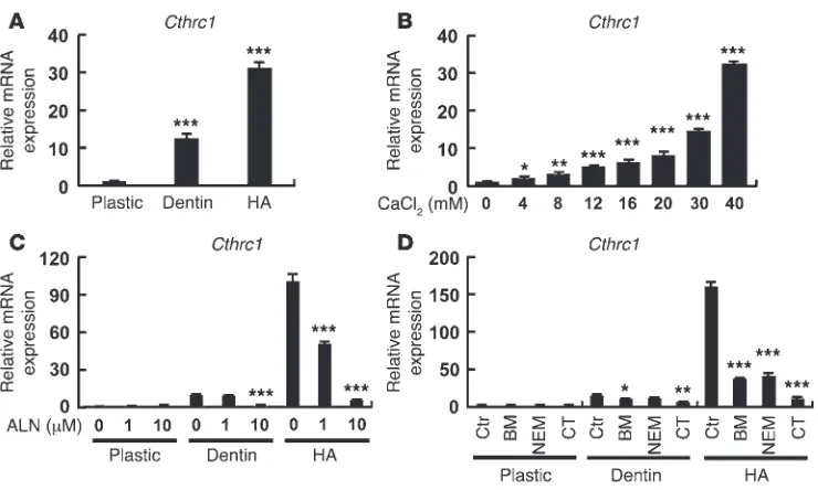
Regulation of Cthrc1 gene expression by contact with hydroxyapatite and calcium. In order to gain further insight into the mechanism of Cthrc1 gene expression in osteoclasts, mature osteoclasts gen-erated from BMMs ex vivo with M-CSF and RANKL were placed on different substrates, including a plastic dish, a dentin slice, or a hydroxyapatite (HA) disc. RT-PCR analysis revealed that Cthrc1
was even more robustly induced on HA than on dentin slices (Figure 2A). Accordingly, increasing extracellular calcium concen-Figure 1
[image:3.585.49.535.79.398.2]trations, as well as phosphate, stimulated Cthrc1 mRNA expres-sion even in inactive mature osteoclasts prepared on plastic dishes (Figure 2B and Supplemental Figure 5). Other divalent cations, such as magnesium and gadolinium, had only modest effects (Supplemental Figure 5).
The upregulated Cthrc1 expression in mature osteoclasts cul-tured on HA or dentin was markedly and dose-dependently inhibited when bone resorption was inhibited by alendronate, by the vacuolar H+ ATPase (V-ATPase) inhibitors bafilomycin A1
or N-ethylmaleimide, and by the direct osteoclast inhibitor cal-citonin (Figure 2, C and D). Collectively, these data suggest that mineral ions such as Ca2+ and PO
43– are important regulators of
Cthrc1 expression in mature osteoclasts and that activation of the bone resorption machinery, especially V-ATPase, is a prerequisite of CTHRC1 production.
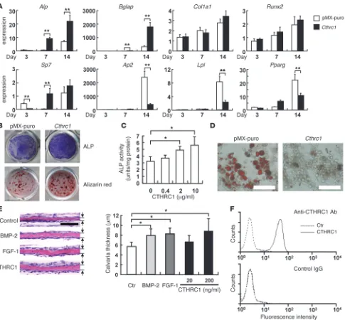
Cthrc1 stimulates osteoblast differentiation. In order to define the function of CTHRC1 in osteoblastogenesis, Cthrc1 was retro-virally expressed in calvaria-derived primary osteoblastic cells. Quantitative RT-PCR analysis revealed that forced Cthrc1 expres-sion stimulated osteogenic differentiation, as demonstrated by increased alkaline phosphatase (Alp) and osteocalcin (Bglap) expression at day 7 and 14 after confluency (Figure 3A). Osteogen-ic induction was confirmed by ALP activity and mineralization, as determined by alizarin red staining (Figure 3B). Treatment of bone marrow stromal ST2 cells with the recombinant CTHRC1 protein also increased ALP activity (Figure 3C). CTHRC1 also suppressed adipogenic marker expression (Figure 3A) and inhibited adipocytic differentiation, as determined by Oil Red-O staining (Figure 3D).
Organ culture experiments in mouse calvaria revealed that recombinant CTHRC1 had bone formation–stimulating activity comparable to BMP-2 and FGF-1 (Figure 3E). In com-bination with WNT3A, a potent bone anabolic protein, CTHRC1 was also capable of stimulating ST2 cell chemotaxis (Supple-mental Figure 6A), consistent with cross-talk between the WNT and CTHRC1 signaling pathways (16). The chemotactic effect of CTHRC1 was also con-firmed in transwell assays using ST2 cells and primary osteo-blasts (Supplemental Figure 6, B and C). In accord with these effects of CTHRC1 on stromal cells, the binding moiety to CTHRC1 was demonstrated on stromal ST2 cells by FACS analysis using the recombinant protein and specific antibody (Figure 3F). Taken together, these findings suggest that CTHRC1 secreted by mature, functional osteoclasts acts on stromal/osteoblastic cells by binding to a putative cell sur-face receptor in order to stimulate osteoblastic differentiation as well as recruitment, thereby promoting bone formation.
Conditional Cthrc1 KO in osteoclasts. In order to gain insight into the physiologic function of osteoclast-derived CTHRC1 in bone remodeling, we generated mice carrying a floxed Cthrc1 allele (Figure 4A). The floxed mice were then mated with Ctsk-Cre (17) or CAG-Cre (18) mice to generate osteoclast-specific conditional
Cthrc1 KO (ΔOC) or systemic Cthrc1 KO (sKO) mice, respectively (Supplemental Figure 7A). Both ΔOC and sKO mice were born with the expected Mendelian frequency, appeared grossly normal, grew normally, and were fertile (data not shown). Specific deletion of Cthrc1 in osteoclasts was verified in mature osteoclasts gener-ated ex vivo (Supplemental Figure 7B). Quantitation of Cthrc1
mRNA expression in bone by RT-PCR demonstrated its near-com-plete absence not only in sKO bone, but also in ΔOC bone, with no difference between them (Figure 4B), which indicates that mature osteoclasts are the major source of CTHRC1 in bone in vivo.
μCT scanning of the proximal tibial metaphysis revealed that
[image:4.585.45.415.79.301.2]ΔOC and sKO mice exhibited a low bone mass phenotype with microstructural abnormalities reminiscent of osteoporosis, such as decreased trabecular number and thickness (Figure 4C and data not shown). Histomorphometric analysis at the proximal tibial metaphysis revealed that osteoid surface and bone formation rate were significantly decreased in ΔOC versus control mice, while the bone resorption parameters (eroded surface and osteoclast sur-face) did not differ between them (Figure 4D). These results sug-gest that osteoclast-produced CTHRC1 carries out an important physiological function in the maintenance of bone mass and tra-becular structure, mainly through regulation of bone formation. Figure 2
Regulation of Cthrc1 gene expression. (A) Cthrc1 mRNA expression was strongly induced in osteoclasts cultured on HA. Osteoclasts were cultured on plastic plates, dentin slices, or Osteologic discs composed of HA crystals, and RNAs were prepared. Real-time RT-PCR for Cthrc1 was performed. (B) Increasing extracellular calcium concentrations increased Cthrc1 transcription in osteoclasts on plastic dishes. Osteo-clasts were cultured at the indicated CaCl2 concentrations for 24 hours. (C) Alendronate (ALN) inhibited the
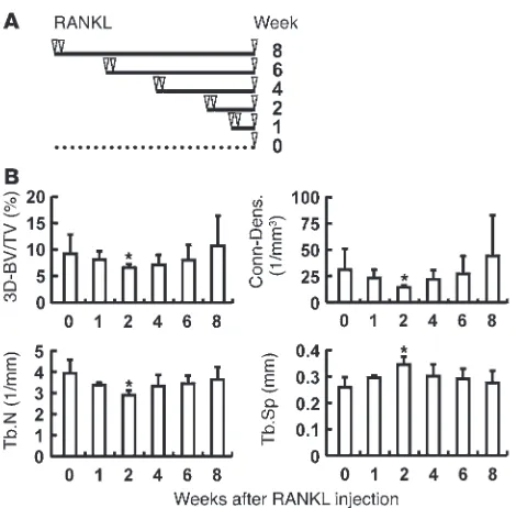
Coupling function in vivo. In order to address the functional importance of CTHRC1 in the coupling process from bone resorption to formation, we developed a mouse model using recombinant RANKL injections (Figure 5A). In this model, robust stimulation of bone resorption by the osteoclast differentiation factor RANKL reportedly takes place as early as 2–3 days (19). μCT
analysis further revealed that the 3D bone volume (BV) decreased rapidly, reaching a nadir at 2 weeks, followed by gradual recovery to baseline at 8 weeks, presumably due to the subsequent stimula-tion of bone formastimula-tion (Figure 5B). We interpret these dynamic changes in bone mass to mean that the catabolic phase of the first 1–2 weeks after RANKL injections triggered subsequent stimula-Figure 3
[image:5.585.55.540.80.530.2]tion of bone formation, resulting in a net transient decrease in bone mass at 2 weeks followed by the anabolic phase and com-plete recovery at 8 weeks.
Cthrc1 expression in bone was markedly increased, together with that of osteoclast markers such as tartrate-resistant acid phosphatase (Acp5), Calcr, and cathepsin K (Ctsk), in control mice 3 days after RANKL administration compared with PBS injection (Figure 6A). In ΔOC mice, induction of Cthrc1 expression after RANKL injections, as well as basal Cthrc1 expression, was sup-pressed almost completely, whereas expression of the mature osteo-clast molecular markers Calcr and Ctsk was increased to the same extent as in control mice (Figure 6A). In fact, the degree of bone loss at 10 days and urinary CTX excretion at 2 days after RANKL injections was indistinguishable from that of the control group
(Figure 6, B and C), which indicates that the presence or absence of CTHRC1 made no difference in the initial phase of osteoclast activation and bone loss induced by RANKL injection. However, the recovery of bone mass was significantly impaired in ΔOC versus control mice (Figure 6B), and this was associated with significantly reduced osteoblast recruitment and matrix synthesis and signifi-cant impairment of bone formation rate and lower serum osteocal-cin concentrations in ΔOC mice (Figure 6, D and E).
Conditional Cthrc1 KO in osteoblasts. In contrast to ΔOC mice, spe-cific Cthrc1 deletion in osteoblasts by crossing the floxed mouse with the Osx1-Cre mouse (ΔOB mice; Figure 7A and ref. 20) had no significant effect on either basal Cthrc1 expression in bone or its upregulation after RANKL injections (Figure 7B), which confirmed that osteoclasts, but not osteoblasts, are the Figure 4
Osteoclast-specific Cthrc1 KO results in a low bone mass phenotype with reduced bone formation. (A) Design of the floxed allele of Cthrc1. (B) Comparable reduction of Cthrc1 expression in bone of ΔOC and sKO mice (generated by crossing Cthrc1fl/fl mice with Ctsk-Cre and CAG-Cre mice,
respectively). (C) Reduced 3D-determined BV relative to tissue volume (3D-BV/TV) and microstructural derangements, by μCT scanning, in ΔOC versus Cthrc1fl/fl control mice. Representative 3D images of proximal tibia are also shown. Tb.N, trabecular number; Tb.Th, trabecular thickness;
major source of CTHRC1 in adult bone. This was further sup-ported by the finding that ΔOB mice exhibited normal bone mass (Figure 7, C and D). Taken together, these results suggest that CTHRC1 produced by mature osteoclasts is required for an adequate anabolic response after the catabolic phase induced by RANKL injections, providing in vivo evidence that the protein has physiologic function in the process coupling bone resorp-tion to formaresorp-tion.
Correlation of Cthrc1 expression with bone turnover states. Finally, in order to determine whether CTHRC1 plays any pathogenic role in age-related bone loss, we examined whether its expression changes with aging. Comparison of mRNA expression in bone among mice of different ages revealed that Cthrc1 expression declined substan-tially with age in both males and females (Figure 8A and data not shown). Alendronate, a potent inhibitor of bone turnover, is widely used for treatment of high-turnover bone disease, such as post-menopausal osteoporosis. However, it has been recognized clini-cally that inhibition of osteoclastic function by alendronate blunts the anabolic action of PTH (4), and the underlying mechanism is beginning to be understood (21). We found that along with a reduction in bone turnover markers after treatment with alendro-nate (Supplemental Figure 8), Cthrc1 expression in bone was sub-stantially reduced, in contrast to the increased expression of Ctsk
and Calcr (Figure 8B). These data suggest that a low bone turnover state associated with aging and alendronate treatment correlates with reduced Cthrc1 expression.
Cathepsin K inhibitors have been developed as a new strategy of inhibiting bone resorption (22). Interestingly, unlike bisphospho-nates, treatment with cathepsin inhibitors has not been associated with suppressed bone formation, a result that is also supported by prior reports of increased, rather than decreased, bone formation in Ctsk-deficient mice (10, 23). Taking advantage of the Ctsk-Cre
knockin construct, we generated Ctsk–/– mice. Unlike the case with
bisphosphonate, no significant reduction in Cthrc1 expression was observed in Ctsk–/– mice (Figure 8C).
Finally, as representative examples of osteoclast-poor and -rich osteopetrosis, Rankl–/– and Src–/– mice were examined (24).
In accordance with the absence of osteoclasts in Rankl–/– mice,
almost no expression of Ctsk and Calcr was detected in Rankl–/–
bone (Figure 8D). In marked contrast, expression of both Ctsk and
Calcr was markedly increased in Src–/– bone (Figure 8E), consistent
with the abundance of inactive osteoclasts in Src–/– bone.
Impor-tantly, Cthrc1 expression was markedly decreased in both Rankl–/–
and Src–/– bone (Figure 8, D and E), consistent with our concept
that the mere presence of inactive osteoclasts is not sufficient for optimal Cthrc1 expression and that the bone-resorbing activity of osteoclasts is a prerequisite for Cthrc1 expression and, therefore, for CTHRC1-mediated coupling of resorption with formation.
Discussion
It has long been recognized that in each remodeling cycle, bone for-mation is tightly coupled to the preceding bone resorption carried out by osteoclasts (25, 26). The fact that bone resorption triggers formation suggests that certain signals derived from osteoclasts recruit osteoblasts or their progenitors to the remodeling site to target bone formation activity, although it is a matter of debate whether the mere presence of osteoclasts is sufficient to initiate the coupling reaction or whether their resorptive activity is required, as well as whether mature osteoclasts or precursor cells represent the source of the coupling factor (27, 28). Here, we demonstrated that mature osteoclasts secreted CTHRC1 only when osteoclasts were placed on dentin or HA, which suggests that CTHRC1 production is closely linked with osteoclast attachment to calcified tissue. This situation was mimicked by placing osteoclasts in an environment high in extracellular calcium and phosphate, which indicates that recognition of these minerals by osteoclasts is essential for CTHRC1 upregulation. Furthermore, our findings that Cthrc1 expression was suppressed in osteoclasts treated with alendronate, calcitonin, or proton pump inhibitors as well as in Src–/– osteoclasts in vivo
sug-gest that establishment of polarity, cytoskeletal reorganization, and/or activation of the secretory function of acid and collageno-lytic enzymes is required for CTHRC1 production. The secreted CTHRC1 protein binds to a putative cell surface receptor on stromal cells, promoting cell differentiation into mature osteoblasts and also chemotaxis along a CTHRC1 pathway, together with WNT proteins. Thus, CTHRC1 may function as a guidance molecule for targeting stromal/osteogenic cells to sites of bone resorption, thus promoting the initiation of subsequent bone-forming activity.
The physiologic importance of osteoclast-derived CTHRC1 in bone homeostasis was evidenced by the low bone mass phenotype with lowered bone formation exhibited by ΔOC mice (in which
Cthrc1 was specifically deleted in osteoclasts by use of Ctsk-Cre
mice; ref. 17), but not by ΔOB mice. Thus, CTHRC1 is a candidate molecule for mediating the coupling of bone resorption with for-mation. However, this does not necessarily exclude a contribution of bone matrix–derived factors, represented by TGF-β1, as TGF-β1 is mobilized and activated during bone resorption and promotes recruitment of osteogenic progenitors to resorption sites (6).
[image:7.585.48.283.79.310.2]S1P, a lipid mediator that is also produced and secreted by osteo-clasts, stimulates both recruitment and survival of osteoblasts (8, 9). S1P is abundant in the circulation, and a recent report suggests that S1P regulates the mobilization of osteoclast precursors to Figure 5
and from bone through the cell surface S1P receptor (29). It has recently been shown that S1P secretion is increased in Ctsk -defi-cient osteoclasts, which is suggested to account for the elevated bone formation in Ctsk–/– mice (10). However, the specific function
of S1P and other secretory proteins, such as WNT10B and BMP6, in the coupling of formation to resorption in vivo remains elusive.
Since the coupling reaction takes place in each individual bone multicellular unit (BMU), and since bone homeostasis is main-tained as a net result of numerous micron-order BMU activities
throughout the entire skeleton, there is no established assay for evaluating coupling efficiency in vivo. We have attempted to syn-chronize osteoclast formation and bone resorption initiation by acute RANKL injections so as to separate the resorption and for-mation phases. It has been reported that 3 RANKL injections at 24-hour intervals induce an acute increase in bone resorption at a time point as early as 50 hours (19). We closely monitored the skel-etal response to acute RANKL challenge up to 8 weeks after injec-tion by 3D μCT scanning and found that the initial catabolic pro-Figure 6
Impaired coupling function in osteoclast-specific Cthrc1 KO mice. (A) Cthrc1, Acp5, Calcr, and Ctsk expression at baseline and after RANKL injection in Cthrc1fl/fl control and ΔOC mice, determined by quantitative RT-PCR. Values were normalized for Gapdh. n = 4. (B) Impaired recovery
of 3D BV at 8 weeks after RANKL injection in ΔOC versus Cthrc1fl/fl control mice. Mice were sacrificed at 10 days and 8 weeks after RANKL
[image:8.585.94.496.78.533.2]cess induced acute bone loss, reaching a nadir in 3D BV at 2 weeks that was followed by a gradual recovery of bone mass due to stimu-lation of bone formation. In ΔOC mice, the initial catabolic phase was indistinguishable from that of the control group, with the same extent of bone loss, which indicates that CTHRC1 deficiency in osteoclasts has no major effect on the bone resorption response to acute RANKL challenge. However, subsequent recovery of bone mass was significantly delayed in the absence of CTHRC1, which suggests that the relay signal from bone resorption to formation is impaired when CTHRC1 production is abrogated in osteo-clasts. Thus, we propose that CTHRC1 is a physiologically impor-tant osteoclast-produced signal that is relayed to bone formation (Supplemental Figure 9).
The reduced Cthrc1 expression that occurs with aging and after alendronate treatment in vivo is thought to dampen the coupling process and leads to insufficient new bone replacement at remod-eling sites (Supplemental Figure 9). Consistent with this concept is a previous report showing that forced Cthrc1 expression in osteo-blasts results in a high bone mass phenotype with stimulated bone formation (14). Thus, although further studies are required to clarify its causative role in age-related bone loss, CTHRC1 may provide an attractive target in the diagnosis and treatment of bone diseases such as osteoporosis, which is considered a coupling dis-order in the bony skeleton.
CTHRC1 was originally identified in the arterial wall, where its gene expression was induced only when the carotid artery and aorta were injured (11). In addition to being responsive to TGF-β1, CTHRC1 is proposed to contribute to vascular remodeling by
regulating matrix deposition and cell migration. During develop-ment, CTHRC1 is expressed in the node/notochord (16) and then the growth plate cartilage (refs. 13, 14, and the present study). However, no apparent abnormality in skeletal development has been observed in mice lacking Cthrc1 (refs. 14, 16, and the pres-ent study), so the physiologic function of CTHRC1 in develop-ment is not yet clear. During adulthood, in contrast, basal Cthrc1
expression is highly restricted to bone and brain (ref. 11 and the present study). In bone, Cthrc1 was predominantly expressed in osteoclasts, which was also supported by quantitative RT-PCR. These results showed that Cthrc1 expression in ΔOC mouse bone was markedly reduced, in fact, to the same level as in global
Cthrc1 KO mice (present study). We speculate that in postnatal life, CTHRC1 functions in skeletal as well as vascular remodel-ing, with dynamic vascular remodeling taking place in response to injury, when Cthrc1 is transiently induced. In this manner, bone is continuously remodeled: bone-resorbing osteoclasts actively pro-duce and secrete CTHRC1 in order to promote bone remodeling.
Methods
Reagents, recombinant proteins, and antibodies. Alendronate sodium hydrate was from Teijin Pharma Ltd.; bafilomycin A1, gadolinium chloride hexahydrate, N-ethylmaleimide, dexamethasone, 3-isobutyl-1-methylxanthine, and salm-on calcitsalm-onin were from Sigma-Aldrich; recombinant BMP-2 and FGF-1 were from R&D Systems Inc.; and Osteologic discs were from BD Biosciences.
[image:9.585.46.539.77.336.2]Recombinant murine M-CSF and GST-RANKL were expressed as described previously (30). Recombinant mouse CTHRC1 proteins were generated using the baculovirus/insect cell system according to standard
Figure 7
Osteoblast-specific Cthrc1 KO mice. (A) Genotyping of the floxed and excised (exon 2) allele (ΔE2) of Cthrc1 and the presence of Osx1-Cre. Genomic DNA was extracted from femurs after bone marrow cells were flashed out, and genomic PCR analysis was performed. (B) Cthrc1 deletion in osteoblasts did not affect Cthrc1 expression at baseline or after RANKL injections. GST-RANKL was injected into Cthrc1fl/fl control or ΔOB mice.
RNAs from long bone were extracted 3 days after 2 RANKL injections, and real-time RT-PCR analysis was performed for Cthrc1, Acp5, Calcr, and Ctsk expression. n = 4 per group. (C) μCT analysis of the trabecular region of the proximal tibial metaphysis of Cthrc1fl/fl control and ΔOB mice. (D)
were transferred to nitrocellulose membranes using a semidry blotter (Bio-Rad) and incubated in blocking solution (5% nonfat dry milk in TBS con-taining 0.1% Tween 20) for 1 hour to reduce nonspecific binding. Mem-branes were then exposed to rabbit anti-CTHRC1 polyclonal antibody overnight at 4°C, washed 3 times, and incubated with secondary rabbit IgG horseradish peroxidase–conjugated antibody for 1 hour. Membranes were washed extensively, and enhanced chemiluminescence detection assay was performed according to the manufacturer’s instructions.
ISH. The femur and tibia from 3-week-old C57BL/6 male mice were dis-sected, fixed, decalcified, embedded in paraffin, and sectioned at 4 μm. Paraffin sections were stained histochemically for TRAP in order to visu-alize osteoclasts.
For ISH, tissue sections were dewaxed with xylene and rehydrated through an ethanol series and PBS. Sections were fixed in 4% paraformaldehyde in PBS for 15 minutes, then washed with PBS. Next, the sections were treated with 7 μg/ml proteinase K in PBS for 30 minutes at 37°C, washed with PBS, refixed with 4% paraformaldehyde in PBS, again washed with PBS, and placed in 0.2 N HCl for 10 minutes. After washing with PBS, sections were acetylated by incubation in 0.1 M tri-ethanolamine-HCl (pH 8.0), 0.25% ace-tic anhydride, for 10 minutes. After washing with PBS, sections were dehy-drated through a series of ethanol solutions. Hybridization was performed with probes at concentrations of 300 ng/ml in Probe Diluent-1 (Genostaff) at 60°C for 16 hours. After hybridization, sections were washed in 5× Hybri-Wash (equal to 5× SSC) at 50°C for 20 minutes, then in 50% formamide with 2× HybriWash at 50°C for 20 minutes, followed by RNase treatment with 50 μg/ml RNase A in 10 mM Tris-HCl (pH 8.0), 1 M NaCl, and 1 mM EDTA for 30 minutes at 37°C. Sections were then washed twice with 2×
HybriWash at 50°C for 20 minutes, twice with 0.2× HybriWash at 50°C for 20 minutes, and once with TBST (0.1% Tween20 in TBS). After treatment with 0.5% blocking reagent (Roche) in TBST for 30 minutes, sections were incubated for 2 hours at room temperature with anti-DIG AP conjugate (Roche) diluted 1:1,000 with TBST. Sections were washed twice with TBST and then incubated in 100 mM NaCl, 50 mM MgCl2, and 0.1% Tween20 in 100 mM Tris-HCl (pH 9.5). Coloring reactions were performed with NBT/ BCIP solution (Sigma-Aldrich) overnight, followed by washing with PBS. Sections were counterstained with Kernechtrot stain solution (Mutoh), dehydrated, and mounted with Malinol (Mutoh).
Retroviral expression. The retroviral vector pMX-puro with Cthrc1 cDNA and/or the pVSVG plasmid were used to transfect the retrovirus packag-ing cells, Plat-E (gift of T. Kitamura, University of Tokyo, Tokyo, Japan) or procedures. In brief, mouse Cthrc1 cDNA containing a His-tag in a pPSC8
vector (Protein Science) was cotransfected with AcNPV DNA (Invitrogen) using CELLFECTIN (Invitrogen) into Sf9 cells, which were then infected into expresSF+ insect cells (Protein Sciences) using recombinant viruses. Cell supernatant was harvested, and the protein was purified using Ni Sepharose High Performance (Amersham Biosciences) and a Tricon 10/20 Column (Amersham Biosciences).
A polyclonal antibody against recombinant His-tagged murine CTHRC1 protein was raised with a standard technique and affinity puri-fied using a CTHRC1 protein–conjugated sepharose column. A mouse monoclonal antibody against mouse CTHRC1, 13B1, was established according to standard procedures.
Isolation of BMMs and osteoclastogenesis. BMMs were prepared from whole bone marrow of 8- to 10-week-old C57BL/6 mice (Clea Japan Inc.) and cul-tured as described previously (31).
TRAP-positive mononuclear pOCs were prepared from a 2-day culture of BMMs in αMEM containing 10% FBS in the presence of M-CSF and RANKL. These pOCs were further cultured in the presence of M-CSF and RANKL to generate either inactive mature osteoclasts prepared on plastic dishes or active mature osteoclasts prepared on dentin slices.
RNA isolation and RT-PCR. Total RNAs were extracted from cells using TRIzol reagent (Invitrogen) and used for microarray and RT-PCR analy-ses. See Supplemental Table 1 for primers used for RT-PCR. Relative gene expression was determined by SYBR green–based real-time PCR using a 7300 fast real-time PCR system (Applied Biosystems). Expression levels were normalized to Gapdh.
Microarray analysis. Total RNAs were processed according to the protocol recommended by Affymetrix. 5 μg labeled cRNA from each sample was hybridized to the mouse genome 430 2.0 Array GeneChip (Affymetrix) representing approximately 38,500 genes. GeneChip data output was nor-malized and analyzed using Affymetrix GeneChip Operating Software ver-sion 1.1. Raw GeneChip data were transformed into log format following the recommendation by Affymetrix. Microarray data were deposited in GEO (accession no. GSE45656).
[image:10.585.50.396.79.285.2]Immunoblotting. Immunoblotting was performed as described previously (32). Culture supernatants of BMMs and of mature osteoclasts prepared on either plastic dishes or dentin slices were immunoprecipitated with an anti-CTHRC1 monoclonal antibody, 13B1, and protein A/G agarose (SantaCruz). Samples were boiled in SDS sample buffer containing 2-mer-captoethanol and subjected to electrophoresis on 12% SDS-PAGE. Proteins
Figure 8
Correlation of Cthrc1 expression with bone turnover states. (A) Cthrc1 expression declined with aging. Cthrc1 mRNA expression in bone was quantitated among male mice of the indicated ages by quantitative RT-PCR. n = 3. (B) Inhibition of Cthrc1 expression after alendronate treat-ment in vivo. (C–E) Cthrc1 expression was examined in bone from Ctsk–/–
(C), Rankl–/– (D), and Src–/– (E)
generate CtskCre/+Cthrc1ΔOC/ΔOC (ΔOC) or Osx1Cre/+Cthrc1ΔOB/ΔOB (ΔOB) mice. Cthrc1fl/fl mice were used as controls. CAG-Cre mice were obtained from M. Okabe (Osaka University, Osaka, Japan) and crossed with Cthrc1fl/fl mice to generate Cthrc1+/+, Cthrc1+/–, and Cthrc1–/– (sKO) mice. Ranklfl/fl mice were generated by inserting a LoxP-FRT-PGKneo-FRT cassette into intron 2 and another LoxP site into intron 4 of Rankl, so that exons 3 and 4 of Rankl would be deleted on crossing with tissue-specific Cre transgenic mice (our unpublished observations). Rankl–/– mice were obtained by crossing Ranklfl/fl and CAG-Cre mice. Src–/– mice were obtained from the Jackson Laboratory.
Mice were raised under standard laboratory conditions at 24°C ± 2°C and 50%∼60% humidity, and allowed free access to tap water and commer-cial standard rodent chow (CE-2) containing 1.20% calcium, 1.08% phos-phate, and 240 IU/100 g vitamin D3 (Clea Japan Inc.). Wild-type C57BL/6 or transgenic mice were treated s.c. with vehicle (saline) or 1 mg/kg BW alendronate 5 times per week for 3 weeks. Wild-type or ΔOC mice were treated with 1 mg/kg BW GST-RANKL twice with a 24-hour interval. Blood samples at sacrifice were centrifuged to obtain the serum.
Bone analysis. Bone histomorphometry was performed on undecalcified sections, with tetracycline and calcein double labeling. The histomorpho-metric parameters were measured at the Niigata Bone Science Institute.
μCT scanning was performed on proximal tibiae using a μCT-40 scanner (SCANCO Medical AG) with a resolution of 12 μm, and 3D microstructure parameters were calculated as described previously (35). The proximal tibia was positioned so as to be scanned craniocaudally using 320 slices with 12-μm increments at 45 kVp and 177 μA. On the original 3D image, morphometric indices — including BV, tissue volume, trabecular thickness, trabecular sepa-ration, and trabecular number — were directly determined from the binarized volume of interest (VOI). Nonmetric parameters, such as the structure model index (SMI) and connectivity density, were also obtained as described previ-ously (35); for SMI, the characteristic form of a 3-dimensionally described structure in terms of the amount of plates and rods composing the structure was quantified, and connectivity density was determined as the number of trabecular connections per cubic millimeter. The nomenclature for μCT and histomorphometry followed previously published guidelines (36, 37).
Biochemical analysis. CTX concentrations in serum and urine were deter-mined using a RatLaps assay according to the manufacturer’s protocols (Immunodiagnostic Systems Ltd.). Serum osteocalcin concentration was measured using a mouse ostocalcin EIA kit (Biomedical Technologies Inc.).
Statistics. Data are expressed as mean ± SD. Statistical analysis was per-formed using paired or unpaired Student’s t test (2-sample, 2-tailed com-parison) or ANOVA followed by Dunnett test or Student-Newman-Keuls test. A P value less than 0.05 was considered significant.
Study approval. All experiments were performed in accordance with NCGG ethical guidelines for animal care, and experimental protocols were approved by the animal care committee.
Acknowledgments
We thank Mie Suzuki for technical assistance, Akemi Ito (Ito Bone Science Institute) for valuable suggestions on bone histomor-phometry, Masaru Okabe for CAG-Cre mice, Takashi Nakamura for Ctsk-Cre mice, Hiroko Meguro and Shogo Yamamoto (Uni-versity of Tokyo) for microarray analysis, and members of NCGG for stimulating discussions. This study was supported in part by a Grant-in-aid for Scientific Research C (no. 18590359; to S. Takeshita) and a Grant-in-aid for Scientific Research on Innova-tive Areas (no. 22118007; to K. Ikeda) from the Ministry of Educa-tion, Science of Japan; by a grant from Promotion of Fundamental Studies in Health Sciences of the National Institute of Biomedical Innovation (NIBIO) of Japan (06-31; to K. Ikeda); and by grants from Ono Foundation (to S. Takeshita), Daiko Foundation (to
GP2-293 (Clontech Co. Ltd.). Calvaria-derived primary osteoblasts and pre-adipocytic 3T3-L1 cells were infected with the retroviral vector pMX-puro expressing CTHRC1 under the control of Mo-MLV LTR, then cultured in the presence of polybrene (8 μg/ml) for 1 day. Infected cells were cultured in the presence of 2 μg/ml puromycin, and ALP and alizarin red staining was performed as described previously (33). Transduced 3T3-L1 cells were cultured in the presence of IBMX and dexamethasone to induce adipogen-esis and were stained with Oil Red-O.
Flow cytometry. ST2 cells were reacted with or without recombinant CTHRC1 protein (10 μg/ml) in PBS for 15 minutes. After washing, cells were stained with rabbit anti-CTHRC1 polyclonal antibody or rabbit IgG as a negative control, and PE-conjugated goat anti-rabbit IgG (Jackson), then analyzed using flow cytometry (FACSCalibur; Becton Dickinson).
Bone formation assay. Organ cultures of mouse calvaria were performed as described previously (34). Briefly, calvariae from 4-day-old pups of C57BL/6 mice were explanted, dissected free of any adjacent connec-tive tissue, placed in BGJ media (Sigma-Aldrich) containing 0.1% BSA (Sigma-Aldrich), and incubated with recombinant CTHRC1 (20 or 200 ng/ml) for 7 days. Recombinant human BMP-2 (100 ng/ml) or FGF-1 (100 ng/ml) was added as a positive control. Media were changed every 3 days. Calvariae were then fixed, decalcified, embedded in paraffin, and sectioned at 4 μm. Sections were placed on coated glass slides and stained with hematoxylin and eosin. Bone-forming activity was assessed by mea-suring calvaria thickness.
In vitro chemotactic assay. EZ-TAXIScan (Effector Cell Institute) was used to detect real-time horizontal chemotaxis of ST2 cells. The EZ-TAXIScan consists of an etched silicon substrate and a flat glass plate, which form 2 compartments with a 5-μm-deep microchannel. In some experiments, Thermanox coverslips (Nalgen Nunc International) were placed onto the glass plates. ST2 cells were placed into the single hole with which the device is held together of the stainless steel holder, and 1 μl recombinant CTHRC1 protein and/or WNT3A (R&D) was placed into the contrahole. A charge-coupled device (CCD) camera was used to record the migration of ST2 cells toward the CTHRC1 and/or WNT3A on the microchannel. Moving cells in a fixed gate were counted using a TAXIScan Analyzer (Effector Cell Institute).
Chemotaxis of ST2 cells or calvaria-derived osteoblasts was also assessed using the Cultrex 96 Well Cell Migration Assay Kit (Trevigen Inc.). In brief, stromal cells at 80% confluence were serum starved in 0.5% FBS for 24 hours. Cells were harvested, and 5 × 10⁴ cells were added to the top chamber. Either CTHRC1, WNT3A, or both was added to the bottom chamber, and cells were cultured for 24 hours. After a washing of each well, the migrating cells were stained with calcein and examined with a reader plate at 485 nm excitation, 520 nm emission.
KO mouse generation and animal experiments. A genomic fragment con-taining Cthrc1 was obtained from the BAC genomic library of the mouse 129SvEv strain and targeted by homologous recombination in ES cells established from blastocysts of BA1 Hybrid (C57BL/6 × 129SvEv) mice. The targeting construct introduced 3 loxP sites into the gene flanking exon 2, which encodes the collagen domain, and the neo cassette was also flanked by 2 FRT sites (Figure 4A). ES cells with a targeted allele were microinjected into blastocysts derived from C57BL/6 mice, and the germ-line chimeras generated by the blastocyst injections were crossed with C57BL/6 female mice. 2 F1 heterozygous mice were born and mated with Flp mice to delete the neo cassette. Mice were backcrossed 6 generations onto the C57BL/6J genetic background.
Address correspondence to: Sunao Takeshita, Department of Bone and Joint Disease, National Center for Geriatrics and Ger-ontology (NCGG), 35 Gengo, Morioka, Obu, Aichi 474-8511, Japan. Phone: 81.562.44.5651, ext. 5047; Fax: 81.562.44.6595; E-mail: sunao@ncgg.go.jp.
K. Ikeda), and Tokyo Biochemistry Research Foundation (to K. Ikeda). Pacific Edit reviewed the manuscript prior to submission.
Received for publication February 22, 2013, and accepted in revised form May 31, 2013.
1. Zaidi M. Skeletal remodeling in health and disease.
Nat Med. 2007;13(7):791–801.
2. Karsenty G, Wagner EF. Reaching a genetic and molecular understanding of skeletal development.
Dev Cell. 2002;2(4):389–406.
3. Hattner R, Epker BN, Frost HM. Suggested sequential mode of control of changes in cell behaviour in adult bone remodelling. Nature. 1965; 206(983):489–490.
4. Black DM, et al. The effects of parathyroid hor-mone and alendronate alone or in combination in postmenopausal osteoporosis. N Engl J Med. 2003; 349:1207–1215.
5. Martin TJ, Sims NA. Osteoclast-derived activity in the coupling of bone formation to resorption.
Trends Mol Med. 2005;11(2):76–81.
6. Tang Y, et al. TGF-beta1-induced migration of bone mesenchymal stem cells couples bone resorp-tion with formaresorp-tion. Nat Med. 2009;15(7):757–765. 7. Zhao C, et al. Bidirectional ephrinB2-EphB4 signal-ing controls bone homeostasis. Cell Metab. 2006; 4(2):111–121.
8. Ryu J, Kim HJ, Chang EJ, Huang H, Banno Y, Kim HH. Sphingosine 1-phosphate as a regulator of osteoclast differentiation and osteoclast-osteoblast coupling. EMBO J. 2006;25(24):5840–5851. 9. Pederson L, Ruan M, Westendorf JJ, Khosla S,
Oursler MJ. Regulation of bone formation by osteoclasts involves Wnt/BMP signaling and the chemokine sphingosine-1-phosphate. Proc Natl
Acad Sci U S A. 2008;105(52):20764–20769.
10. Lotinun S, et al. Osteoclast-specific cathepsin K deletion stimulates S1P-dependent bone formation.
J Clin Invest. 2013;123(2):666–681.
11. Pyagay P, et al. Collagen triple helix repeat contain-ing 1, a novel secreted protein in injured and dis-eased arteries, inhibits collagen expression and pro-motes cell migration. Circ Res. 2005;96(2):261–268. 12. LeClair R, Lindner V. The role of collagen triple helix repeat containing 1 in injured arteries, col-lagen expression, and transforming growth fac-tor beta signaling. Trends Cardiovasc Med. 2007; 17(6):202–205.
13. Durmus T, LeClair RJ, Park KS, Terzic A, Yoon JK,
Lindner V. Expression analysis of the novel gene collagen triple helix repeat containing-1 (Cthrc1).
Gene Expr Patterns. 2006;6(8):935–940.
14. Kimura H, et al. Cthrc1 is a positive regulator of osteo-blastic bone formation. PLoS One. 2008;3(9):e3174. 15. Kadowaki T, Yamauchi T. Adiponectin and
adipo-nectin receptors. Endocr Rev. 2005;26:439–451. 16. Yamamoto S, et al. Cthrc1 selectively activates the
planar cell polarity pathway of Wnt signaling by sta-bilizing the Wnt-receptor complex. Dev Cell. 2008; 15(1):23–36.
17. Nakamura T, et al. Estrogen prevents bone loss via estrogen receptor alpha and induction of Fas ligand in osteoclasts. Cell. 2007;130(5):811–823. 18. Matsumura H, Hasuwa H, Inoue N, Ikawa M,
Okabe M. Lineage-specific cell disruption in liv-ing mice by Cre-mediated expression of diphtheria toxin A chain. Biochem Biophys Res Commun. 2004; 321(2):275–279.
19. Tomimori Y, et al. Evaluation of pharmaceuticals with a novel 50-hour animal model of bone loss.
J Bone Miner Res. 2009;24(7):1194–1205.
20. Rodda SJ, McMahon AP. Distinct roles for Hedge-hog and canonical Wnt signaling in specification, differentiation and maintenance of osteoblast pro-genitors. Development. 2006;133(16):3231–3244. 21. Wu X, et al. Inhibition of Sca-1-positive skeletal
stem cell recruitment by alendronate blunts the anabolic effects of parathyroid hormone on bone remodeling. Cell Stem Cell. 2010;7(5):571–580. 22. Khosla S. Odanacatib: location and timing are
everything. J Bone Miner Res. 2012;27(3):506–508. 23. Pennypacker B, et al. Bone density, strength, and
formation in adult cathepsin K (–/–) mice. Bone. 2009;44(2):199–207.
24. Henriksen K, Bollerslev J, Everts V, Karsdal MA. Osteoclast activity and subtypes as a function of physiology and pathology — implications for future treatments of osteoporosis. Endocr Rev. 2011; 32(1):31–63.
25. Takahashi H, Epker B, Frost HM. Resorption precedes formative activity. Surg Forum. 1964; 15:437–438.
26. Howard GA, Bottemiller BL, Turner RT, Rader JI,
Baylink DJ. Parathyroid hormone stimulates bone formation and resorption in organ culture: evi-dence for a coupling mechanism. Proc Natl Acad Sci
U S A. 1981;78(5):3204–3208.
27. Karsdal MA, et al. Acidification of the osteoclastic resorption compartment provides insight into the coupling of bone formation to bone resorption.
Am J Pathol. 2005;166(2):467–476.
28. Lee SH, et al. v-ATPase V0 subunit d2-deficient mice exhibit impaired osteoclast fusion and increased bone formation. Nat Med. 2006;12(12):1403–1409. 29. Ishii M, et al. Sphingosine-1-phosphate mobilizes
osteoclast precursors and regulates bone homeo-stasis. Nature. 2009;458(7237):524–528. 30. Takeshita S, et al. SHIP-deficient mice are severely
osteoporotic due to increased numbers of hyper-resorptive osteoclasts. Nat Med. 2002;8(9):943–949. 31. Takeshita S, Kaji K, Kudo A. Identification and char-acterization of the new osteoclast progenitor with macrophage phenotypes being able to differenti-ate into mature osteoclasts. J Bone Miner Res. 2000; 15(8):1477–1488.
32. Ishii KA, et al. Coordination of PGC-1beta and iron uptake in mitochondrial biogenesis and osteoclast activation. Nat Med. 2009;15(3):259–266. 33. Tatsumi S, et al. Targeted ablation of osteocytes
induces osteoporosis with defective mechanotrans-duction. Cell Metab. 2007;5(6):464–475.
34. Mundy G, et al. Stimulation of bone formation in vitro and in rodents by statins. Science. 1999; 286(5446):1946–1949.
35. Ito M, et al. Multi-detector row CT imaging of vertebral microstructure for evaluation of fracture risk. J Bone Miner Res. 2005;20(10):1828–1836. 36. Bouxsein ML, Boyd SK, Christiansen BA, Guldberg
RE, Jepsen KJ, Muller R. Guidelines for assessment of bone microstructure in rodents using micro-computed tomography. J Bone Miner Res. 2010; 25(7):1468–1486.
37. Dempster DW, et al. Standardized nomenclature, symbols, and units for bone histomorphometry: a 2012 update of the report of the ASBMR Histo-morphometry Nomenclature Committee. J Bone