Cardiac-specific ablation of ARNT leads to
lipotoxicity and cardiomyopathy
Rongxue Wu, … , Gary Lopaschuk, Hossein Ardehali
J Clin Invest.
2014;124(11):4795-4806. https://doi.org/10.1172/JCI76737.
Patients with type 2 diabetes often present with cardiovascular complications; however, it is
not clear how diabetes promotes cardiac dysfunction. In murine models, deletion of the
gene encoding aryl hydrocarbon nuclear translocator (ARNT, also known as HIF1
b
) in the
liver or pancreas leads to a diabetic phenotype; however, the role of ARNT in cardiac
metabolism is unknown. Here, we determined that cardiac-specific deletion of
Arnt
in adult
mice results in rapid development of cardiomyopathy (CM) that is characterized by
accumulation of lipid droplets. Compared with hearts from ARNT-expressing mice, ex vivo
analysis of ARNT-deficient hearts revealed a 2-fold increase in fatty acid (FA) oxidation as
well as a substantial increase in the expression of PPAR
a
and its target genes.
Furthermore, deletion of both
Arnt
and
Ppara
preserved cardiac function, improved survival,
and completely reversed the FA accumulation phenotype, indicating that PPAR
a
mediates
the detrimental effects of
Arnt
deletion in the heart. Finally, we determined that ARNT
directly regulates
Ppara
expression by binding to its promoter and forming a complex with
HIF2
a
. Together, these findings suggest that ARNT is a critical regulator of myocardial FA
metabolism and that its deletion leads to CM and an increase in triglyceride accumulation
through PPAR
a
.
Research Article
Cardiology
Find the latest version:
The Journal of Clinical Investigation
R e s e a R c h a R t i c l eIntroduction
Diabetes mellitus is a common disorder, with approximately 300 million people worldwide believed to be affected (1). Type 2 dia-betes is associated with cardiovascular complications, including an increased incidence of heart failure after myocardial infarc-tion or even in the absence of coronary artery disease (2). The mechanism linking diabetes to heart dysfunction is not entirely clear, and several hypotheses have been proposed, including microvascular disease, glucose toxicity, mitochondrial dysfunc-tion, and lipid toxicity. The latter mechanism is supported by studies showing that lipid accumulation and excessive fatty acid oxidation (FAO) contribute to cardiomyocyte dysfunction (3, 4). Furthermore, the hearts of diabetic animals and patients cannot fully utilize glucose, resulting in increased fatty acid uptake (FAU) and FAO and lipid deposition (3). The cardiomyopathy (CM) that is associated with these features is termed lipotoxic CM.
Aryl hydrocarbon nuclear translocator (ARNT), also known
as hypoxia-inducible factor 1β (HIF1β), belongs to the basic helix
-loop-helix (bHLH) family of transcription factors and heterod-imerizes with several other bHLH family members, including
HIF1α, HIF2α, and the aryl hydrocarbon receptor (AHR) (5–7). In
response to hypoxia, ARNT binds to HIF1/2α and interacts with
hypoxia response elements (HREs) to activate the transcription of genes needed for adaptation to oxygen deprivation and for cell survival under hypoxia. ARNT also dimerizes with AHR, and this heterodimer plays a role in the development and detoxification of
a wide array of xenobiotics (8). The homozygous deletion of Arnt in mice leads to embryonic lethality due to defects in angiogene-sis and an impaired response to glucose deprivation and hypoxia (9). Interestingly, reduced ARNT expression has been observed in the pancreas and liver of humans with type 2 diabetes, and Arnt deletion in the pancreas and liver of mice leads to a condition that mimics type 2 diabetes (10, 11). These studies suggest that ARNT deficiency plays an important role in the altered metabolic func-tion associated with diabetes, however, the mechanism for this phenomenon is not well characterized. Furthermore, the role of ARNT in the metabolism of other insulin-responsive tissues, such as the heart, has not been studied.
Peroxisome proliferator–activated receptors (PPARs) are ligand-activated transcription factors that belong to the nuclear receptor superfamily and play an important role in the regulation
of cellular metabolism (12). PPARα controls the expression of a
wide range of proteins involved in both the transport and β
oxi-dation of free FAs in liver, kidney, heart, muscle, and adipose tis-sue (13, 14) and is activated by FA moieties and fibrates (15). In the
vasculature, PPARα exerts antiinflammatory effects in addition
to having a positive influence on vessel reactivity and remodeling
and macrophage lipid handling within the plaque (16–18). PPARα
is also known to play a role in the development of hypertension and the cardiac response to ischemia-reperfusion and pressure
overload (12), and the levels of PPARα and its target genes are
ele-vated in the hearts of mouse models of diabetes (19). Ppara–/– mice
are viable and have normal cardiac function at baseline, how-ever, they exhibit lipid accumulation in their tissues in response to inhibition of mitochondrial FA import (20). Transgenic (Tg)
mice that overexpress PPARα in their hearts display
pheno-typic features similar to those observed in diabetic hearts (19),
Patients with type 2 diabetes often present with cardiovascular complications; however, it is not clear how diabetes promotes cardiac dysfunction. In murine models, deletion of the gene encoding aryl hydrocarbon nuclear translocator (ARNT, also known as HIF1β) in the liver or pancreas leads to a diabetic phenotype; however, the role of ARNT in cardiac metabolism is unknown. Here, we determined that cardiac-specific deletion of Arnt in adult mice results in rapid development of cardiomyopathy (CM) that is characterized by accumulation of lipid droplets. Compared with hearts from ARNT-expressing mice, ex vivo analysis of ARNT-deficient hearts revealed a 2-fold increase in fatty acid (FA) oxidation as well as a substantial increase in the expression of PPARα and its target genes. Furthermore, deletion of both Arnt and Ppara preserved cardiac function, improved survival, and completely reversed the FA accumulation phenotype, indicating that PPARα mediates the detrimental effects of Arnt deletion in the heart. Finally, we determined that ARNT directly regulates Ppara expression by binding to its promoter and forming a complex with HIF2α. Together, these findings suggest that ARNT is a critical regulator of myocardial FA metabolism and that its deletion leads to CM and an increase in triglyceride accumulation through PPARα.
Cardiac-specific ablation of ARNT leads to lipotoxicity
and cardiomyopathy
Rongxue Wu,1 Hsiang-Chun Chang,1 Arineh Khechaduri,1 Kusum Chawla,1 Minh Tran,1 Xiaomeng Chai,1 Cory Wagg,2
Mohsen Ghanefar,1 Xinghang Jiang,1 Marina Bayeva,1 Frank Gonzalez,3 Gary Lopaschuk,2 and Hossein Ardehali1
1Division of Cardiology, Department of Medicine, Northwestern University School of Medicine, Chicago, Illinois, USA. 2Cardiovascular Research Centre, Mazankowski Alberta Heart Institute, University of
Alberta, Alberta, Edmonton, Canada. 3Laboratory of Metabolism, Center for Cancer Research, National Cancer Institute (NCI), Bethesda, Maryland, USA.
Conflict of interest: Hossein Ardehali receives speaking honoraria from Merck and
consulting honoraria from Cubist Pharmaceuticals and Takeda.
Submitted: April 23, 2014; Accepted: September 8, 2014.
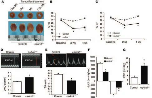
persisting at least up to 4 weeks (Figure 2, B–D). csArnt–/– mice also
displayed a reduction in the early to late ventricular filling veloci-ties (E/A) ratio that was measured across the mitral valve (Figure 2E). Invasive hemodynamic measurements revealed increased end-diastolic pressure (EDP), while the maximum and minimum rates of pressure change in the left ventricle (+dP/dt and –dP/dt) were significantly reduced with Arnt deletion (Figure 2, F and G), suggesting a combination of systolic and diastolic dysfunction
in the hearts of csArnt–/– mice. Consistent with cardiac
dysfunc-tion, csArnt–/– mice also displayed higher heart and lung weights
normalized to tibia length (Supplemental Figure 4, A–C). Finally, the levels of heart failure markers atrial natriuretic protein (ANP) and brain natriuretic protein (BNP) were significantly higher in
csArnt–/– mice than in controls (Supplemental Figure 5, A and B).
These results suggest that Arnt deletion in the heart leads to the development of spontaneous dilated CM.
Arnt deletion in the heart leads to excess lipid accumulation. We then studied the mechanism for the CM associated with Arnt
deletion. H&E staining showed myofiber architectural distortion
and markedly increased interstitial fibrosis in csArnt–/– hearts 4
weeks after tamoxifen administration (Figure 3A). We also noted
evidence of enhanced apoptosis in csArnt–/– hearts compared with
that seen in control hearts, as assessed by TUNEL staining of ven-tricular tissue (Figure 3B). Electron microscopic analysis revealed the presence of large vacuoles scattered throughout the heart
tissue of csArnt–/– mice (Figure 3C), bearing structural similarity
to lipid vacuoles (26). These results suggest that Arnt deletion in the heart may lead to lipid accumulation; thus, we performed
fur-ther analysis of the lipid levels in csArnt–/– hearts 4 weeks after
initiation of tamoxifen treatment. Oil red O staining revealed
enhanced dye accumulation in csArnt–/– hearts (Figure 3, D and
E), confirming increased lipid content. Moreover, we found that
triacylglycerol (TAG) levels were higher in the hearts of csArnt–/–
mice (Figure 3F), while serum TAG levels were similar compared with those in control mice (Supplemental Figure 6A). Consistent with these results, we also found an increase in TAG levels in iso-lated neonatal rat cardiomyocytes (NRCMs) in response to ARNT knockdown with siRNA (Supplemental Figure 6, B and C). Taken together, these results show that Arnt deletion causes significant lipid accumulation in the heart.
Arnt deletion in the heart leads to an increase in FA metabolism. We next assessed the effects of Arnt deletion on cardiac FA and glucose metabolism in isolated working hearts using radioiso-tope-labeled substrates. Deletion of Arnt in the heart resulted in an approximately 2-fold increase in FAO (as assessed by
oxida-tion of 3H-palmitate) (Figure 4A). csArnt–/– mice also displayed a
decrease in glucose oxidation (Supplemental Figure 7A) and a sig-nificant reduction in genes involved in glucose metabolism (Sup-plemental Figure 7B). To confirm that the observed changes in
glucose and FA metabolism in the csArnt–/– hearts were specific to
the absence of cardiac ARNT and not secondary to the CM associ-ated with Arnt deletion, we performed in vitro studies in NRCMs. Consistent with the in vivo data, ARNT knockdown in NRCMs led to increased FA uptake (FAU) (Figure 4B) and FAO (Figure 4C). Knockdown of ARNT in NRCMs also resulted in a significant reduction in glucose uptake and the extracellular acidification rate (ECAR) (Supplemental Figure 7, C and D). These results suggest including increased myocardial FAU and FAO, reduced glucose
utilization, and CM (21). They also exhibit FA deposits throughout their heart, which is believed to be due to an imbalance between FAU and FAO, both of which are increased to different degrees.
Furthermore, the CM seen in PPARα Tg mice is exacerbated by a
high-fat diet, suggesting that FA overload contributes to the lipo-toxicity and cardiac dysfunction (22). However, it is not known
whether an increase in PPARα through non-Tg models would still
result in the same phenotype.
Here, we demonstrate that deletion of Arnt in the heart leads to CM and lipid accumulation. Our mechanistic studies
deter-mined that PPARα mediates the deleterious effects and metabolic
changes of Arnt deletion in the heart, as deletion of PPARα reverses
the phenotype associated with Arnt knockout. Furthermore, we
demonstrate that ARNT regulates PPARα at the transcriptional
level by binding to its promoter and by forming a complex with
HIF2α. We believe that these findings provide the first evidence for
a functional link between the HIF/ARNT pathway and PPARα in
the heart and demonstrate that deletion of Arnt leads to CM and
cardiac lipotoxicity through PPARα activation.
Results
Cardiac deletion of Arnt leads to CM. ARNT levels are reduced in the liver and pancreas in diabetes, and its deletion in those tis-sues leads to a diabetic phenotype (10, 11). However, the function of ARNT in the heart is not known. We first assessed the levels of ARNT in hearts from diabetic mice. The levels of ARNT were sig-nificantly decreased in db/db mice at 32 weeks of age compared with those detected in age-matched WT animals (Figure 1A). To study the role of ARNT in the heart, we generated tamoxifen-
inducible cardiac-specific ARNT-knockout mice (csArnt–/–) by
crossing mice with loxP sequences flanking exon 8 of Arnt (Arntfl/fl)
(23) with mutated estrogen receptor (MER)-Cre-MER (MCM)
Tg mice under the α-myosin heavy chain (Myh6) promoter
(24) (Figure 1, B and C, and Supplemental Figure 1A; supple-mental material available online with this article; doi:10.1172/ JCI76737DS1). Intraperitoneal injection of tamoxifen in these mice resulted in transient but severe CM independent of Arnt deletion, consistent with previous work (25). Thus, we adopted a 2-week protocol of oral tamoxifen administration (Supplemental Figure 1B), which did not result in any changes in body weight (Supplemental Figure 1C). Deletion of Arnt in the heart was confirmed at the genomic DNA (Figure 1D), mRNA (Figure 1E and Supplemental Figure 2A), and protein levels (Figure 1F). We found that deletion of Arnt was specific to the heart, as its mRNA and protein levels in other tissues were comparable to those in the control mice (Supplemental Figure 2, B and C).
We then assessed the effects of Arnt deletion on cardiac
func-tion. A dilated left ventricle in csArnt–/– mice was observed at 4
weeks after tamoxifen treatment, while all control group mice displayed normal cardiac size and function (Figure 2A and
Sup-plemental Figure 3, A–E). For subsequent studies, we used Arntfl/fl
mice treated with tamoxifen as controls. Echocardiographic
anal-ysis of the hearts of csArnt–/– mice revealed decreased fractional
The Journal of Clinical Investigation
R e s e a R c h a R t i c l eHIF binding sites) (Figure 5A) or a control plasmid into HEK293 cells, along with control or ARNT siRNA. ARNT siRNA resulted in effective knockdown in HEK293 cells (Supplemental Figure 9A) and led to a significant increase in luciferase activity (Figure 5B),
suggesting that ARNT likely regulates PPARα at the mRNA level.
We then assessed which of the HREs is responsible for the inhibi-tory effects of ARNT on the Ppara promoter by making truncated forms of the promoter to sequentially remove HREs (Figure 5A). A truncated construct with removal of the second HRE upstream of the initiation site abolished the inhibitory effect of ARNT on the Ppara promoter, suggesting that this site is likely involved in the regulation of the gene (Figure 5B). To confirm this observation, we performed ChIP analysis on this HRE. Negative and positive con-trols for the ChIP are shown in Supplemental Figures 9, B and C, and ChIP analysis of the second HRE upstream of the Ppara initia-tion site confirmed that ARNT binds to this sequence (Figure 5C). To determine which ARNT partner mediates its effects on
PPARα expression, we then downregulated HIF1α, HIF2α, and
AHR in NRCMs and assessed PPARα levels. A reduction in HIF1α
or AHR did not alter the levels of PPARα significantly, while HIF2α
knockdown, similar to the effects of ARNT reduction, resulted in
an increase in PPARα levels (Supplemental Figure 10, A–D, and
Figure 5D). These results suggest that the effects of Arnt
dele-tion on PPARα are, at least partially, through its association with
that Arnt deletion in the heart results in an increase in FA metabo-lism and a decrease in glucose utilization.
The PPARα pathway is activated in the hearts of csArnt–/– mice.
We next studied the mechanism for increased lipid
accumula-tion in the hearts of csArnt–/– mice. PPARs are known to regulate
FA metabolism (12), thus, we examined the effects of Arnt dele-tion on the expression of PPAR proteins. Although Arnt deledele-tion in the heart did not alter the mRNA levels of Pparg or Pparb/d, it did result in a significant increase in Ppara mRNA (Figure 4D) and protein (Figure 4E) levels. Similarly, knockdown of ARNT with
siRNA in NRCMs resulted in a significant increase in PPARα
lev-els (Supplemental Figure 8, A and B). In addition, PPARα target
genes involved in mitochondrial β oxidation, such as carnitine
palmitoyltransferase 1 (Cpt1) and fatty acyl-CoA synthetase (Acs),
were significantly increased in csArnt–/– hearts. Diacylglycerol
acyltransferase (Dgat), the key enzyme involved in the esterifica-tion of FA to TAG, was also significantly elevated in the hearts of
csArnt–/– mice (Figure 4F).
These results suggest that ARNT inhibits PPARα expression.
The HIF pathway generally causes activation of genes; however, recent studies suggest that HIF may also inhibit promoter activity
in certain genes (27–29). To assess whether ARNT regulates PPARα
[image:4.585.53.543.57.344.2]at the transcriptional level, we first transfected a plasmid construct of the human Ppara promoter luciferase reporter (which has 5
Deletion of Ppara prevents cardiac metabolic derangements
associated with Arnt deletion. We next assessed whether PPARα is
increased in the hearts of diabetic mice and showed that its mRNA levels were higher in hearts from db/db mice (Supplemental Figure 13). These findings are consistent with studies by Finck et al., who
demonstrated that PPARα mRNA and protein levels are increased
in streptozotocin-treated hearts and that PPARα target genes (but
not its own mRNA levels) are increased in db/db hearts (19). To
bet-ter assess the role of PPARα in the metabolic effects of ARNT in the
heart, we knocked down ARNT or PPARα individually or together
in NRCMs, then measured the genes involved in FA metabolism. Deletion of Ppara either completely or partially reversed the effects of Arnt deletion on genes involved in FA metabolism (Figure 6A). Furthermore, we found that the increase in FAO (as assessed by oxygen consumption in the presence of palmitate) with ARNT knockdown was completely reversed with Ppara deletion (Figure
6B). Also, csArnt–/– hearts displayed higher levels of UCP2 and
UCP3 (Supplemental Figure 14), suggesting that the hearts of these mice have more uncoupling and inefficient fuel use.
To confirm that myocardial lipid accumulation and FAO
associated with Arnt deletion in the heart are PPARα
depen-dent, we generated csArnt–/– and Ppara–/– double-knockout mice
by crossing Myh6-MCM Arntfl/fl mice with Ppara–/– mice, followed
HIF2α and through the binding of this complex to the second HRE
upstream of the Ppara initiation site. To confirm these results, we
then performed sequential ChIP studies using HIF2α and ARNT
antibodies on the second HRE sequence. As shown in Figure 5E,
ARNT and HIF2α bound to this HRE sequence as a complex.
Finally, we either deleted or mutated the second HRE in the full-length Ppara promoter attached to luciferase and showed that both of these changes resulted in a reversal in the increase in luci-ferase activity with ARNT kno(Figure 5, F and G). These results provide evidence that the second HRE mediates the effects of
ARNT/HIF2α on the Ppara promoter.
[image:5.585.49.536.56.370.2]We next assessed whether the effects of ARNT knockdown on the Ppara promoter occurs through changes in the activity of histone deacetylases (HDACs). We showed that treatment of cells with the HDAC inhibitor trichostatin A (TSA) did not alter Ppara mRNA levels in the presence or absence of ARNT knockdown (Supplemental Figure 11). Furthermore, we found that HDAC1–4 levels were not changed with ARNT knockdown (Supplemental Figure 12, A and B), and ChIP studies showed that the levels of acetylated histones H3 and H4 were not increased on the Ppara promoter after ARNT knockdown (Supplemental Figure 12, C–E). Thus, the effects of ARNT on the Ppara promoter are independent of HDACs and histone deacetylation.
The Journal of Clinical Investigation
R e s e a R c h a R t i c l eprovide in vitro and in vivo data showing that PPARα mediates
the lipid accumulation and increase in FAO associated with ARNT knockdown.
Cardiac function is restored with Ppara deletion in csArnt–/–
mice. Finally, we sought to determine whether the CM observed
in csArnt–/– mice is due to PPARα activation. We assessed
car-diac function by echocardiography in the csArnt–/– and csArnt–/–
Ppara–/– double-knockout mice 1, 2, and 4 weeks after tamoxifen
administration. While csArnt–/– mice displayed CM 4 weeks after
tamoxifen treatment, csArnt–/– Ppara–/– double-knockout mice
showed a complete reversal of CM, as determined by cardiac output, EF, and FS (Figure 7, A–D, and Supplemental Table 1). Moreover, the molecular markers of heart failure, ANP and BNP,
were also reduced upon PPARα deletion (Supplemental Figure
16, A and B). Finally, Kaplan-Meier analysis showed that csArnt–/–
Ppara–/– double-knockout mice had no mortality by 2 months of
age, while csArnt–/– mice had approximately 15% mortality by that
time (Figure 7E). These results suggest that Arnt deletion leads to
cardiac dysfunction, which is mediated through PPARα-mediated
lipid accumulation (Figure 7F). by tamoxifen treatment (Supplemental Figure 15A). The
geno-type of the double-knockout mice was confirmed by PCR, and deletion of the genes was confirmed by measuring the levels of both proteins in the hearts of double-knockout mice with dou-ble immunofluorescence (Supplemental Figure 15, B and C). We then measured substrate metabolism in the hearts of these
mice using a working heart model and 3H-labeled palmitate. As
expected, FAO was significantly higher in csArnt–/– hearts
com-pared with that in control hearts. Deletion of PPARα in csArnt–/–
hearts reversed the increase in myocardial TAG content (Figure
6C), FAO (Figure 6D), and gene targets of PPARα, including
Cpt1 and Acs (Figure 6E). Moreover, csArnt–/– Ppara–/–
double-knockout mice displayed significantly lower lipid accumulation
compared with csArnt–/– mice, as assessed by oil red O
[image:6.585.59.532.56.381.2]stain-ing (Figure 6F). Since myocardial cell death is a consequence of lipotoxicity in the heart, we assessed myocardial cell death and interstitial fibrosis. Histologic examination of left ventric-ular tissue revealed that deletion of Ppara normalized myocar-dial cytoarchitecture and reversed cell death, as determined by TUNEL staining (Figure 6, F and G). Collectively, these results
Figure 3. csArnt–/–hearts display increased lipid accumulation. (A) Representative cardiac histological sections of control (Arntfl/fl littermates) and csArnt–/– mice 4 weeks after initiation of tamoxifen treatment. MT, Masson’s trichrome. Scale bars: 1 mm (H&E-stained ×4 images) and 100 μm (H& E-stained ×200 and MT-E-stained images). Experiments were repeated 3 times. (B) TUNEL staining of hearts from control and csArnt–/– mice. TUNEL+ cells
Discussion
Here, we showed that csArnt–/– mice display enlarged left
ven-tricular size and reduced cardiac function, consistent with CM.
csArnt–/– hearts also exhibited lipid accumulation, as assessed by
electron microscopy, oil red O staining, and TAG measurements. Using an ex vivo working heart system, we demonstrated that
csArnt–/– hearts have an approximately 2-fold increase in FAO
and a decrease in glucose metabolism. In addition, we showed
that PPARα and its target genes involved in FAU and FAO are
significantly increased in the csArnt–/– hearts. To confirm that
the effects of ARNT downregulation on cardiac metabolism
are through PPARα, we crossed csArnt–/– with Ppara–/– mice and
showed that knockout of Ppara reverses the pathologic changes associated with ARNT reduction. Finally, we showed that ARNT inhibits Ppara promoter activity by forming a complex with
HIF2α. We conclude that ARNT is a critical regulator of
myocar-dial FA metabolism and that a reduction in ARNT levels leads to cardiac dysfunction at baseline by increasing TAG accumulation
through a PPARα-mediated pathway.
The mechanism for an increased incidence of heart dysfunc-tion in diabetes is not totally clear, and several hypotheses have been proposed. Lipid toxicity has been suggested as a potential mechanism, an idea that is supported by studies showing that excessive FA accumulation and oxidation contribute to cardio-myocyte dysfunction. Furthermore, glucose uptake and utilization are reduced in diabetic hearts, while there is a concurrent increase
in FAU and metabolism, leading to lipotoxic CM. Overexpression
of PPARα results in a phenotype similar to that of diabetic cardiac
dysfunction, and PPARα levels are increased in the hearts of
dia-betic mice (19), raising the possibility that PPARα plays a major role
in the pathogenesis of lipotoxicity associated with diabetic CM. In
our studies, we also noted an increase in PPARα with Arnt
dele-tion, which was associated with lipid accumulation in the heart.
It is important to point out that the extent of the PPARα increase
in the csArnt–/– mice was only approximately 5- to 6-fold at the
protein level (Figure 4E), which was associated with a significant reduction in cardiac function. This phenotype is not totally
consis-tent with what was observed in PPARα Tg mice, which displayed
a greater than 15-fold increase in PPARα levels in the heart and
a milder form of CM compared with csArnt–/– mice (19). Thus,
in addition to a PPARα-dependent pathway, other mechanisms
(independent of PPARα) may contribute to the pathogenesis of
CM associated with Arnt deletion. Nevertheless, our findings
pro-vide epro-vidence that even an indirect increase in the levels of PPARα
(as opposed to direct overexpression in Tg animals) contributes to
lipotoxicity and CM, providing a further link between PPARα and
the diabetic CM phenotype.
The active HIF complex is composed of 2 subunits: ARNT
and either HIF1α or HIF2α. In the ischemic heart, the HIF/ARNT
complex participates in the cardiomyocyte response to low oxy-gen tension by upregulating glucose transporters and glycolytic enzymes (30). Global Hif1a knockout results in embryonic
lethal-Figure 4. csArnt–/–hearts display increased FAO and PPARα levels. (A) Myocardial FAO in control and csArnt–/– hearts measured in isolated working hearts using 3H-labeled palmitate (n = 5–6 independent experiments). (B) FAU in NRCMs treated with control or ARNT siRNA (n = 6 independent experiments).
[image:7.585.49.526.53.331.2]The Journal of Clinical Investigation
R e s e a R c h a R t i c l eity with failure of neural tube closure, defective vascularization, and increased thickness of the heart in embryos (31, 32). Cardiac- specific Hif1a knockout leads to mild cardiac hypertrophy with reductions in contractility, vascularization, and high-energy phos-phate content (33), but no evidence of lipid accumulation. How-ever, inducible knockout of Hif1a after birth in the heart does not
cause cardiac dysfunction (34), suggesting that baseline HIF1α
function may be more important during development than in the maintenance of adult cardiac function. Mice with global Hif2a knockout display changes in different organs, including lipid accu-mulation in skeletal muscle, cardiac muscle, and liver (35). These mice also have cardiac hypertrophy, although cardiac function was not assessed directly (35). We showed that deletion of Arnt in the adult heart leads to spontaneous CM due to lipid accumulation,
which is distinct from what is observed with HIF1α deletion, but
is more similar to that of Hif2a-knockout mice. In the background of Arnt deletion, it is not technically possible to reconstitute the
activity of each of its partners (i.e., HIF1α and -2α and AHR).
However, we assessed the effects of ARNT partners in PPARα
regulation and demonstrated that a reduction in HIF2α increases
PPARα levels in vitro and that ARNT forms a complex with HIF2α
to inhibit the Ppara promoter. Although this effect is likely inde-pendent of HDACs and histone deacetylation, it is possible that
the binding of ARNT/HIF2α to the Ppara promoter blocks the
access of other transcription factors to their binding sites.
The link between the HIF pathway and PPARs was previously
studied in the heart. Krishnan et al. showed that HIF1α activates
PPARγ, which in turn, activates FAU, glucose-to-lipid conversion
via the glycerol-3-phosphate pathway, apoptosis, and contractile
dysfunction (36). They also demonstrated that HIF1α activates
PPARγ by binding to its promoter and that deletion of Hifa
atten-uates stress-induced cardiac hypertrophy. These studies focused
on HIF1α, which is only one of the partners of ARNT and which
[image:8.585.59.533.57.362.2]was shown in our studies not to play a role in the CM associated with Arnt deletion. Furthermore, our studies focused on CM rather than the cardiac hypertrophy studied by Krishnan et al. (36).
In summary, our results demonstrate that deletion of Arnt in
the heart leads to CM, lipid accumulation, and PPARα activation.
The mechanism for PPARα activation occurs at the
transcrip-tional level and through the regulation of its promoter by ARNT/
HIF2α. These results demonstrate a functional link between the
HIF/ARNT pathway and lipid metabolism in the heart through
PPARα activation and provide a novel potential target for the
treatment of cardiac lipotoxicity.
Methods
Animal models. csArnt–/– mice were generated by crossing Arntfl/fl mice
with Myh6-MCM (tamoxifen-inducible heart-specific Cre) Tg mice (purchased form The Jackson Laboratory), followed by oral
adminis-Nevertheless, these 2 studies provide evidence for a link between the HIF pathway and FA metabolism in the heart in response to stress.
In addition to providing substrates for oxidative phosphory-lation and ATP generation, FA and TAG stores serve as important signaling molecules in the cell. Importantly, free FAs are known to
bind to nuclear receptors such as PPARα and stimulate their
translo-cation into the nucleus. It is conceivable that the activation of PPARα
signaling we observed with Arnt deletion was secondary to the
bind-ing of free FAs to PPARα and its subsequent activation. However,
this mechanism is unlikely to explain all of our findings, since our luciferase studies of the Ppara promoter showed regulation of this receptor at the transcriptional level. Nevertheless, higher TAG
[image:9.585.56.524.56.461.2]lev-els in Arnt-knockout mice may further amplify PPARα signaling.
Figure 6. PPARα knockdown reverses the metabolic changes associated with Arntdeletion. (A) mRNA levels of genes involved in FAU and FAO in NRCMs treated with control, ARNT, PPARα, and ARNT plus PPARα siRNA. MCAD, medium-chain acyl-CoA dehydrogenase (n = 6 independent experiments). (B) FAO as determined by oxygen consumption with the exogenous addition of palmitate in NRCMs treated with control, ARNT, PPARα, and ARNT plus PPARα
siRNA (n = 6 independent experiments). (C) Myocardial TAG levels in control, csArnt–/–, and csArnt–/–Ppara–/– mice (n = 6–8 independent experiments). (D) Myocardial FAO determined in isolated working hearts of csArnt–/–, csArnt–/–Ppara–/–, or littermate controls using 3H-labeled palmitate (n = 5 hearts). (E)
mRNA levels of gene targets of Ppara in control, csArnt–/–, and csArnt–/–Ppara–/– hearts (n = 6 independent experiments). (F) Oil red O, TUNEL, MT, and H&E staining of hearts from control, csArnt–/–, and csArnt–/–Ppara–/– mice. Scale bar: 100 μm. Experiments were repeated in triplicate. (G) Summary of TUNEL+
The Journal of Clinical Investigation
R e s e a R c h a R t i c l eand 4 weeks after oral administration of tamoxifen or normal chow. Parasternal short- and long-axis views were used to obtain 2D and M-mode images. At least 10 independent cardiac cycles per experi-ment were obtained.
Cardiac hemodynamic measurement. Hemodynamic studies were
performed 2 weeks after tamoxifen treatment was completed, as described previously (39). A high-fidelity transducer-tipped pres-sure-volume catheter (Scisense Inc.) was introduced from the right carotid artery into the left ventricle of the anesthetized mouse to determine hemodynamics. Signals were digitized using a data trans-lation series analog-digital converter and then stored and analyzed.
Histological analysis. Hearts were fixed in 10% formalin (PBS
buffered), dehydrated, and embedded in paraffin. Heart architec-ture was determined using transverse 5-μm deparaffinized sections stained with H&E. Fibrosis was detected with Masson trichrome staining. TUNEL positivity in the hearts from csArnt–/– and control
mice was determined using an in situ detection kit according to the manufacturer’s instructions (Roche Diagnostics). Oil red O staining was performed on sections of unfixed, freshly frozen heart samples (6-μm in thickness).
Electron microscopy. Cardiac tissue from the left ventricle free wall
was fixed with 2% glutaraldehyde in sodium cacodylate buffer at 4°C. Fixed tissues were embedded in epoxy resin, processed for thin (70-nm) cutting with a Leica UC6 ultramicrotome, and examined with a FEI Tecnai Spirit transmission electron microscope.
Heart TAG content. Heart TAG was extracted as previously
reported (40) and quantified using a Triglyceride Quantification Kit (Abcam) following the manufacturer’s instructions. To measure TAG levels in isolated cells, NRCMs were homogenized in 5% NP-40 solu-tion. Samples were solubilized by slowly heating to 80°C to 100°C for 5 minutes and then cooling down to room temperature. They were tration of tamoxifen (30 mg/kg) for 2 weeks as described previously
(37). Three sets of sex-matched littermate mice (Myh6-MCM with tamoxifen; Arntfl/fl with tamoxifen; and Myh6-MCM Arntfl/fl without
tamoxifen) were used as controls. db/db mice were purchased from The Jackson Laboratory and studied at 32 weeks of age. All groups of mice included a mix of males and females.
Genotyping. Genomic DNA was prepared from tail clips using
the PureGene DNA isolation kit (Gentra Systems) according to the manufacturer’s protocol. Approximately 10 ng of the genomic DNA was used for PCR. Genotyping of Myh6-MCM mice was performed using 2 sets of primers. The first primer set was designed to amplify the Cre Tg construct (forward, 5′-ATACCGGAGATCATGCAA GC-3′; reverse, 5′-AGGTGGACCTGATCATGGAG-3′). The second primer set was used to amplify a mouse WT internal positive control (forward, 5′-CTAGGCCACAGAATTGAAAGATCT-3′; reverse, 5′-GTAGGT-GGAAATTCTAGCATCATCC-3′). Genotyping of Arntfl/fl mice was
performed by PCR amplification (forward, 5′-CACCTGAGCTAAAT-TACCAGGCC-3′; reverse, 5′-GCATGCTGGCACATGCCTGTCT-3′). The 2 sets of primers used for genotyping of Ppara–/– mice were:
for-ward, 5′-GAGAAGTTGCAGGAGGGGATTGTG-3′ (served as com-mon); reverse: 5′-CCCATTTCGGTAGCAGGTAGTCTT-3′ (WT); and reverse, 5′-GCAATCCATCTTGTTCAATGGC-3′ (mutant).
Primary cell culture. NRCMs were isolated from 1- to 2-day-old
Sprague–Dawley rats as previously described (38). Cardiomyocytes were cultured in DMEM/M199 (3:1), supplemented with 5% FBS, 1.5 mM vitamin B12, and 1 mM penicillin-streptomycin (Invitrogen). To prevent the proliferation of nonmyocytes, 100 μM BrdU was added to the culture media (Sigma-Aldrich).
Echocardiography. Cardiac function was noninvasively
[image:10.585.56.534.60.269.2]moni-tored by transthoracic echocardiography using a VisualSonics Vevo 770 high-resolution imaging system with a 30 MHz scanhead 1, 2, 3,
Figure 7. Pparadeletion reverses the cardiac dysfunction of csArnt–/–mice. (A) Representative m-mode echocardiographic
HDAC3, and HDAC4 were purchased from Cell Signaling Technol-ogy. GAPDH and tubulin antibodies were purchased from Santa Cruz Biotechnology Inc. Samples were run on an SDS-PAGE gel and trans-ferred to a nitrocellulose membrane (Invitrogen). The protein bands were developed with an enhanced chemiluminescence substrate kit. Quantification of blots was performed using ImageJ software (NIH). (Full, uncut gels are shown in the Supplemental Material.)
Heart isolation and perfusion conditions. Hearts from csArnt–/– and
littermate control mice were perfused in the working mode. Mice were anesthetized with an intraperitoneal injection of Avertin (400 mg/kg body weight), and the hearts were subsequently excised and immersed in ice-cold Krebs-Henseleit bicarbonate solution (118 mM NaCl, 25 mM NaHCO3, 5.9 mM KCl, 5 mM EDTA [pH = 7.4], 1.2 mM MgSO4·7H2O, 2.5 mM CaCl2·2H2O, and 5 mM glucose). The aorta was cannulated and perfused with Krebs-Henseleit solution (37°C) initi-ated at a hydrostatic pressure of 60 mmHg. The hearts were trimmed of excess tissue, and the opening to the left atrium was cannulated. After equilibration in the Langendorff mode, the hearts were switched to the working mode by clamping off the aortic inflow line from the Langendorff reservoir and opening the left atrial inflow line. Oxygen-ated Krebs-Henseleit solution consisting of 0.4 mM palmitate bound to 3% FA-free BSA, 5 mM glucose, and 1 mM lactate in the presence of 100 mU/ml insulin was delivered to the left atrium at a preload pres-sure of 11.5 mmHg. Perfusate was ejected from spontaneously beat-ing hearts into a compliance chamber and into the aortic outflow line against a hydrostatic afterload pressure of 50 mmHg. The perfusate was recirculated, and the pH was adjusted to 7.4 by gassing the perfus-ate in a glass oxygenator with a gas mixture containing 95% O2 and 5% CO2. At the end of perfusion, the hearts were immediately frozen in liquid N2 with Wollenberger tongs and stored at –80°C.
Determination of glucose and palmitate oxidation. Glucose
oxi-dation was measured by perfusing hearts with [U-14C] glucose and
unlabeled lactate and palmitate (42, 43). Palmitate oxidation mea-surements were performed by perfusing the hearts with glucose and [9,10-3H]palmitate. The total myocardial 3H
2O production and 14CO2
production were determined at 10-minute intervals during the 40- or 60-minute aerobic perfusion period. To measure the rates of palmi-tate oxidation, 3H
2O in perfusate samples was separated from [3H]
palmitate hearts using a vapor transfer method (42, 43). This method consisted of adding 500 μl of water into a 5-ml scintillation vial, then placing a lidless 1.5-ml microcentrifuge tube inside the scintillation vial. A 200-μl perfusate sample was then added to the microcen-trifuge tube, and the scintillation vial was capped. Scintillation vials were then stored initially at 50°C for 24 hours and then at 4°C for 24 hours. Following storage, the microcentrifuge tube was removed, scintillation fluid (EcoLite; Fisher Scientific) was added, and the radioactivity was counted in a liquid scintillation counter. The glucose oxidation rate was determined by quantitative measurement of 14CO
2
production from 14C-glucose–perfused hearts released as a gas in the
oxygenation chamber and 14CO
2 dissolved as H14CO3– in perfusate.
The gaseous 14CO
2, which exits the perfusion system via an exhaust
line, was trapped in hyamine hydroxide solution. The dissolved 14CO 2
as H14CO
3– was released and trapped on filter paper saturated with
hyamine hydroxide in the central well of 25-ml stoppered flasks after perfusate samples were acidified by the addition of 1 ml 9N H2SO4.
Luciferase studies. H9EK293 cells were cotransfected using
Lipo-fectamine Plus Reagent (Invitrogen) in a serum-free medium with both centrifuged to remove insoluble material and then diluted 10-fold in
ddH2O. TAG levels were determined using the Triglyceride Quantifi-cation Kit from BioVision. All samples and standards were run in trip-licate, and values were normalized to protein content.
Measurement of cellular glycolysis (ECAR) and FAO. Cellular
gly-colysis was determined by measuring the ECAR in NRCMs using a Seahorse Bioscience XF24 extracellular flux analyzer (Billerica) as described previously (41). An increased oxygen consumption rate (OCR) immediately following the addition of palmitate substrate to the cells was used as an indicator for FAO in the XF24 Analyzer. Sixty thousand cells were seeded per well, and 24 hours later were treated for 2 subsequent days with siRNA. Forty-eight hours later, cells were used for oxygen consumption studies using unbuffered DMEM plus 1 mM sodium pyruvate plus 1 g/l glucose. Oxygen con-sumption readings were normalized to total protein per well.
2-Deoxyglucose uptake. Neonatal cardiomyocytes were grown
in 6-well plates. Following serum depletion, cells were rinsed with HEPES buffer and incubated with 1 μl Ci 3H-2-deoxy-glucose and 5
mM glucose per well for 10 minutes. Cells were then rinsed with ice-cold PBS and lysed in a buffer containing 0.1% SDS. 2-Deoxyglucose uptake was measured for 30 seconds. Nonspecific uptake was deter-mined in the presence of 10 μM cytochalasin B, and this value was sub-tracted from all values. Cell-associated radioactivity was determined by lysing the cells with 0.05 M NaOH, followed by measuring radio-activity using a liquid scintillation counter. Total cellular protein was determined by the Bradford method.
FAU. BSA-conjugated palmitate was freshly prepared with
Krebs-Henseleit buffer at a final concentration of 0.0425 mM for BSA and 0.25 mM for palmitate. Cells were plated in a 6-well plate, rinsed with PBS buffer with 1% BSA, and incubated with 2 ml BSA-palmitate in each well for 2 minutes at 37°C. After washing 5 times with ice-cold PBS, cells were lysed with RIPA buffer, followed by liquid scintilla-tion counting. Total cellular protein was determined by the Bradford method and used for normalization.
RNA isolation and quantitative real-time PCR. Total RNA from
cultured NRCMs was isolated using the RNA Stat-60 reagent (Tel-Test Inc.), and RNA from mouse heart was isolated using an RNeasy Fibrous Tissue Mini Kit from QIAGEN according to the manufactur-er’s instructions. Samples of total RNA (500 ng) were reverse tran-scribed using the TaqMan reverse transcription reagents PCR Kit (Applied Biosystems), and the resulting cDNA was used as a PCR template. The mRNA levels of Arnt, Ppara, Pparg, Pparb, Cd36, Acs,
Cpt1, Mcad, Hk2, Glut1, and Glut4 were determined by quantitative
real-time PCR (qRT-PCR) with SYBR Green using the 7500 Fast Real-Time PCR system (Applied Biosystems) according to the manufactur-er’s instructions. 18S RNA was amplified as an internal control. Rela-tive gene expression levels were calculated using the comparaRela-tive Ct method formula 2−ΔΔCt.
siRNA treatment of NRCMs. siRNA was transfected into NRCMs
using a TransMessenger Transfection Kit (QIAGEN). Forty-eight hours later, cells were lysed and used for Western blot analysis or for RNA isolation by qRT-PCR.
Western blot analysis. For Western blotting of total protein, hearts
The Journal of Clinical Investigation
R e s e a R c h a R t i c l eThe positive control genes were identified in earlier studies, and the primer sequences for those genes have been validated as described previously (45). For ChIP against acetylated histones, sodium butyrate was added to all buffers. Anti-HIF2α antibody was purchased from Novus, and acetylated histone H3 and acetylated histone H4 antibod-ies were purchased from Millipore.
Statistics. Data are expressed as the mean ± SEM. Statistical
significance was assessed with ANOVA. For multiple group com-parisons, a post-hoc Tukey’s test was performed when ANOVA reached significance. A P value of less than 0.05 was considered statistically significant.
Study approval. All animal studies were reviewed and approved by
the IACUC of Northwestern University.
Acknowledgments
We would like to thank Konrad Sawicki for careful review of the manuscript. Imaging work was performed at the Northwest-ern University Cell Imaging Facility, which is supported by NCI grant CCSG P30 CA060553, awarded to the Robert H. Lurie Comprehensive Cancer Center. H. Ardehali is supported by NIH grants K02 HL107448, R01 HL104181, and 1PO1 HL108795. R. Wu is supported by American Heart Association (AHA) grant 13SDG17270046.
Address correspondence to: Hossein Ardehali, Tarry 14-733, 303 E. Chicago Ave., Chicago, Illinois 60611, USA. Phone: 312.503.2342; E-mail: h-ardehali@northwestern.edu.
the recombinant firefly luciferase plasmids containing a human PPARα promoter-luciferase reporter (200 ng) and the Renilla luciferase plas-mid (25 ng), along with 25 nM of either ARNT siRNA or control siRNA. Forty-eight hours after transfection, cells were collected, and the luci-ferase signal intensity was measured with a dual-luciluci-ferase reporter assay kit (Promega) on a Berthold Lumat LB 9570 luminometer. Firefly luciferase expression was corrected by Renilla luciferase expression in the same well to normalize for variations in transfection efficiency.
Mutagenesis of the HRE site was performed using PCR-based site-directed mutagenesis. Briefly, primers containing the desired muta-tions were used to amplify the Ppara promoter-luciferase reporter plasmid using the HiFi PCR Kit (Kapa Biosystems), followed by DpnI (New England Biolabs Inc.) digestion to remove the original template. The following primers were used for the HRE sequence, with the mutation indicated in bold letters: sense primer,GGGCCGAGGGG-CGGTTATGCTCGCGGGGGCGCGGC; anti-sense primer, GCCG-CGCCCCCGCGAGCATAACCGCCCCTCGGCCC. The sequence of the resulting plasmid was verified before subsequent experiments.
ChIP studies. Confluent HepG2 culture was harvested with trypsin
and cross-linked with 1.1% formaldehyde. ChIP was performed using anti-ARNT antibody (Cell Signaling Technology) as described previ-ously (44). After reverse cross-linking and proteinase K and RNAse A digestion, DNA was purified with a QIAGEN PCR purification kit. Purified DNA was used as input for qRT-PCR. For sequential ChIP, chromatin from the first IP was eluted with elution buffer containing 5 mM DTT and diluted 10-fold before being used as input for the sec-ond IP. The secsec-ond IP was performed as described previously (44).
1. Wild S, Roglic G, Green A, Sicree R, King H. Global prevalence of diabetes: estimates for the year 2000 and projections for 2030. Diabetes Care. 2004;27(5):1047–1053.
2. Battiprolu PK, Lopez-Crisosto C, Wang ZV, Nemchenko A, Lavandero S, Hill JA. Diabetic cardiomyopathy and metabolic remodeling of the heart. Life Sci. 2013;92(11):609–615. 3. Dirkx E, Schwenk RW, Glatz JF, Luiken JJ, van
Eys GJ. High fat diet induced diabetic cardiomy-opathy. Prostaglandins Leukot Essent Fatty Acids. 2011;85(5):219–225.
4. van de Weijer T, Schrauwen-Hinderling VB, Schrauwen P. Lipotoxicity in type 2 diabetic car-diomyopathy. Cardiovasc Res. 2011;92(1):10–18. 5. Ema M, et al. Two new members of the murine
Sim gene family are transcriptional repres-sors and show different expression patterns during mouse embryogenesis. Mol Cell Biol. 1996;16(10):5865–5875.
6. Rowlands JC, Gustafsson JA. Aryl hydrocarbon receptor-mediated signal transduction. Crit Rev Toxicol. 1997;27(2):109–134.
7. Semenza GL. HIF-1: mediator of physiological and pathophysiological responses to hypoxia. J Appl Physiol. 2000;88(4):1474–1480. 8. Bugger H, Abel ED. Molecular mechanisms
for myocardial mitochondrial dysfunction in the metabolic syndrome. Clin Sci (Lond). 2008;114(3):195–210.
9. Maltepe E, Schmidt JV, Baunoch D, Bradfield CA, Simon MC. Abnormal angiogenesis and responses to glucose and oxygen deprivation in mice lacking the protein ARNT. Nature.
1997;386(6623):403–407.
10. Gunton JE, et al. Loss of ARNT/HIF1β mediates
altered gene expression and pancreatic-islet dysfunction in human type 2 diabetes. Cell. 2005;122(3):337–349.
11. Wang XL, et al. Ablation of ARNT/HIF1β in
liver alters gluconeogenesis, lipogenic gene expression, and serum ketones. Cell Metab. 2009;9(5):428–439.
12. Robinson E, Grieve DJ. Significance of perox-isome proliferator-activated receptors in the cardiovascular system in health and disease. Pharmacol Ther. 2009;122(3):246–263. 13. Fruchart JC. Peroxisome proliferator-activated
receptor-alpha (PPARα): at the crossroads of
obesity, diabetes and cardiovascular disease. Atherosclerosis. 2009;205(1):1–8.
14. Madrazo JA, Kelly DP. The PPAR trio: regulators of myocardial energy metabolism in health and disease. J Mol Cell Cardiol. 2008;44(6):968–975. 15. Bortolini M, Wright MB, Bopst M, Balas B.
Examining the safety of PPAR agonists — current trends and future prospects. Expert Opin Drug Saf. 2013;12(1):65–79.
16. Chinetti G, et al. Activation of proliferator-ac-tivated receptors α and γ induces apoptosis of human monocyte-derived macrophages. J Biol Chem. 1998;273(40):25573–25580.
17. Chinetti G, et al. PPAR-α and PPAR-γ activators induce cholesterol removal from human mac-rophage foam cells through stimulation of the ABCA1 pathway. Nat Med. 2001;7(1):53–58. 18. Delerive P, et al. Peroxisome proliferator-activated
receptor alpha negatively regulates the vascular
inflammatory gene response by negative cross-talk with transcription factors NF-κB and AP-1. J Biol Chem. 1999;274(45):32048–32054. 19. Finck BN, et al. The cardiac phenotype induced
by PPARalpha overexpression mimics that caused by diabetes mellitus. J Clin Invest. 2002;109(1):121–130.
20. Djouadi F, et al. A gender-related defect in lipid metabolism and glucose homeostasis in perox-isome proliferator- activated receptor alpha- defi-cient mice. J Clin Invest. 1998;102(6):1083–1091. 21. Park SY, et al. Cardiac-specific overexpression
of peroxisome proliferator-activated receptor-α
causes insulin resistance in heart and liver. Dia-betes. 2005;54(9):2514–2524.
22. Finck BN, et al. A critical role for PPARα-mediated
lipotoxicity in the pathogenesis of diabetic car-diomyopathy: modulation by dietary fat content. Proc Natl Acad Sci U S A. 2003;100(3):1226–1231. 23. Doenst T, Bugger H, Schwarzer M, Faerber G,
Borger MA, Mohr FW. Three good reasons for heart surgeons to understand cardiac metabo-lism. Eur J Cardiothorac Surg. 2008; 33(5):862–871.
24. Sohal DS, et al. Temporally regulated and tis-sue-specific gene manipulations in the adult and embryonic heart using a tamoxifen-inducible Cre protein. Circ Res. 2001;89(1):20–25. 25. Koitabashi N, et al. Avoidance of transient
car-diomyopathy in cardiomyocyte-targeted tamox-ifen-induced MerCreMer gene deletion models. Circ Res. 2009;105(1):12–15.
26. Son NH, et al. PPARγ-induced cardiolipotoxicity
despite increases in fatty acid oxidation. J Clin Invest. 2010;120(10):3443–3454.
27. Eltzschig HK, et al. HIF-1-dependent repression of equilibrative nucleoside transporter (ENT) in hypoxia. J Exp Med. 2005;202(11):1493–1505. 28. Krishnan J, et al. Dietary obesity-associated
Hif1α activation in adipocytes restricts fatty acid oxidation and energy expenditure via
sup-pression of the Sirt2-NAD+ system. Genes Dev.
2012;26(3):259–270.
29. Wang Y, et al. Regulation of endocytosis via the oxygen-sensing pathway. Nat Med. 2009;15(3):319–324.
30. Kaelin WG Jr. How oxygen makes its presence felt. Genes Dev. 2002;16(12):1441–1445. 31. Iyer NV, et al. Cellular and developmental control
of O2 homeostasis by hypoxia-inducible factor 1
α. Genes Dev. 1998;12(2):149–162.
32. Ryan HE, Lo J, Johnson RS. HIF-1α is required for
solid tumor formation and embryonic vasculari-zation. EMBO J. 1998;17(11):3005–3015.
33. Huang Y, et al. Cardiac myocyte-specific HIF-1α
deletion alters vascularization, energy
availabil-ity, calcium flux, and contractility in the nor-moxic heart. FASEB J. 2004;18(10):1138–1140. 34. Sano M, et al. p53-induced inhibition of Hif-1
causes cardiac dysfunction during pressure over-load. Nature. 2007;446(7134):444–448. 35. Scortegagna M, et al. Multiple organ pathology,
metabolic abnormalities and impaired homeo-stasis of reactive oxygen species in Epas1–/– mice.
Nat Genet. 2003;35(4):331–340.
36. Krishnan J, et al. Activation of a HIF1α-PPARγ
axis underlies the integration of glycolytic and lipid anabolic pathways in pathologic cardiac hypertrophy. Cell Metab. 2009;9(6):512–524. 37. Ichikawa Y, et al. Disruption of ATP-binding
cas-sette B8 in mice leads to cardiomyopathy through a decrease in mitochondrial iron export. Proc Natl Acad Sci U S A. 2012;109(11):4152–4157. 38. Ardehali H, O’Rourke B, Marban E.
Cardiopro-tective role of the mitochondrial ATP-binding cassette protein 1. Circ Res. 2005;97(8):740–742. 39. Wu R, et al. Reduction in hexokinase II levels
results in decreased cardiac function and altered remodeling after ischemia/reperfusion injury.
Circ Res. 2011;108(1):60–69.
40. Marsili A, et al. Mice with a targeted deletion of the type 2 deiodinase are insulin resistant and susceptible to diet induced obesity. PLoS One. 2011;6(6):e20832.
41. Guo S, et al. A cell-based phenotypic assay to identify cardioprotective agents. Circ Res. 2012;110(7):948–957.
42. Barr RL, Lopaschuk GD. Direct measurement of energy metabolism in the isolated work-ing rat heart. J Pharmacol Toxicol Methods. 1997;38(1):11–17.
43. Lopaschuk GD, Barr RL. Measurements of fatty acid and carbohydrate metabolism in the isolated working rat heart. Mol Cell Biochem. 1997;172(1–2):137–147.
44. Huang Z, et al. GATA-2 reinforces megakaryocyte development in the absence of GATA-1. Mol Cell Biol. 2009;29(18):5168–5180.

![Figure 3. csArntcsmagnification, ×100 and ×500 [insets]). Experiments were repeated 3 times](https://thumb-us.123doks.com/thumbv2/123dok_us/8146610.801163/6.585.59.532.56.381/figure-csarntcsmagnification-insets-experiments-repeated-times.webp)


