http://dx.doi.org/10.4236/ojoph.2016.62016
How to cite this paper: Saatci, A.O., Ayhan, Z. and Engin, C.D. (2016) Simultaneous Intravitreal Ranibizumab and Dexame-thasone Implant Administration at the Same Setting in Eyes with Severe Diabetic Macular Edema. Open Journal of Oph-thalmology, 6, 112-118. http://dx.doi.org/10.4236/ojoph.2016.62016
Simultaneous Intravitreal Ranibizumab and
Dexamethasone Implant Administration at
the Same Setting in Eyes with Severe
Diabetic Macular Edema
Ali Osman Saatci*, Ziya Ayhan, Ceren Durmaz EnginDepartment of Ophthalmology, Dokuz Eylul University, Izmir, Turkey
Received 14 April 2016; accepted 27 May 2016; published 31 May 2016
Copyright © 2016 by authors and Scientific Research Publishing Inc.
This work is licensed under the Creative Commons Attribution International License (CC BY).
http://creativecommons.org/licenses/by/4.0/
Abstract
Aim: To share our experience in eyes with severe DME (exhibiting serous retinal detachment or large cysts) treated with simultaneous intravitreal ranibizumab and dexamethasone implant ad-ministration at the same setting as the first treatment step. Subjects and Results: Five eyes of three patients with DME who were either treatment naiveor relatively undertreated were presented in this report. As optical coherence tomography exhibited serous retinal detachment or severe cys-toid edema with large cysts, intravitreal ranibizumab and dexamethasone implant were simulta-neously employed at the same setting as the first treatment step in those eyes. Panretinal photo-coagulation was also commenced bilaterally a week after the start of injections when at least one eye had retinal neovascularization. Subsequent treatments of intravitreal ranibizumab and/or dexamethasone implant were administered. Patients were followed up for seven, eight and 13 months respectively. All five eyes achieved a relative anatomic stability and experienced visual improvement at the end of follow-up. Conclusion: In some cases with severe DME with or without proliferative diabetic retinopathy, simultaneous intravitreal ranibizumab and dexamethasone implant administration at the same setting may be a better option to initiate the treatment over mono ranibizumab treatment. A randomized study comparing the mono anti-VEGF therapy and mono dexamethasone implant administration with simultaneous treatment may outline the place of this type of therapy in the treatment armamentarium of severe DME.
Keywords
Dexamethasone Implant, Diabetic Macular Edema, Optical Coherence Tomography, Ozurdex, Ranibizumab
*
A. O. Saatci et al.
1. Introduction
Diabetic macular edema (DME) can be classified into three morphological types as diffuse retinal thickening, cystoid macular edema and serous retinal detachment (SRD) with the help of optical coherence tomography (OCT) [1]. Presence of SRD was noted in up to 36.5% of eyes with DME [2] and the prevalence of SRD was directly proportional to the severity of the diabetic retinopathy [3].
Inflammatory factors besides the VEGF play an important role in the pathogenesis of diabetic retinopathy [4]. Study of 27 aqueous humor cytokines in patients with type 2 diabetes with or without retinopathy demonstrated that VEGF, interleukin-1β (IL-1β), interleukin 6 (IL-6), interleukin 8 (IL-8), monocyte chemoattractant pro-tein-1 (MCP-1) and interferon induced protein 10 (IP-10) levels in the aqueous humor were increased in accor-dance with the severity of diabetic retinopathy [5]. In another study, IL-6 was found to be the pivotal factor as-sociated with the presence of SRD in DME [2]. It was also shown that intravitreal bevacizumab injection could only reduce the aqueous VEGF levels whereas intravitreal triamcinolone acetonide injection could reduce not only the VEGF but also the IL-6, MCP-1, IP-10 and platelet derived growth factor AA (PDGF-AA) levels [6].
Therefore, it may be reasonable to administer intravitreal ranibizumab and dexamethasone implant simulta-neously in eyes with severe DME in order to suppress both the VEGF and inflammatory cytokines at least at the initiation of pharmacological therapy. We hereby report five eyes of three patients that were treated with simul-taneous intravitreal ranibizumab and dexamethasone implant at the same setting as the initial therapeutic proce-dure.
2. Report of the Cases
2.1. Case 1A 51-year-old man with type 2 diabetes of eight known years of disease duration was examined by us for bila-teral marked visual deterioration. He had also hypertension and microalbuminuria. On our examination, best- corrected visual acuity was 4/10 in OD and 3/10 in OS. Slit-lamp evaluation was unremarkable bilaterally. Fun-dus examination and fluorescein angiography revealed severe macular edema OU and the presence of NVE in OD (Figures 1(a)-(d)). OCT examination demonstrated the presence of serous retinal detachment in OS and large cysts with a cystic cavity in OS (Figure 1(e) and Figure 1(f)). The option of simultaneous intravitreal ra-nibizumab and dexamethasone implant administration was explained to the patient as we felt that the visual outcome might be better over monotherapy. Both eyes underwent uneventful simultaneous intravitreal ranibi-zumab and dexamethasone implant administration one week apart at the same setting. A week later, panretinal photocoagulation together with macular laser was commenced and completed in three separate sessions. Three more bilateral ranibizumab injections were given in a timespan of four months. While the right eye received the fifth ranibizumab injection alone, left eye received once again simultaneous ranibizumab and dexamethasone implant simultaneously at the same setting due to recurrence of macular edema again with very large cysts. Ap-proximately eight months after the initial presentation best-corrected visual acuity was 6/10 in OD and 4/10 in OS and both posterior poles looked relatively stable (Figures 2(a)-(d)).
2.2. Case 2
A 62-year-old woman with type 2 diabetes of six known years’ duration was referred to us as she decided to have a second opinion after having laser photocoagulation elsewhere. On our examination, best-corrected visual acuity was 1/10 in OD and counting fingers at 3 meters in OS. Slit-lamp examination demonstrated +2 nuclear sclerosis and +1 posterior subcapsular opacity OU. Intraocular pressure was 14 mm Hg bilaterally.
Figure 1. Composite color fundus pictures of the right (a) and the left eye (b) depicting the severe diabetic macular edema. Composite fluorescein angiographic picture of the right eye showing the diffuse macular leakage, leakage related to NVE nasal to the disc peripheric retinal capillary nonperfusion areas (c) and composite fluorescein angiographic picture of the left eye showing the diffuse macular leakage and retinal peripheric capillary nonperfusion areas (d) OCT exhibiting serous subretinal fluid with severe macular thickening in OD (e) and large retinal cysts with severe macular thickening in OS (f).
[image:3.595.162.468.464.676.2]A. O. Saatci et al.
Figure 3. Composite color fundus pictures of the right (a) and the left eye (b) depicting the severe diabetic macular edema with extensive exudation. Composite fluorescein angiographic pictures of the right (c) and left (d) eye demonstrating the neovascularization and severe macular leakage. OCT scan delineating the severe foveal thickening in OD (e) and marked foveal thickening with serous subfoveal retinal fluid in OS (f).
Best-corrected visual acuity was 3/10 in OD and 2/10 in OS two weeks after the last cataract surgery and the re-tinopathy status looked much better (Figures 4(a)-(d)). Unfortunately the patient suffered a serious stroke and was unable to return for further treatment as she became hemiplegic.
2.3. Case 3
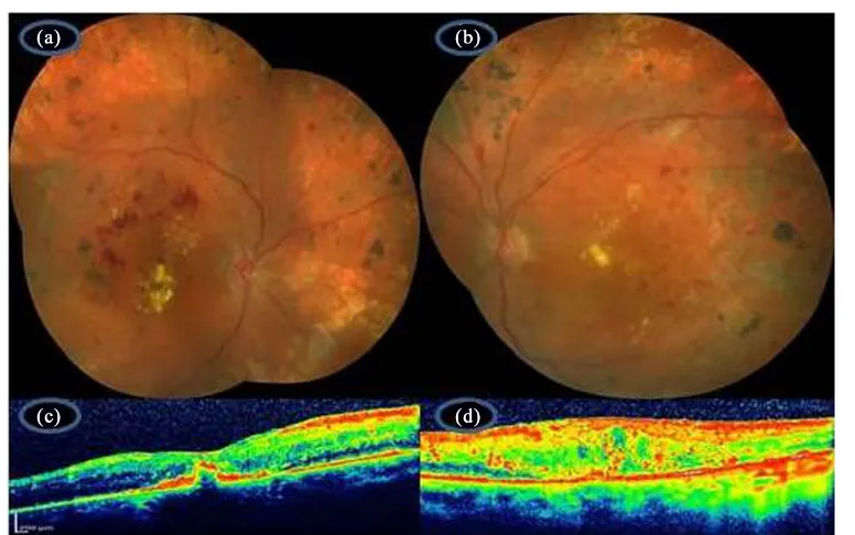
Figure 4. Composite color fundus pictures of the right (a) and left (b) eye showing the scattered laser scars 360 degree and markedly reduced macular hard exudates. OCT scan showing the relatively thinned macula with a small scar formation in OD (c) and relatively improved foveal contour in OS (d).
Figure 5. Right eye. Composite color fundus picture showing the severe circinate exudation temporal to the fovea (a), composite fluorescein angiographic frame demonstrating the severe macular leakage and leakage in association with NVE (b) and the OCT scan depicting the subfoveal serous fluid accumulation (c).
first injection panretinal photocoagulation was commenced and completed in three sessions. Four additional bi-lateral ranibizumab injection were given in a timespan of six months OU. Seven months after the start of therapy, best-corrected visual acuity was 3/10 in OD and 2/10 in OS with a significantly improved fundus appearance (Figure 6(a) and Figure 6(b)).
3. Discussion
[image:5.595.89.540.371.510.2]A. O. Saatci et al.
Figure 6. Right eye. Composite color fundus picture showing the almost dry macula with full panretinal photocoagulation (a); OCT scan showing the normal looking foveal contour (b).
and it was suggested that intravitreal treatment with bevacizumab alone and combined bevacizumab and triam-cinolone acetonide administration resulted in similar visual improvement but to some degree the combined treatment protocol appeared to offer a marginal advantage over patients treated with only bevacizumab injection [8].
Clinicians still continue to elucidate which clinical clues present at the baseline examination are meaningful to predict the success of anti-VEGF monotherapy in eyes with DME. Some authors documented that morphologi-cal edema type might predict the therapeutic outcome and help the clinicians to choose the best treatment alter-native [9]-[12]. In a group of 65 eyes of 48 patients treated with three monthly 1.25 mg bevacizumab injections, the treatment effect was assessed according to baseline OCT pattern of the edema [11]. Eyes with SRD and cys-toid macular edema responded less when compared to eyes with diffuse macular thickening in regard to central foveal thickening and visual outcome at the sixth month of treatment. Seo et al. [12] prospectively evaluated the outcome of intravitreal ranibizumab treatment in 55 eyes with DME based on the morphologic pattern of edema. Best-corrected visual acuity was significantly worse in eyes with SRD after 12 months than that of other types of macular edema. Moreover, disruption of the photoreceptor integrity at the baseline was more frequently ob-served in eyes with SRD. Thereby visual outcome was much poorer.
im-plant for the same purpose. However, only a prospective study will ascertain the place of simultaneous treatment with anti-VEGF agents and dexamethasone implant over mono anti-VEGF agents in eyes with severe DME at least for the kickoff of therapy.
References
[1] Otani, T., Kishi, S. and Maruyama, Y. (1999) Patterns of Diabetic Macular Edema with Optical Coherence Tomogra-phy. American Journal of Ophthalmology, 127, 688-693. http://dx.doi.org/10.1016/S0002-9394(99)00033-1
[2] Sonoda, S., Sakamoto, T., Yamashita, T., et al. (2014) Retinal Morphologic Changes and Concentrations of Cytokines in Eyes with Diabetic Macular Edema. Retina, 34, 741-748. http://dx.doi.org/10.1097/IAE.0b013e3182a48917
[3] Alkuraya, H., Kangave, D. and Abu El-Asrar, A.M. (2005) The Correlation between Optical Coherence Tomographic Features and Severity of Retinopathy, Macular Thickness, and Visual Acuity in Diabetic Macular Edema. International Ophthalmology, 26, 93-99. http://dx.doi.org/10.1007/s10792-006-9007-8
[4] Noma, H.,Mimura, T., Yasuda, K. and Shimura, M. (2014) Role of Inflammation in Diabetic Macular Edema. Oph-thalmologica, 232, 127-135. http://dx.doi.org/10.1159/000364955
[5] Dong, N., Xu, B., Chu, L. and Tang, X. (2015) Study of 27 Aqueous Humor Cytokines in Type 2 Diabetic Patients with or without Macular Edema. PloS One, 10, e0125329. http://dx.doi.org/10.1371/journal.pone.0125329
[6] Sohn, H.J., Han, D.H., Kim, I.T., et al. (2011) Changes in Aqueous Concentrations of Various Cytokines after İntravitreal Triamcinolone versus Bevacizumab for Diabetic Macular Edema. American Journal of Ophthalmology,
152, 686-694. http://dx.doi.org/10.1016/j.ajo.2011.03.033
[7] Bressler, S.B., Ayala, A.R., Bressler, N.M., et al. (2016) Persistent Macular Thickening after Ranibizumab Treatment for Diabetic Macular Edema with Vision İmpairment. JAMA Ophthalmology, 134, 278-285.
http://dx.doi.org/10.1001/jamaophthalmol.2015.5346
[8] Liu, X., Zhou, X., Wang, Z., et al. (2014) Intravitreal Bevacizumab with or without Triamcinolone Acetonide for Di-abetic Macular Edema: A Meta-Analysis of Randomized Controlled Trials. Chinese Medical Journal, 127, 3471- 3476. [9] Wu, P.C., Lai, C.H., Chen, C.L. and Kuo, C.N. (2012) Optical Coherence Tomographic Patterns in Diabetic Macula
Edema Can Predict the Effects of İntravitreal Bevacizumab İnjection as Primary Treatment. Journal of Ocular Phar-macology and Therapeutics, 28, 59–64. http://dx.doi.org/10.1089/jop.2011.0070
[10] Kim, M., Kim, Y. and Lee, S.J. (2015) Comparison of Aqueous Concentrations of Angiogenic and Inflammatory Cy-tokines Based on Optical Coherence Tomography Patterns of Diabetic Macular Edema. Indian Journal of Ophthal-mology, 63, 312-317. http://dx.doi.org/10.4103/0301-4738.158069
[11] Kim, M., Lee, P., Kim, Y., et al. (2011) Effect of İntravitreal Bevacizumab Based on Optical Coherence Tomography
Patterns of Diabetic Macular Edema. Ophthalmologica, 226, 138-144. http://dx.doi.org/10.1159/000330045
[12] Seo, K.H., Yu, S.Y., Kim, M. and Kwak, H.W. (2016) Visual and Morphologic Outcomes of Intravitreal Ranibizumab for Diabetic Macular Edema Based on Optical Coherence Tomography Patterns. Retina, 36, 588-595.