http://dx.doi.org/10.4236/ijcm.2013.412099
Antimicrobial Assay of Chlorhexidine-Wetted Textile
Napkins for Surgical Site Disinfection in Ocular Surgery
Amir Reza Daneshmand Eslami
Department of Ophthalmology, National Medical Academy of Postgraduate Education, Kiev, Ukraine. Email: danshmann@gmail.com
Received October 29th, 2013; revised November 25th, 2013; accepted December 6th, 2013
Copyright © 2013 Amir Reza Daneshmand Eslami. This is an open access article distributed under the Creative Commons Attribution License, which permits unrestricted use, distribution, and reproduction in any medium, provided the original work is properly cited.
ABSTRACT
Background: As a new intraoperative disinfection method, chlorhexidine-wetted textile napkins have been employed in order to cover the upper and lower eyelid edges, eyelid skin, eyelashes, lid margins and palpebral conjunctiva during phacoemulsification cataract extraction. This study was conducted to compare the antimicrobial activity of textile nap- kins before and after their use. Methods: This study evaluated 80 textile napkins wetted with 0.02% aqueous solution of chlorhexidine. All textile napkins were divided into groups. The study group consisted of 60 used textile napkins which were collected from 29 patients (30 eyes) at the end of phacoemulsification, and the control group included 20 unused sterile textile napkins. Antimicrobial assay was performed by means of measuring the growth inhibition zones of the standard or clinical isolate strains under the textile napkins on the surface of agar media. Results: The number of textile napkins and the diameter of the growth inhibition zones (mm) in the study group and in the control group relat-ing to gram-positive, gram-negative, and fungi were: 24/31 vs. 8/31, 32/30 vs. 8/30, and 4/30 vs. 4/30. The diameter of the growth inhibition zones of gram-positive bacteria was more than other investigated microorganisms. In the growth inhibition zones, exogenous microorganism colonies were not found. Conclusion: Antimicrobial activity of textile nap-kins wetted with 0.02% aqueous solution of chlorhexidine against gram-positive bacteria is more than gram-negative bacteria and fungi, and is preserved to the end of the phacoemulsification.
Keywords: Chlorhexidine; Endophthalmitis; Levofloxacin; Phacoemulsification; Surgical Site Disinfection
1. Introduction
Post-operative endophthalmitis after cataract surgery is a rare but serious and potentially blinding complication [1,2]. The sources of ocular bacterial contamination are commonly the conjunctival sac and the eyelids [3,4]. Different methods of antimicrobial prophylaxis are used to prevent the post-operative infectious complications. The main idea of these methods is to eliminate the tran-sient organism and reduce the resident flora to as low level as possible [5].
Since 2005, a new disinfection method in order to prevent microbial contamination of surgical wound dur-ing intraocular surgery has been employed in Ukraine. The components of the method are sterile textile napkins, and an antiseptic for instance, 0.02% aqueous solution of chlorhexidine (CHG) [6].
The purpose of this work is to compare the antimicro- bial activity of textile napkins wetted with 0.02% CHG
before and after their use during phacoemulsification cataract extraction.
2. Materials and Methods
2.1. Patients
standard eye drops to dilate the pupil. Retrobulbar anes- thesia was performed in addition to the preoperative ap- plication of ocular compression.
2.2. Surgical Site Disinfection
During the preoperative procedure used chlorhexidine formulations were two-fold as follows. Firstly, 0.05% alcohol-based chlorhexidine solution which was prepared by 1:400 dilution of 20% CHG (Chlorhexidine, 1,6-di(4’- chlorophenyl-diguanido)hexane) using 70% ethanol for disinfection. Secondly, 0.02% CHG which was prepared by 1:1000 dilution of 20% CHG using sterile purified water. The solutions were prepared in the hospital labo-ratory with the potential concerns about quality control and safety for these solutions as their sterility, stability of pH, and shelf life. Antiseptic eye skin surface treatment was carried out twice with two fresh sterile cotton swabs soaked with 0.05% alcohol-based chlorhexidine in 2 min interval by circular movements outwards (for the left eye—clockwise, for the right—on the contrary). The brow, upper and lower eyelids, eyelashes, and adjacent forehead, nose, cheek and temporal orbital area were scrubbed for 5 min before surgery and meticulous drap- ing was used to isolate the effected eye. Prior to begin- ning of surgery, the upper and lower eyelid edges, eyelid skin, eyelashes, lid margins and palpebral conjunctiva were covered with two textile napkins (Calico fabric), prepared by a punch press and sterilized by autoclave in the hospital laboratory by flushing them with 1 ml of 0.02% CHG.After two min the ocular surface was vig- orously rinsed with 5 ml balanced salt solution of So- dium Chloride (BSS).
Preventing CHG toxicity carried out according to the following approaches: firstly, choosing Calico fabric which CHG has ability to connect with [5], secondly, preparing the textile napkin diameter 30 mm, thickness 1 mm that totally absorbs 6 drops of 0.02% CHG, thirdly, meticulous batting, showering the ocular surface and the textile napkins just before beginning the surgery with 5 ml BSS and lastly, using the constant flow of BSS from an anterior chamber maintainer during phacoemulsifica- tion. Thereby the expected residue of 0.02% CHG in the operative area is much lower than toxicological dose of CHG [7,8] likewise, the emergence of entry residual CHG into the anterior chamber is avoided.
2.3. Surgical Technique
Phacoemulsification cataract extraction was performed through a superior clear corneal incision with implanta- tion intraocular lens using an injector system. During the operations additional antimicrobial treatment was not given. On completion of the surgery after sealing corneal incisions, two textile napkins were withdrawn from each
eye by sterile forceps and placed in separate sterile Petri dish with slightly opened lid. Then the lid was secured with scotch tape and immediately sent to the Microbiol- ogy Laboratory. After a subconjunctival injection of 0.5 ml dexamethasone and ceftriaxone, the conjunctival cav- ity was washed with 2 ml BSS, this was followed by in- stilling one drop of 0.5% levofloxacin ophthalmic solu- tion into the inferior culs-de-sac and the eye was closed with aseptic eye gauze pad. The duration of operation ranged from 20 ± 5 min. Operations were completed without surgery-related complications.
2.4. Study Sample
60 used textile napkins collected at the end of the surgery were employed as study group. 20 unused sterile textile napkins wetted with 6 drops of 0.02% CHG and em- ployed as control group. Microorganism strains were used to compare antimicrobial activity of unused textile napkins with used ones wetted with 0.02% CHG. The source of the microorganism strains for this work was formed by pure clinical patterns isolated from ocular post-operative infection, laboratory strains and standard culture collection types of microorganisms. The micro-organism strains were employed for the successful ac-complishment of the study viz. gram-positive bacteria, gram-negative bacteria and fungal species.
2.5. Antimicrobial Activity Assay
growth inhibition zones of corresponding microorganism under the textile napkins and around them in mm. A fur- ther step was the identification of possible appearance of microbial colonies in the growth inhibition zones of the microorganisms. For this purpose the textile napkins from the surface of nutrient media were removed, and each Petri dish was re-incubated in thermostat at 37˚C during 24 h (Petri dishes with Candida species for 48 h at 35˚C).
2.6. Ethics Approval
Approval for accessing the patient health records was obtained from the local research ethics committee. In- formed consent was obtained from each patient. The study protocol and the safety and efficacy of the inter- ventions were explained to all of the participants prior to their enrolment.
2.7. Statistical Analysis
The Mann-Whitney U-test (for small samples) and the Z-test were used to compare the studied variables. A P value less than 0.05 was considered statistically signifi- cant.
3. Results
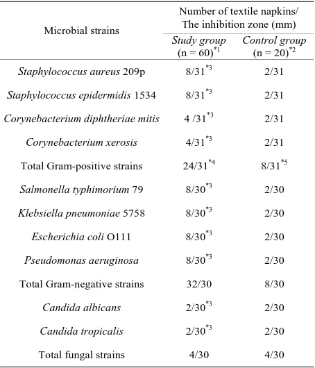
The results of this study are summarised in Table 1. The diameter of the growth inhibition zones of micro- organism strains under the textile napkin and around them varies for different microorganisms after incubation. When removing the textile napkins from the surface of nutrient agar media, no colony was seen on their inner side. As well as after re-incubation of these media in the field of the growth inhibition zones of the microorganism strains identified at the first stage, no colonies of micro- organism strain and exogenous microbial contamination were formed. No ocular toxicity associated with CHG was noted.
4. Discussion
[image:3.595.310.537.111.376.2]Local preoperative antibiotic prophylaxis, sterile prepara- tion of the skin surrounding the surgical eye with Povi- done-Iodine 10%, meticulous draping of the lids and eye- lashes [10], and instillation of Povidone-Iodine 5% onto the ocular surface at least 3 - 5 min prior to surgery are widely used in many countries. Many authors believe these measures have the most long-standing and highest quality which is the evidence of their efficacy [11-14]. With this approach, bacteria were isolated in the conjunc- tival sac or in the anterior chamber at the beginning of cataract extraction and on its completion [15,16]. Thus the duration of action of these preoperative regimens is disputed. Numerous studies have reported that after ap-
Table 1. Comparison of antimicrobial activity of textile napkins wetted with 0.02% aqueous chlorhexidine.
Number of textile napkins/ The inhibition zone (mm) Microbial strains
Study group
(n = 60)*1 Control group(n = 20)*2
Staphylococcus aureus 209p 8/31*3 2/31
Staphylococcus epidermidis 1534 8/31*3 2/31
Corynebacterium diphtheriae mitis 4 /31*3 2/31
Corynebacterium xerosis 4/31*3 2/31
Total Gram-positive strains 24/31*4 8/31*5
Salmonella typhimorium 79 8/30*3 2/30
Klebsiella pneumoniae 5758 8/30*3 2/30
Escherichia coli O111 8/30*3 2/30
Pseudomonas aeruginosa 8/30*3 2/30
Total Gram-negative strains 32/30 8/30
Candida albicans 2/30*3 2/30
Candida tropicalis 2/30*3 2/30
Total fungal strains 4/30 4/30
*1
Used textile napkins, *2Unused textile napkins, *3P = 1 compared with the control group. *4P = 0 compared with the total Gram-negative strains, *4P = 0 compared with the total fungal strains. *5P = 0 compared with the total Gram-negative strains; *5P = 0.001 compared with the total fungal strains.
plying the various invasive techniques, including the addition of antibiotics in the balanced salt solution, or their infusion into the anterior chamber, the material taken from anterior chamber at the conclusion of opera- tions or postoperative endophthalmitis cases may release bacteria sensitive to these antibiotics [17]. A disadvan- tage of the intracameral antibiotics has potential risks, such as toxic anterior segment syndrome secondary to dilution errors [18].
cavity. Thus, for a certain period of time, the oral cavity becomes a CHG reservoir, which prolongs its chemical activity in preparations [22]. The ocular surface (cornea and conjunctiva) is also negatively charged [23,24], and the paracellular space is more permeable to cations than to anions at physiological pH [25,26] consistently the ocular surface may also become a CHG reservoir, which prolongs its chemical activity in preparations. This phe- nomenon depathogenizes the ocular surface flora intra- operatively and postoperatively but bacteriological cul- ture from the ocular surface may continue to be positive.
Results showed that the use of BSS during cataract surgery did not totally decrease the antimicrobial activity of the textile napkins due to CHG absorbed in the fibers of certain textile, particularly cotton, and consistently re- sisted removal by washing [5]. Thereby, the textile nap- kin virtually serves as a sustained release reservoir of CHG during phacoemulsification. Moreover, equal di- ameter of the growth inhibition zones before and after phacoemulsification cataract extraction also indicates that 0.5% levofloxacin ophthalmic solution instilled pre- operatively is not an antagonist of 0.02% CHG. Addi- tionally, antimicrobial activity of both unused textile nap- kins and used textile napkins, wetted with 0.02% CHG against gram-positive bacteria is more than gram-nega- tive (Table 1), that is comparable to certain reports [5, 20]. Since microbial flora under the textile napkins mixed with flora of the conjunctival sac and the lid mar- gin, the result of microbial culture from the conjunctival sac after withdrawing the textile napkins on completion of the surgery is disputed. Test strains colonies were not seen after incubation of Petri dishes on the surface and inner side of used textile napkins. Likewise in the growth inhibition zones after re-incubation, exogenous microor- ganism colonies that could contaminate used textile nap- kins during surgery were not seen.
5. Conclusion
The antimicrobial activity assay of CHG-wetted textile napkins indicates a persistent antimicrobial effect of a residue of CHG in the textile napkins during phacoe- mulsification cataract extraction. Intraoperatively, the isolation of the lid edges with 0.02% CHG-wetted textile napkins in combination with a preoperative antibacterial prophylaxis for instance 0.5% levofloxacin ophthalmic solution reliably prevents microbial surface contamina- tion during ophthalmic surgery.
6. Acknowledgements
The author appreciates Ms. Sepideh Elahi of INOVA Fairfax Hospital, VA, USA for assistance with statistical analysis. This paper was derived from the author’s doc- toral dissertation.
REFERENCES
[1] G. A. Peyman, J. T. Paque and H. I. Meisels, “Post- operative Endophthalmitis: A Comparison of Methods for Treatment and Prophylaxis with Gentamicin,” Ophthal-
mic Surgery, Vol. 6, No. 1, 1975, pp. 45-55.
[2] A. Pathengay, M. Khera, T. Das, S. Sharma, D. Miller and H. W. Flynn Jr., “Acute Postoperative Endophthal- mitis Following Cataract Surgery: A Review,” Asia-Pa-
cific Journal of Ophthalmology, Vol. 1, No. 1, 2012, pp.
35-42. http://dx.doi.org/10.1097/APO.0b013e31823e574b
[3] C. B. Walker and C. M. Claoue, “Incidence of Conjunc- tival Colonisation by Bacteria Capable of Causing Post- Operative Endophthalmitis,” Journal of the Royal Society
of Medicine, Vol. 79, No. 9, 1986, pp. 520-521.
[4] M. G. Speaker, F. A. Milch, M. K. Shah, W. Eisner and B. N. Kreiswirth, “Role of External Bacterial Flora in the Pathogenesis of Acute Postoperative Endophthalmitis,”
Ophthalmology, Vol. 98, No. 5, 1991, pp. 639-649.
http://dx.doi.org/10.1016/S0161-6420(91)32239-5
[5] G. W. Denton, “Chlorhexidine,” In: S. S. Block, Ed.,
Disinfection, Sterilization, and Preservation, 5th Edition.
Lippincot Williams & Wilkins, Philadelphia, 2001, pp. 321-336.
[6] N. M. Sergienko, Y. N. Kondratenko, N. V. Chumak and A. Daneshmand, “Results of Prophylaxis of Bacterial Endophthalmitis in Cataract Surgery,” Proceedings of
The Joint Congress of SOE/AAO, Geneva, June 2011, p.
51.
[7] K. Green, V. Livingston, K. Browman and D. S. Hull, “Chlorhexidine Effects on Corneal Epithelium and Endo- thelium,” Archives of Ophthalmology, Vol. 98, No. 7, 1980, pp. 1273-1278.
http://dx.doi.org/10.1001/archopht.1980.01020040125020
[8] N. L. Burstein, “Preservative Cytotoxic Threshold for Benzalkonium Chloride and Chlorhexidine Digluconate in Cat and Rabit Corneas,” Journal Investigative Oph-
thalmology & Visual Science, Vol. 19, No. 3, 1980, pp.
308-313.
[9] A. W. Bauer, W. M. Kirby, J. C. Sherris and M. Turck, “Antibiotic Susceptibility Testing by Standardized Single Disc Method,” American Journal of Clinical Pathology, Vol. 45, No. 4, 1966, pp. 493-496.
[10] L. D. Perry and C. Skaggs, “Preoperative Topical Anti- biotics and Lash Trimming in Cataract Surgery,” Oph-
thalmic Surgery, Vol. 8, No. 5, 1977, pp. 44-48.
[11] S. J. Isenberg, L. Apt, R. Yoshimori, C. Pham and N. K. Lam, “Efficacy of Topical Povidone-Iodine during the First Week after Ophthalmic Surgery,” American Journal
of Ophhalmology, Vol. 124, No. 1, 1997, pp. 31-35.
[12] T. J. Liesegang, “Use of Antimicrobials to Prevent Post- operative Infection in Patients with Cataracts,” Current
Opinion in Ophthalmology, Vol. 12, No. 1,2001, pp. 68-
74.
http://dx.doi.org/10.1097/00055735-200102000-00012
http://dx.doi.org/10.1016/S0161-6420(01)00899-5
[14] A. Safar and M. C. Dellimore, “The Effect of Povidone Iodine Flush versus Drops on Conjunctival Colonization before Intravitreal Injections,” International Ophthal-
mology, Vol. 27, No. 5, 2007, pp. 307-312.
http://dx.doi.org/10.1007/s10792-007-9073-6
[15] D. S. Hughes and R. J. Hill, “Infectious Endophthalmitis after Cataract Surgery,” British Journal of Ophthalmol- ogy, Vol. 78, No. 3, 1994, pp. 227-232.
http://dx.doi.org/10.1136/bjo.78.3.227
[16] M. N. Shi, H. J. Guan, Y. J. Ye, M. S. Chen and C. T. Yu, “Comparative Study of Gentamycin and Povidone-Iodine on Eliminating the Conjunctival Bacteria,” Chinese Jour-
nal of Ophthalmology, Vol. 44, No. 12, 2008, pp. 1098-
1102.
[17] J. Feys, A. Salvanet-Bouccata, J. P. Emond and A. Dub- lanchet, “Vancomycin Prophylaxis and Intraocular Con- tamination during Cataract Surgery,” Journal of Cataract
Refractive Surgery, Vol. 23, No. 6, 1997, pp. 894-897.
http://dx.doi.org/10.1016/S0886-3350(97)80250-7
[18] J. C. Lloyd and R. Braga-Mele, “Incidence of Postopera- tive Endophthalmitis in a High-Volume Cataract Sur- gicentre in Canada,” Canadian Journal Ophthalmology, Vol. 44, No. 3, 2009, pp. 288-292.
http://dx.doi.org/10.3129/i09-052
[19] G. A. Peyman, P. L. Lee and D. V. Seal, “Endophthal- mitis,” Martin Dunitz, London, 2004.
[20] M. B. Hamill, M. S. Osato and K. R. Wilhelmus, “Ex- perimental Evaluation of Chlorhexidine Gluconate for Ocular Antisepsis,” Antimicrobial Agents and Chemo-
therapy, Vol. 26, No. 6, 1984, pp. 793-796.
http://dx.doi.org/10.1128/AAC.26.6.793
[21] M. T. Suller and A. D. Russell, “Antibiotic and Biocide Resistance in Methicillin-Resistant Staphylococcus Aur- eus and Vancomycin-Resistant Enterococcus,” Journal of
Hospital Infection, Vol. 43, No. 4, 1999, pp. 281-291.
http://dx.doi.org/10.1016/S0195-6701(99)90424-3
[22] E. Dabrowska, M. Letko, W. Roszkowska-Jakimiec, R. Letko and J. Sadowski, “Effect of Chlorhexidine Mou- thrinse on Cathepsin C Activity in Human Saliva,” Ad-
vances in Medical Sciences, Vol. 51, No. 1, 2006, pp. 96-
99.
[23] T. Gershanik, S. Benzeno and S. Benita, “Interaction of a Self-Emulsifying Lipid Drug Delivery System with the Everted Rat Intestinal Mucosa as a Function of Droplet Size and Surface Charge,” Pharmaceutical Research, Vol. 15, No. 6, 1998, pp. 863-869.
http://dx.doi.org/10.1023/A:1011968313933
[24] S. Tamilvanan, “Oil-in-Water Lipid Emulsions: Implica- tions for Parenteral and Ocular Delivering Systems,” Pro-
gress in Lipid Research, Vol. 43, No. 6, 2004, pp. 489-
533. http://dx.doi.org/10.1016/j.plipres.2004.09.001
[25] Y. Rojanasakul and J. R. Robinson, “Transport Mecha- nisms of the Cornea: Characterization of Barrier Permse- lectivity,” International Journal of Pharmaceutics, Vol. 55, No. 2-3, 1989, pp. 237-246.
http://dx.doi.org/10.1016/0378-5173(89)90047-1
[26] J. Liaw, Y. Rojanasakul and J. R. Robinson, “The Effect of Drug Charge Type and Charge Density on Corneal Transport,” International Journal of Pharmaceutics, Vol. 88, No. 1-3, 1992, pp. 111-124.