Journal of Chemical and Pharmaceutical Research, 2015, 7(7):1013-1019
Research Article
CODEN(USA) : JCPRC5
ISSN : 0975-7384
Evaluation the effect of different types of separating medium on color stability
of heat-cure acrylic resin “A comparative study”
Ola Al-Jubouri
1,2and Abbas Azari
3*1Prosthodontics Techniques Department, College of Health & Medical Technology, Middle Technical University, Baghdad, Iraq
2Department of Prosthodontics Technology, School of Dentistry, Tehran University of Medical Sciences, Tehran-Iran
3
Department of Removable Prosthodontics, School of Dentistry, Tehran University of Medical Sciences, Tehran, Iran
_____________________________________________________________________________________________
ABSTRACT
During acrylic resin processing, the mold must be separated from the surface of the gypsum to prevent liquid resin from penetrating into the gypsum, and water from the gypsum seeping into the acrylic resin. Also, the relatively rough surface of gypsum mould may be penetrated by acrylic denture base resin and adhere to it, to prevent this, a separating medium must be employed. In this study, cold mold seal, tinfoil &olive oil were used as separating medium, and evaluated the effect of these materials on color stability of clear hot cure acrylic resin. Twenty one acrylic resins samples are prepared falling in three main groups (seven samples of each group) according to the separating medium that used and compared the effect this materials on color stability of heat clear acrylic curing by conventional method, the first group used cold mold seal, the second group used tin-foil, and third group used olive oil as separating medium. From the result obtained by t-test shows that there is a statistically no-significant difference (p> 0.05) have been noticed between cold-mold seal, tin- foil, and olive oil samples on color stability of heat cure acrylic resin. It can be concluded that olive oil can be considered as a satisfactory separating medium of clear heat cured acrylic denture base resins, especially because it is easy to get, easy to use and cheap. Also the results have shown that tin foil is still the best separating medium that is used due to easily open the flask during defklasking procedure and removed the samples from stone (most of failure was adhesive ) ,followed by cold mold seal and finally olive oil (most of failure was cohesive ) .
Keywords: Denture base, Hot cures acrylic resin, color stability, Separating medium.
_____________________________________________________________________________________________
INTRODUCTION
Separating medium is a coat layer applied to a surface to prevent a second surface from adherence to the first
surface, or a material, usually applied on an impression to facilitate cast. [1]
If the mold surface is not coated by a separating material, a layer of gypsum will penetrate with polymer remains
attached to the surface of the denture, it will be very difficult to remove [2], improperly denture’s contouring and
produce ugly, poorly fitting denture base [3]. So that, the resin surface must be carefully protected from the gypsum
surface during it’s processing in the mold cavity for two reasons:
1-Any water impregnated into the resin from the gypsum during processing will absolutely affect the rate of polymerization and the color of the resin, the denture produced will defect readily because of the stresses formed by the evaporation of water after the processing, particularly if the resin is not cross-linked.
after polymerization process, with the result that it will be substantially impossible to separate the investing material
from the resin [2, 4].
Processing mold cavity with a liquid tin-foil substitute to seal the porosity present in the investing material (artificial stone) represent as successful method of preventing the gypsum surface from absorbing the liquid of acrylic resin. Tin-foil substitute is available and used successfully if the residual wax are thoroughly cleaned from the mold cavity
before applying a tin-foil, while processing of tin-foiling a waxed denture is tedious and time consuming [4, 5].
The objective of this study is evaluating and comparing the effect of different types of separating medium (alginate mold seal (cold-mold seal), tin foil olive oil) on color stability of the processed acrylic resin denture base and evaluating the mode of failure “adhesive & cohesive” between acrylic resin &stone during remove the acrylic from the stone mold when use this types of separating medium and the null hypothesis is there is no significant
differences among the tested groups.
Pilot study:
A pilot study concerning laboratory procedures had been conducted to eliminate as much as possible the limitations and problems that could be faced during the study. The color stability is tested by spectrophotometer machine in order to evaluate and compare of the color stability of the acrylic denture base materials and to find any changes occurred during the procedure. One specimen (2cm X 3cm X0.2mm) width, length and thickness respectively is constructed of the clear heat cure acrylic resin processed by a conventional method is used in color stability test
according to [6].
Three methods are present to produce this size of specimen as follow: 1- Use base plate wax.
2- Glass or metal pattern.
3- Make sample with suitable dimension by mixing 5ml of monomer with 0.001 g of polymer then immersion this of dioxin solution to dissolve this sample and obtain correct dimension.
In this study the second method was used because the first method need more time (wax elimination for each sample),the third method also need more time, expensive, and the sample in the end have flexible properties may be effect in produce the correct thickness of the sample.
Methods:
General Preparation of the Acrylic Resin Denture Base Samples:
Four different glasses patterns are constructed with correct dimensions to save time and effort. Dimensions and shape of each glass pattern are made according to the required tests. Twenty one samples were prepared and divided into three groups (according to the types of separating medium that used in curing process), seven samples for each group; group 1 (G1) used cold-mold seal (U.K by MR. Dental supplies LTD., England), group 2 (G2) used tin foil (Zinnfoile, Dentaurum Pforzheim, Starke: 0.01mm, Germany), group 3 (G3) used olive oil (Zer, Zer YAG SAN Vetic .A.S. TS 341, Turkey), Fig. (1).
Figure (1): Diagram illustrates the distribution of the samples
The conventional flasking technique was followed in the mold preparation. Each glass pattern was coated with the separating medium (cold mold seal). Slurry stone (type III model, Elite Model thixortropic, Zermack- Italy) was prepared according to the manufacturers’ instruction (W/P ratio is 25 ml/100g) and poured into the lower half of the dental flask, then immerse the glass pattern in the slurry stone. After setting of the stone, a layer of cold mold seal
21
Samples from clear heat cure acrylic resin
7 samples GROUP1 Cold mold seal
G1
7 samples GROUP 2 Tin-foil
G2
7 samples GROUP 3 Olive oil
separating medium was applied on the stone surface and another layer of stone was poured into the second half of the flask. The lid was adapted in its place and the flask was allowed to stand for one hour, after that the flask was opened and the glass pattern was removed. Then the separating media was applied, in case of using cold mold seal or olive oil, (2cc) was measured with a disposable syringe and applied onto the stone surface in each half of the flask, using a fine brush. While when tin-foil separating medium was used, it was adapted to the stone surface in
each half of the flask, with fingers then the mould was ready for packing with acrylic dough [7, 8]. Clear heat cured
acrylic resin (Vertex, Germany) was mixed according to manufacture’s instructions (3:1) by volume. The liquid was poured in a clean and dry mixing vessel followed by slow addition of powder, stirred the mixture with wax knife and left at room temperature in a closed container until reaching the dough stage. The acrylic resin dough was packed into the mould, and then the two halves of the flask were closed together and placed under press with gradual application of pressure to allow even flow of the dough throughout the mold space. The pressure was then released, the flask was opened and removed the excess material (flash) surrounding the mould space with a sharp knife. A second trial closure was performed, the two halves of the flask were finally closed under pressure to intimate metal
contact had been established and left under press (20 bars) for 5 min. before clamping was done [9] then the flasks
were left for curing.
Curing was accomplished by inserting the clamped flask in a water bath (Maestra, Spain) and processed by heating at 74ºC for about an hour and half. The temperature was then a raised to the boiling point for 30 minutes according to [6]. After complete curing process, the flask was allowed to cool slowly at room temperature for 30 minutes.
Followed by, complete cooling of the flask with tap water for 15 minutes before deflasking. The acrylic patterns were then removed from the mold, remove all flashes of acrylic by an acrylic bur followed by 120-grain size sand
paper with continuous water-cooling (to prevent over heating) according to [10] in order to get correct sample.
Polishing was performed using bristle brush and rag wheel with pumice using dental lathe polishing machine with low speed (1500 rpm) (Degauss, Germany), the final measurements of the samp les were obtained using the ruler and metal caliper device. The specimen were immersed in distilled water until were tested for about 24 hour in darker container to prevent the effect of this specimen by any external light that may be effect on polymerization of
acrylic and lead to effect on physical and chemical properties of acrylic [5].
Color stability (Testing Procedure):
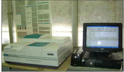
The color stability is usually tested by the spectrophotometer (UV-visible, EL 04113001, varian, U.S.A.), it is an ideal instrument in studying color stability of acrylic resins, since this instrument give analysis measurement that can be calculated and compared statically. In this study, the surface of the specimens has been measured by
spectrophotometer which appeared in computer monitor [11], Fig. (2).
Figure (2): UV-visible –spectrophotometer (EL 04113001, varian, U.S.A.)
[image:3.595.195.407.466.589.2]For the sake of testing, a carton slides was constructed with the size of (5 X 8cm) width and length respectively, a window was made in the center of the slide with the dimensions of (1.5 X 2.5cm) width and length, Fig (3); specimens were placed inside the carton slide and tested for color stability by spectrophotometer which consists of two parts the first part is for specimens testing, and the second part is the computer monitor was the readings can be seen, Fig. (2). after that the number of color stability in each specimens were measured and the means were recorded
[12].
The color stability data obtained were recorded and submitted to statistical analysis.
The mode of failure of each sample was evaluated, two methods were followed:
1 -Visual examination (unaided eye) by two persons in order to establish the percentage of residual stone on acrylic specimens (ten persons).
2- Under traveling light microscope (TM 10 Measuring Microscope, Titan, Buffalo, NY, USA), (4X magnification)
[13].
In this study, the first method was used because the sample was large and can be detected by visual examination.
RESULTS
1-Color stability test
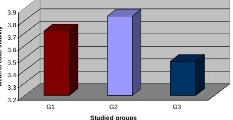
Table (1) mentioned the mean, stander deviation, stander error and maximum &minimum value for each group.
Table (1): Mean distribution of color stability among studied groups
Groups Mean Stander deviation Stander error Maximum Minimum Cold mold Seal (G1) 3.7157 0.13587 0.5136 3.85 3.55
Tinfoil (G2) 3.8357 0.46882 0.17720 4.53 3.29
Olive oil(G3) 3.4729 0.17997 0.06802 3.76 3.27
Table (2) represented t-test for comparing between each two groups together.
Table (2): T-test for comparison between each two groups
Study groups t-test Correlation Sig. G1 G2 -0.665 0.079 No sig. P(>0.5)
G3 2.674 -0.140 No sig. P(>0.5)
G2 G3 2.670 0.728 No sig. P(>0.5)
Graphical presentation by bar chart between the mean color stability of the three groups, shown in Fig (4).
Figure (4): Mean distribution of color stability among studied groups
2- Mode of Failure
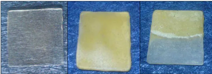
After completion deflasking procedure, all the specimens were examined visually in an effort to determine the type of bonding failure. Three types of bonding failure were presented at the debonding site (between acrylic samples& stone):
• Adhesive failure: Failure occurred at the interface between acrylic resin and stone, Fig. (5).
• Cohesive failure: Failure occurred in the bulk of the stone, Fig. (6).
• Cohesive / adhesive failure (mixed failure): A combination of both (adhesive and cohesive failure), Fig. (7), the
percentage of mode of failure of three groups shown in table (5).
3.2 3.3 3.4 3.5 3.6 3.7 3.8 3.9
M
e
a
n
o
f
c
o
lo
r
s
ta
b
il
it
y
G1 G2 G3
[image:4.595.181.416.485.606.2]Fig. (5): Adhesive failure Fig. (6): Cohesive failure Fig. (7): Mixed failure
Table (3): The percentage of Mode of failure of three groups
Studies groups Mode of failure % Adhesive Cohesive Mixed
Cold mold seal G1 71.43 0 28.57 Tin –foil G2 100 0 0 Olive oil G3 0 85.71 14.29
DISCUSSION
Among other factors, separating medium represented as one of the important factors in dental prosthesis success, due to its effect on the physical and mechanical properties of the processed acrylic denture base materials. Porosity can be prevented by controlling both the heating rate and maintaining pressure on the specimen during polymerization. Monomers lose may be occurred by: (a) Evaporation during mixing. (b) Absorption into the plaster during packing. Both can be prevented, by first mixing in narrow vessels with lids and second by painting all plaster
surfaces with some suitable separating medium [14 ,15] .
Keng et al. 1979, pointed out that the choice of separating medium is the most important factor influencing the fairness of the resin. The cured denture resin may show some opacity, depending upon the type of resin used
because of the alginate mold seal film is not completely water-eliminator [16]. Pure methyl methacrylate is almost as
transparent when cured against the alginate mold seal film as against the tin foil. However, some slight opacity may
be found with some acrylic copolymers [4].
For separating a dental stone, many materials are used when dentures are being invested in a flask, petroleum jelly is
one of commonly used separating medium [17, 18].
Neillet al. 1968 [19] stated that although tin-foil has been largely superseded by the substitute sodium alginate this is
a matter of expediency, a layer of tin-foil is to be preferred for the following reasons:
1) It is impermeable to water-the layer of water vapor between the acrylic and the plaster during processing leads to
a lack of a clear surface in the acrylic.
2) It allows a complete polymerization of the surface of the acrylic; this may be inhibited by the sodium alginate
layer.
The routine use of tin-foil as a separating medium may minimize some of the discomfort that some patients may feel
from the slight movement of a rough surface on the mucosa ensure dentures with smoother fitting surface [15]. Unlike
tin-foil, the alginate mould seals do not seal the pores in the mold surface sufficiently to stop permeability of water
vapor from the plaster into the denture [2].
However; the process of tin-foiling is tedious and time-consuming unless a technician has had extensive practical
experience with it, many authorities still consider that tin-foil is the best separating medium [3].
Mohammed [20] shown that tin-foil is still the best separating medium that is used due to the best properties obtained
when using tin-foil as a separating medium.
Craig and Powers stated that for many years tin-foil considers as an acceptable separating medium, and the researchers found out by visual examination which is done by the spectrophotometer that the absorption of color
[image:5.595.125.467.71.190.2]Muhsin and Abboud found that tin-foil is the best separating medium concerning porosity in heat and cold-cure acrylic specimens, while the porosity are comparable in both olive oil and cold mould seal lined specimens of heat
and cold-cure acrylic [21]. And Kadir et al., show that tin foils provide the best smooth surface (lowest surface
roughness) of acrylic after curing than detry isolate and cold mold seal with a high statistically difference during
studying the effect of separating medium on the roughness of acrylic denture base [22].
While Al-Khayyat found that the surface roughness and water solubility of both base materials (acrylic and nylon
denture base) are significantly higher when using cold mold seal than glycerin [23].
In this study, tin –foil, cold mold seal and olive oil were used as a separating medium in the process of curing clear heat acrylic resins denture base.
1- Color stability Examination
From table (2), the results obtained showed statistically no significant difference between samples processed using cold-mold seal separating medium; tin foil medium and those samples processed using olive oil separating media.
This results disagreement with [8, 24] they found high values of hardness was obtained in heat – cured acrylic resin
and cold cure resin processed using tin foil separating media when compared with those samples processed using
cold – mold seal and olive oil separating media, also disagreement with [6] found that tin foil produces extremely
high clarity, but alginate mold separator causes surface blanching.
Also the main disadvantage when used the olive oil during the processing the acrylic denture base material that this
materials need more time for drying, this result agreement with [25, 26] mentioned that, colloidal tin foil substitutes,
liquid soap and mineral oil are also used, a tin foil substitute and liquid soap take considerable time to dry and oil never dries, leaving on oily film.
The results of this study accepted the null hypothesis that there is no significant difference among the tested groups and that concluded the tin foil, cold mold seal, and olive oil can be used safely as separating medium for clear hot cure acrylic resin against stone during flasking.
2- Mode of Failure
In table (3) and Fig. (5),(6),(7) ,the results showed the most adhesive failure that found in acrylic denture base resin processed against tin-foil as separating medium (100%) followed by cold mold seal (71.43%) adhesive failure, and (28.57%) mixed failure ,but no showed adhesive failure in group 3 ,the most failure was cohesive, this mean the best result that obtain when used the tin –foil as separating medium, followed by cold mold seal, because when used olive oil as separating medium the stone material was adhesive with all sample and difficult open the flask and
difficult removed the stone from the specimen. This finding agreed with [7, 16, 20, 21, 22].
They stated that examination of the specimens revealed that acrylic resin when processed against tin foil substitute showed blanching and fogging and in some cases adherence of plaster particles. This may be related to that tin foil substitutes films which are permeable to water allowing it to pass from the gypsum mold and enter the acrylic resin
denture base during the process, this explanation agreed with [7, 27].
CONCLUSION
On the basis of the results, the following conclusions can be drawn:
1- Statistically no significant difference in mean color stability value was observed of different groups with different types of separating medium and that accepted the null hypothesis and that concluded the tin foil, cold mold seal, and olive oil can be used safely as separating medium for clear hot cure acrylic resin against stone during flasking . 2 - In general ,the mode of failure are mostly adhesive in tin –foil group ,followed by cold mold seal group ,but the mode of failure are mostly cohesive in olive oil group ,this mean the best out come from tin-foil groups because it easily open the flask and easily remove the specimen from stone .
REFERENCES
[1] Glossary of Prosthodontic terms, J prosthet. Dent. Mosby Co., 8 ed., 94 (1), 1-83; 2005.
[2] Anderson JN., Applied dental materials, Mosby Co., London, St. Louis, 5th, 1972.
[3] Rahan AO; Heartwell CM., Textbook of complete dentures, 1993.
[6] ADA., American Dental Association Specification No.12 for denture base polymer, council on dental materials and devices, Chicago, 1999.
[7] AL-Musawi RM., Evaluation of glycerin as a separating medium for processing acrylic denture base materials,
(Comparative study).M. tech. Thesis, Dental Technologies, College of Health and Medical Technology, Iraq. 2005.
[8] Al-taai A.Z., Evaluation of olive oil as separating medium and its effect on some physical and mechanical
properties of processed acrylic resin denture base (A comparative study), M. tech. Thesis, Dental Technologies,
College of Health and Medical Technology. Iraq. 2006.
[9] Abdul- Karim JF., Evaluation of some mechanical properties of acrylic denture base materials relined with
different denture reline materials, M. Sc. Thesis, University of Baghdad, College of Dentistry, Iraq. 2001.
[10] ADA., American Dental Association Specification No.12 for denture base polymers, Council on dental
materials and devices, Chicago, 1975.
[11] Addy M. & Prayiton S.W., J Periodontal, 51:39-43, 1980.
[12] Sheen S.R. & Harrison A., J Prosthetic Dent, 84(6), 594-601, 2000.
[13] Galindo D.F., Ercoli C., Crafer G.N., Tallents R.H. and Moss M.E., J Prosthet Dent, 85: 88-98, 2001.
[14] Harcourt JK., Lautenschlager EP. and Molnar EJ., J Dent Res, 48:61-66, 1969.
[15] Osborne J., Wilson HJ. and Mansfield MA., “Dental technology and materials for students”', Black well
scientific publications, 1979.
[16] Keng SB., Cruickshanks-Boyd DW. and Davies EH., J.oral Rehabil., 6(4): 327-35, 1979.
[17] Martinelli N., Dental laboratory, Mosby Co., London, St Louis, 1975.
[18] Asher ES., Evans JH. and Wright RF., J Prosthet Dent, 85(4): 415-417, 2001.
[19] Neill DJ., Nairn RI., Bristol JW. and Sons LT., Complete denture prosthetics, 1968.
[20] Mohammed N. H., Iraqi national journal of nursing specialties, 24 (2): 94-108, 2011.
[21] Muhsin S.A. and Abboud E.Z.”Kufa Med. Journal, 11(2): 267-73, 2008.
[22] Kadir S. K., Hama-Saeed N. H., Hanna B. A. Iraqi national journal of nursing specialties, 25 (1): 89- 94, 2012.
[23] Al-Khayyat F. N. M. T. Karbala J. Med, 6 (2): 1675- 83, Dec 2013.
[24] Parr G. R., Rueggebery F. A., J. Prosth. Dent., 88(2): 139-144, 2002.
[25] Renner Rp., Blackeslee RW. and Shiu A., Dental technology, theory and practice, Mosby, London, St. Louis,
134, 1980.
[26] Craig R G., Restorative dental material, Mosby Co., London, St. Louis, 1989.