Left Atrial Function Following Surgical Ablation
of Atrial Fibrillation: Prospective Evaluation Using Dual-Source
Cardiac Computed Tomography
Joon Bum Kim,
1* Dong Hyun Yang,
2* Joon-Won Kang,
2Sung-Ho Jung,
1Suk Jung Choo,
1Cheol Hyun Chung,
1Jae-Kwan Song,
3and Jae Won Lee
1Departments of 1Thoracic and Cardiovascular Surgery, 2Radiology, and 3Cardiology, Asan Medical Center, University of Ulsan College of Medicine, Seoul, Korea.
Received: March 20, 2014 Revised: July 31, 2014 Accepted: August 13, 2014
Corresponding author: Dr. Jae Won Lee, Department of Thoracic and
Cardiovascular Surgery, Asan Medical Center, University of Ulsan College of Medicine, 88 Olympic-ro 43-gil, Songpa-gu, Seoul 138-736, Korea.
Tel: 82-2-3010-3584, Fax: 82-2-3010-6966 E-mail: jwlee@amc.seoul.kr
*Joon Bum Kim and Dong Hyun Yang contributed equally to this work.
∙ The authors have no financial conflicts of interest.
© Copyright:
Yonsei University College of Medicine 2015 This is an Open Access article distributed under the terms of the Creative Commons Attribution Non-Commercial License (http://creativecommons.org/ licenses/by-nc/3.0) which permits unrestricted non-commercial use, distribution, and reproduction in any medium, provided the original work is properly cited.
Purpose: The Maze procedure has shown excellent efficacy in the elimination of
atrial fibrillation (AF); however, little is known about the quality of functional re-covery in the left atrium (LA) following successful sinus rhythm conversion by the Maze procedure. Materials and Methods: We prospectively enrolled 12 pa-tients (aged 52.5±10.1 years, 1 female) with valvular AF undergoing mitral valve surgery combined with the Maze procedure. Parameters of LA function in three anatomic compartments [anterior, posterior, and LA appendage (LAA)] were eval-uated using electrocardiography-gated dual-source cardiac CT at one month and at six months after surgery. Twelve subjects matched by age, gender, and body sur-face area served as controls. Results: At one month after surgery, ejection fraction (EF) and emptying volume (EV) of the LA were 14.9±7.4% and 21.3±9.7 mL, re-spectively, and they were significantly lower than those of the control group (EF, 47.9±11.2%; EV, 46.0±10.7%; p<0001). These values did not significantly change throughout late periods (p=0.22 and 0.21, respectively). Functional contributions of the anterior, posterior, and appendage compartments (EV of each compartment/ overall EV) were 80.4%, -0.9%, and 20.5%, respectively, for those with LAA preservation (n=6); 100.1%, -0.1%, and 0% for those with LAA resection (n=6;
p<0.05); and 62.2%, 28.2%, and 9.7% in the control subjects (p<0.001).
Conclu-sion: Contractile functions of the LA significantly decreased after the Maze
proce-dure. Functional contributions of three compartments of the LA were also altered. The influence of LAA preservation on postoperative LA functions needs to be evaluated through studies of larger populations.
Key Words: Atrial fibrillation, ablation, surgery, left atrial systolic function
INTRODUCTION
reduc-general health checkup at the Asan Medical Center during the same study period.
This study was approved by our Institutional Review Board, and informed consent was obtained from all patients.
Surgical procedure
The AF ablation was performed using an argon-based flexi-ble CryoAblation system, SurgiFrost (Medtronic, Minneap-olis, MN, USA), in all patients. After snaring down the su-perior and inferior vena cavae, right atrial ablation was preformed through an oblique right atriotomy on a beating heart. Endocardial ablation comprised a cavo-tricuspid isth-mus isolation lesion and a line from the isthisth-mus lesion to the superior vena cava.
After aortic cross-clamping, MV exposure was obtained through a LA incision. The LA ablation was done endocar-dially before the MV procedure, which included a single box lesion for pulmonary veins isolation, a line from the pulmonary isolation lesion to the LAA, and another line from the pulmonary isolation lesion to the MV annulus, posteriorly. Additional epicardial coronary sinus ablation was done on the opposite side of the MV annular lesion. LA size was reduced by resection of redundant atrial tissue between the inferior pulmonary veins and the posterior mi-tral annulus.12 In cases of concomitant tricuspid valve re-pair, 26‒30-mm commercially available rings (Edwards MC3 Ring, Edwards Lifesciences, Irvine, CA, USA) were used for annuloplasty.
CT evaluations of LA functional parameters
At one and six months after surgery, assessment of LA functional parameters was performed using ECG-gated second generation dual source cardiac CT (Somatom Defi-nition Flash, Siemens, Erlangen, Germany) for those with restored sinus rhythm (expected number of patients at each point=10). Tube voltage and tube current-time product were adjusted by body size, and scan parameters were as follows: tube voltage, 80‒120 kV; tube current-time prod-uct, 185‒380 mAs; collimation, 128×0.6 mm; spatial reso-lution, 0.3 mm; gantry rotation time, 280 sec; and a tempo-ral resolution of 75 ms. A bolus of 70‒90 mL of contrast agent was administered at a rate of 4.0 mL/s, followed by 40 mL of saline chaser. The scan delay was determined by the bolus-tracking method (region of interest, ascending aorta; attenuation threshold level, 100 HU; scan delay, 8 seconds). Retrospective ECG-gated spiral scan was done, and ECG-based tube current modulation was applied to re-tion in thromboembolic events, and even improved
surviv-al.1-7 However, little is known about how much of atrial contractile functional recovery can be achieved by success-ful sinus rhythm conversion with the Maze procedure. This is partly attributable to difficulties in evaluating atrial func-tions with conventional imaging modalities, such as echo-cardiography, in which the complex three-dimensional morphology of the atria hinders comprehensive evaluation of contractile functions. Furthermore, deciding between whether to preserve or amputate the left atrial appendage (LAA) during surgical ablation of AF remains controver-sial, and due to a lack of reliable scientific evidence, man-agement thereof is left to the discretion of the surgeon. Re-cently, multi-slice cardiac computed tomography (CT) has been reported as a promising tool for the assessment of left atrium (LA) volume and function in various clinical set-tings,8-11 making comprehensive evaluation of the atria pos-sible in the setting of surgical ablation of AF.
In this study, we aimed to evaluate postoperative left atri-al function using duatri-al-source cardiac CT in patients with valvular atrial fibrillation undergoing the Maze procedure. For secondary analysis, we sought to evaluate the impact of LAA resection versus preservation during the surgery on postoperative LA function.
MATERIALS AND METHODS
This prospective observational study included patients with valvular AF who were scheduled for mitral valve (MV) sur-gery combined with the Maze procedure. Exclusion criteria were as follows: 1) contraindicated to receive radiocontrast media; 2) severe LV dysfunction [LV ejection fraction (EF) <40%]; 3) valvular dysfunction≥a moderate degree, other than functional tricuspid insufficiency; 4) presence of LA thrombus; 5) requirement of multi-vessel coronary bypass, aortic replacement, or correction for congenital heart de-fects; and 6) history of previous cardiac surgery.
ually on the volume rendering image.
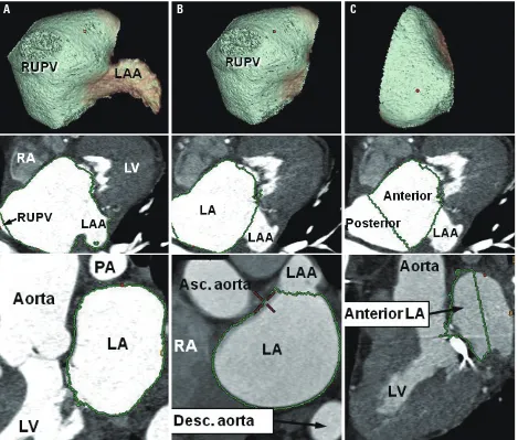
The LAA was defined as the volume from the tip of the LAA to the ridge between the left superior pulmonary vein and the LAA.8,13 The LA proper was defined by subtracting the LAA volume from the total LA volume (Fig. 1B). The boundary that divides the LA proper into the anterior and posterior compartments was defined as follows: the anterior border of the ostia of the four pulmonary veins was identi-fied, and the curvilinear line that connected them was delin-eated. The anterior compartment was defined as the volume from the anterior wall of the LA to the boundary of the os-tia of the four pulmonary veins (Fig. 1C). The posterior compartment was defined by subtracting the anterior com-partment from the LA proper. The following parameters were evaluated in each compartment of the LA, pre- and post-operatively: 1) maximum LA volume, 2) minimum duce radiation doses. Four-dimensional multiphase cardiac
CT data were transferred to an external workstation (Ad-vantage Workstation Server, GE, Milwaukee, MI, USA) for post-processing.
We obtained three-dimensional volumetric data for the entire LA (Fig. 1A). The total LA volume was defined as the volume from the MV to the posterior wall of the LA, and it was divided into the anterior, posterior, and LAA compartments (Fig. 1). To divide each component of LA, we generated volume rendering images of LA using a dedi-cated semi-automatic tool (Auto Ejection Fraction, GE, Milwaukee, MI, USA) in the workstation. This software provides a threshold-based LA segmentation function. By rotating the volume rendering images of LA, anatomic landmarks of each LA component were evaluated, and LAA and posterior compartments of LA were clipped
man-Fig. 1. Three-dimensional measurement of LA volumes using volume-rendering imaging. Green demarcation lines indicate the extents of measurement for each compartment including (A), entire LA (right-anterior view); (B) LAA-extracted LA (right-anterior view); and (C) anterior compartment of the LA (right lat-eral view). RUPV, right upper pulmonary vein; LA, left atrium; LAA, left atrial appendage; RA, right atrium; LV, left ventricle; PA, pulmonary artery.
[image:3.595.58.527.302.701.2]the patients are summarized in Table 1. There was no oper-ative mortality or morbidity.
Overall postoperative rhythm statuses during the study period are detailed in Table 2. Overall, 10 patients (83.3%) were free of AF and off anti-arrhythmic medications after 6 months of surgery. The incidences of postoperative brady-arrhythmia (1 vs. 1) and AF (2 vs. 2), as well as require-ment of anti-arrhythmic medications (1 vs. 2), were not dif-ferent between the LAA resection and preservation groups. The baseline demographic variables of the control group were well matched with those of the study group, including age (53.2±11.4 years vs. 52.5±10.1 years, p=0.88), BSA (1.90±0.14 m2 vs. 1.83±0.14 m2, p=0.30), and gender distri-bution (females: 1/12 vs. 1/12, p=1.0).
LA functional parameters
Except for two patients who showed AF, 10 patients (with sinus rhythm) underwent cardiac CT evaluation at one month after surgery. EF of the entire LA chamber was 16.8±6.3%, and this value was significantly lower than that in the control subjects (47.9±11.2%, p<0.001). An EV of 21.3±9.7 mL was also significantly lower than that in the control subjects (46.0 control subjects 10.7 mL, p<0.001). At six months after surgery, CT data from 11 patients who showed restored sinus rhythm were obtained. CT data at 6 months were available in 11 patients who showed restored sinus rhythm, and all LA functional parameters showed similar results as those at one month (Figs. 2 and 3). Details of LA functional parameters at six months for each com-partment are shown in Table 3. EV, EF, and functional con-tribution (EV of a compartment/EV of total LA chamber) for the anterior compartment were significantly higher than those for the posterior compartment (p<0.001 for all val-ues), in which the contributions of the posterior compart-ment to overall LA contraction were trivial (-0.9 to -0.1% of overall function) (Fig. 4). Although LAA contributed a significant proportion to overall LA contractile function (20.5±14.2%) in patients with LAA preservation, overall EV and EF of the LA were not significantly different be-tween LAA preservation and resection groups (p=0.58 and 0.86, respectively).
During the postoperative period, echocardiographic eval-uation revealed LV EF of 53.5±4.6% (range, 53‒64%; p= 0.86), LV systolic dimension of 35.6±4.5 mm (range, 30‒44 mm; p=0.007), and LV diastolic dimension of 53.8±4.4 mm (range, 45‒61 mm; p=0.38), showing compa-rable LV systolic function with that in preoperative settings. LA volume, 3) EV, and 4) EF. The maximum LA volume
was defined as the volume at the end of atrial diastole and immediately before MV opening. The minimum LA vol-ume was defined as the volvol-ume after atrial emptying at the end of atrial systole. The EV was calculated as the differ-ence between the maximum and minimum LA volume. The EF was calculated as the EV divided by the maximum LA volume.14
Rhythm follow-up
The rhythm follow-up protocol in this study was as fol-lows: 1) continuous monitoring during intensive care unit stay, 2) daily ECG and electroatriography during hospital stay, 3) a 24-hr Holter monitoring before hospital discharge if the patient maintained “free-of-AF status,” and 4) snap EKGs at 1 month, 3 months, and 6 months post-surgery.
Statistical analysis
A power calculation estimated that approximately 10 pa-tients were required to achieve a minimum power of 90% to detect a 20% difference in LA EF between the study and normal subjects with a 90% confidence interval, given that normal LA EF is 42.9±6.6% (42.89%).8 Assuming a study dropout rate of 15%, including those who did not restore si-nus rhythm or were contraindicated to undergo postopera-tive CT evaluation (i.e., postoperapostopera-tive renal failure), around 12 study subjects were required.
Categorical variables are presented as frequencies and percentages, and were compared using Fisher’s exact test. Continuous variables are expressed as mean±SD or medi-ans with ranges, and were compared using the Wilcoxon signed rank test or the Mann-Whitney U test, as appropri-ate. Changes in maximal and minimal LA volumes were in-vestigated using repeated-measures ANOVAs.
The authors had full access to and take full responsibility for the integrity of the data. All authors have read and agree to the manuscript as written.
RESULTS
Baseline profiles of patients and postoperative rhythm status
Maze procedure was effective in restoring sinus rhythm in valvular AF patients; however, even after successful recov-ery of sinus rhythm, functional recovrecov-ery of the LA was sig-nificantly lower than that in normal subjects. Additionally,
DISCUSSION
[image:5.595.57.526.147.572.2]In this prospective observational study, we found that the
Table 1. Baseline Characteristics of Patients
Variables, n (%) or mean±SD All patients (n=12) LAA preservation (n=6) LAA resection (n=6)
Age, yrs 52.5±10.1 55.7±8.8 49.3±11.1
Female gender 1 (8.3) 1 0
Diabetes mellitus 2 (16.7) 2 0
Hypertension 1 (8.3) 1 0
Previous stroke 1 (8.3) 1 0
NYHA functional class III or IV 4 (33.3) 2 2
AF type
Paroxysmal 1 (8.3) 1 0
Persistent 4 (33.3) 2 2
Longstanding persistent 7 (58.3) 3 4
Fine (<1 mm) AF wave pattern 3 (25.0) 2 1
MV disease
Rheumatic 7 (58.3) 3 4
Degenerative 5 (41.7) 3 2
Echocardiography
LV EF, % 54.2±7.8 53.7±7.2 54.7±9.0
LVIDs, mm 39.3±5.4 38.0±4.1 40.7±6.6
LVIDd, mm 56.6±7.9 53.7±6.2 59.5±9.0
LA diameter, mm 57.4±9.7 54.3±8.6 60.5±10.5
MV pathology
Predominant regurgitation 8 (66.7) 4 4
Predominant stenosis 3 (25.0) 1 2
Mixed steno-regurgitation 1 (8.3) 1 0
TR>mild 5 (41.7) 1 4
Peak TR pressure gradient, mm Hg 49.0±18.4 41.8±14.9 55.0±20.1
MV surgery
Repair 3 (25.0) 3 0
Replacement* 9 (75.0) 3 6
TV ring annuloplasty 7 (58.3) 3 4
Minimally invasive approach 6 (50.0) 3 3
Aortic clamping time, min 133.4±57.1 147.2±73.5 119.7±36.2
CPB time, min 157.2±33.8 164.8±31.6 149.5±37.1
NYHA, New York Heart Association; AF, atrial fibrillation; MV, mitral valve; LV, left ventricle; EF, ejection fraction; LVIDs, left ventricular systolic dimension; LVIDd, left ventricular diastolic dimension; LA, left atrium; TR, tricuspid regurgitation; CPB, cardiopulmonary bypass; LAA, LA appendage.
*All were mechanical prosthetic implantation.
Table 2. Postoperative Cardiac Rhythm Status
Variables, n (%) Intraoperative rhythm Baseline rhythm (hospitalization) (hospitalization)*Episode of AF Rhythm at 1 month 1 month AAD at Rhythm at 6 months 6 monthsAAD at
NSR 10 (83.3) 10 (83.3)
4 (33.3)
10 (83.3) 3 (25.0) 11 (91.7) 1 (8.3)
Junctional 1 (8.3) 1 (8.3) 0 0
CAVB 1 (8.3) 1 (8.3) 0 0
AF 0 0 2 (16.7) 1 (8.3)
[image:5.595.55.526.633.709.2]The LA is a complex three-dimensional structure that comprises three anatomic compartments of three different embryologic origins: the anterior LA, the posterior or venous LA, and the LAA.13,20 Since the structure of the LA differs in each compartment, and, consequently, the functional contri-bution of each compartment may also differ, it is important to understand the functional nature of each LA compartment during AF ablation. Although several research groups report-ed on the roles of the LAA as a contractile component and as a main source of thrombus formation in AF patients,21,22 functional assessments of the other compartments have not been reported for surgical ablation of AF.
In the present study, LA booster function, represented by EF and EV, was much lower than that of normal subjects re-ported in the literature.8 This finding may be attributable to several reasons: first, since cryoablation does not produce sharp injuries, and typically tends to freeze surrounding tis-sue together, transient or permanent collateral cryo-damages may have occurred in adjacent atrial tissue, resulting in func-tional damage to the atrium. Furthermore, as the funcfunc-tional contribution of the posterior compartment was nearly zero in this study, isolation of the posterior compartment by cre-we found that LAA contributed to a significant proportion
of LA contractile function after the Maze procedure, al-though a valid comparison between preservation and resec-tion procedures could not be made because of the small number of patients in this study.
[image:6.595.73.540.67.219.2]Although surgical ablation of AF is primarily targeted to-ward the elimination of AF, its benefits secondarily extend to improving hemodynamic performance by the incorporation of synchronous atrial kicking into the cardiac output.1,15,16 To date, however, the extent of functional recovery of the atria by a successful Maze procedure is poorly understood. This is mainly attributed to a limited ability to precisely evaluate LA geometry and booster function with the use of conven-tional two-dimensional echocardiography due to the com-plex three-dimensional shape of the LA.17-19 Furthermore, since diastolic inflow measurements, such as E/A ratio, are heavily affected by several factors,17,18 assessment by echo-cardiography holds intrinsic limitations in evaluating LA contractile function. In these regards, multi-slice cardiac CT has been reported to be a precise tool for the assessment of LA volume and booster functions in various clinical set-tings.8-11
Fig. 2. Changes in overall LA volume at each time point. (A) Maximal volumes. (B) Minimal volume. LA, left atrium; LAA, left atrial appendage.
Fig. 3. LA contractile function at one and six months after surgery. (A) LA emptying volume. (B) LA ejection fraction. LA, left atrium; LAA, left atrial appendage. 0
0 0
0 100
10 10
100 200
20
20 200 300
30 400
40 30
300
Maximal LA volume, mL
LA emptying volume, mL LA ejection fraction, %
Minimal LA volume, mL
Baseline 1 month Baseline
1 month 1 month
1 month 6 months
6 months 6 months
6 months A
A B
B
p>0.99
p=0.63
p=0.75
p=0.77
p=0.63
p=0.23
p=0.80
p=0.61
Trend: p=0.021
1 mon vs. 6 mon: p=0.22 1 mon vs. 6 mon: p=0.22 Trend: p=0.035 LAA preservation
LAA resection
LAA preservation
LAA resection LAA preservation LAA resection LAA preservation LAA resection
[image:6.595.74.540.247.398.2]through which to better preserve posterior compartment function of the LA.
Finally, but most importantly, the differences in LA func-tion between the subject patients and normal subjects most likely stem from differences in baseline pathologic features of the atrial tissue. Longstanding volume and/or pressure overload to the LA secondary to MV diseases results in substrate changes in the atrium, the pathologic process of ating a box lesion may have adversely affected LA
[image:7.595.56.528.82.401.2]func-tion. For instance, an EF of the posterior compartment of -0.9 to -0.1% is quite distinctive, compared with normal populations, in which it is reported to be around 20‒40% (28.2% for control subjects in this study).8 In these regards, creating separate left and right pulmonary-vein isolation le-sions connected by another linear lesion, rather than a large posterior box lesion, may be a more reasonable option
Table 3. Postoperative Left Atrial Functional Parameters at 6 Months (11 Patients with Sinus Rhythm)
Variables, mean±SD Study group (n=11) Control group (n=12) p value
Preservation (n=6) Resection (n=5) p value Total LA chamber
Maximum volume, mL 144.9±29.4 167.6±61.6 0.47 97.6±19.0 <0.001
Minimum volume, mL 125.6±31.7 144.0±65.8 0.58 51.6±18.2 <0.001
EV, mL 19.4±7.4 23.6±12.5 0.58 46.0±10.7 <0.001
EF, % 13.9±6.8 16.1±8.6 0.86 47.9±11.2 <0.001
Anterior compartment
Maximum volume, mL 70.0±25.6 97.2±57.9 0.23 58.9±13.1 0.074
Minimum volume, mL 54.5±24.7 78.0±57.0 0.47 30.4±10.3 0.001
EV, mL 15.5±5.6 20.2±5.9 0.27 28.5±7.6 0.002
EF, % 23.2±8.9 24.8±12.6 >0.99 48.9±10.6 <0.001
Functional contribution, %* 80.4±12.1 100.1±7.3 0.020 62.2±8.5 <0.001
Posterior compartment
Maximum volume, mL 58.4±11.6 70.5±18.6 0.27 30.0±7.7 <0.001
Minimum volume, mL 58.6±11.6 70.5±16.8 0.47 17.1±6.6 <0.001
EV, mL -0.2±0.8 0.0±14.4 0.58 12.9±5.3 0.001
EF, % -0.3±1.4 0.0±17.1 0.86 43.8±14.8 <0.001
Functional contribution, %* -0.9±4.5 -0.1±7.3 0.86 28.2±9.9 0.001
LA appendage
Maximum volume, mL 16.5±7.1 0 0.004 8.7±2.7 0.88
Minimum volume, mL 12.5±7.0 0 0.004 4.2±3.2 0.95
EV, mL 4.0±3.3 0 0.004 4.5±2.2 0.028
EF, % 26.5±20.8 0 0.004 54.0±20.6 0.001
Functional contribution, %* 20.5±14.2 0 0.004 9.7±3.7 0.35
EV, emptying volume; EF, ejection fraction; LA, left atrium.
*Functional contribution is calculated as 100%×(EV of each compartment)/(EV of total LA chamber).
Fig. 4. Booster functional contribution of each left atrial chamber (anterior, posterior, and LAA) at one and six months after surgery. LAA, left atrial append-age.
-0.2 -0.2 -0.2
0.0 0.0
0.2
0.0 0.2 0.3
0.4 0.4
0.6 0.6 0.6
0.8 0.8 0.8
1.3 1.2 1.2
1.1 1.0 1.0
Functional contribution Functional contribution Functional contribution
1 month 6 months 1 month 6 months 1 month 6 months
p=0.037
p=0.34
p=0.040
p=0.020
p=0.80
p=0.018
LAA preservation LAA resection
LAA preservation
LAA resection LAA preservation LAA resection
[image:7.595.60.527.437.590.2]diomyopathy with resultant LV systolic dysfunction.26,27 Tachycardia-induced cardiomyopathy is commonly thought to be associated with ventricular rates of 100 to 120/min in chronic phases. Even in patients with AF and well-con-trolled resting heart rates, minimal activity may frequently induce rapid ventricular beating, which can be associated with the development of tachycardia-induced cardiomyopa-thy.28 Meanwhile, elimination of AF has been suggested to restore LV function by blocking this pathological process. For instance, a group reported that surgical AF ablation in patients with lone AF improved LV ejection and reduced LV end-diastolic diameter, which appeared immediately after surgery and was maintained through late periods.29 Recently, Nedios showed via the evaluation of 69 patients with im-paired LV function undergoing catheter AF ablation that both the elimination of AF and baseline heart rate following the procedure were independent predictors of a marked im-provement in LV function.
Although the number of patients enrolled in this study was calculated based on pertinent statistical assumptions, it was relatively small, limiting the drawing of robust conclu-sions. Also, since the subject population in this study was confined to patients with valvular AF, the study results may not be generalizable to patient with AF of other etiologies. The heterogeneity of surgical procedures in this study (e.g., sternotomy vs. mini-thoracotomy and valve replacement vs. repair) may be another limitation of this study. Also, the control subjects were fundamentally different from those receiving MV surgery in the presence of AF, and therefore, the comparisons of outcomes between these two groups may not be fair. Nevertheless, the study design was set as such because the primary aim of this study was to describe the nature of functional recovery in the LA after the Maze procedure in comparison to normal LA function. Finally, while the sample size was too small to evaluate the effects of resection versus preservation of the LAA in this study, our results could be of use as references for further studies to determine sample sizes required to fairly compare the ef-fects of LAA resection in the setting of Maze surgery.
In conclusion, LA function was significantly decreased after successful sinus rhythm conversion by the Maze pro-cedure in patients with valvular AF undergoing MV sur-gery, which persisted throughout late postoperative period. Conclusions regarding preservation versus resection of the LAA in terms of clinical, cardiac rhythm-, and atrial func-tional outcomes await further studies involving larger popu-lations.
which involves atrophy of atrial myocytes, excessive inter-stitial fibrosis, and overall thinning and dilatation of the atri-al watri-all.23,24 These pathologic changes very likely limit the extent of atrial booster function, even with optimal preserva-tion of the atrial contractile component during the Maze pro-cedure. This hypothesis is supported by a previous study that quantitatively assessed LA booster function after the Maze procedure.9 In the cited study, the authors examined 14 consecutive patients undergoing concomitant MV sur-gery and the Maze procedure, and found out that LA EF was only 15% following the Maze procedure, which was significantly smaller than that of patients with sinus rhythm who underwent coronary bypass (p=0.004). The EF of 15% corresponds well to the result of the present study.
Management of LAA during surgical ablation of AF is an-other important issue to address. Since ectopic foci of AF may arise from the LAA,25 its resection may improve the ef-ficacy of AF elimination. Furthermore, given that a signifi-cant proportion of patients receiving the Maze procedure fail to achieve AF elimination and that the LAA is the most com-mon site of thrombus formation and embolization, amputa-tion of the LAA may reduce the risk of thromboembolic complications or the requirement for long-term anticoagula-tion therapy.27 Preservation of the LAA, on the other hand, may benefit patients by conserving LA transport function, since the LAA has been reported to play significant roles in the contribution of overall atrial contractile function.8
The secondary objective of the present study, therefore, was to compare resection versus preservation of the LAA in regards to postoperative LA function. Since the number of patients for this study had been determined based on the as-sessment of overall LA function in comparison with a nor-mal reference value, the sample size was too snor-mall to draw a statistically meaningful comparison between the two groups (resection vs. preservation of LAA). Nevertheless, we found out that the contribution of the LAA to overall LA booster function was 21.5±16.1% for patients who underwent LAA preservation, which was significantly higher than that of the posterior compartment (p<0.001). We believe that these findings open the possibility for superior functional restora-tion of the LA by preservarestora-tion of the LAA, and this hypoth-esis needs to be tested by further studies. LA functional val-ues presented in this study may help calculate sample sizes required to compare LA functions in prospective random-ized studies.
car-atrial fibrillation. Ann Thorac Surg 2011;92:1397-404.
13. Kerut EK. Anatomy of the left atrial appendage. Echocardiogra-phy 2008;25:669-73.
14. Abhayaratna WP, Seward JB, Appleton CP, Douglas PS, Oh JK, Tajik AJ, et al. Left atrial size: physiologic determinants and clini-cal applications. J Am Coll Cardiol 2006;47:2357-63.
15. Middlekauff HR, Stevenson WG, Stevenson LW. Prognostic sig-nificance of atrial fibrillation in advanced heart failure. A study of 390 patients. Circulation 1991;84:40-8.
16. Pritchett EL. Management of atrial fibrillation. N Engl J Med 1992;326:1264-71.
17. Feinberg MS, Waggoner AD, Kater KM, Cox JL, Lindsay BD, Pérez JE. Restoration of atrial function after the maze procedure for patients with atrial fibrillation. Assessment by Doppler echo-cardiography. Circulation 1994;90(5 Pt 2):II285-92.
18. Yashima N, Nasu M, Kawazoe K, Hiramori K. Serial evaluation of atrial function by Doppler echocardiography after the maze procedure for chronic atrial fibrillation. Eur Heart J 1997;18:496-502.
19. Jessurun ER, van Hemel NM, Kelder JC, Defauw JA, Brutel de la Rivière A, Ernst JM, et al. The effect of maze operations on atrial volume. Ann Thorac Surg 2003;75:51-6.
20. Anderson RH, Cook AC. The structure and components of the atrial chambers. Europace 2007;9 Suppl 6:vi3-9.
21. Cox JL. The central controversy surrounding the interventional-surgical treatment of atrial fibrillation. J Thorac Cardiovasc Surg 2005;129:1-4.
22. Lönnerholm S, Blomström P, Nilsson L, Oxelbark S, Jideus L, Blomström-Lundqvist C. Effects of the maze operation on health-related quality of life in patients with atrial fibrillation. Circulation 2000;101:2607-11.
23. Henry WL, Morganroth J, Pearlman AS, Clark CE, Redwood DR, Itscoitz SB, et al. Relation between echocardiographically deter-mined left atrial size and atrial fibrillation. Circulation 1976;53: 273-9.
24. Bailey GW, Braniff BA, Hancock EW, Cohn KE. Relation of left atrial pathology to atrial fibrillation in mitral valvular disease. Ann Intern Med 1968;69:13-20.
25. Lin WS, Tai CT, Hsieh MH, Tsai CF, Lin YK, Tsao HM, et al. Catheter ablation of paroxysmal atrial fibrillation initiated by non-pulmonary vein ectopy. Circulation 2003;107:3176-83.
26. Shinbane JS, Wood MA, Jensen DN, Ellenbogen KA, Fitzpatrick AP, Scheinman MM. Tachycardia-induced cardiomyopathy: a re-view of animal models and clinical studies. J Am Coll Cardiol 1997;29:709-15.
27. Umana E, Solares CA, Alpert MA. Tachycardia-induced cardio-myopathy. Am J Med 2003;114:51-5.
28. Stulak JM, Dearani JA, Daly RC, Zehr KJ, Sundt TM 3rd, Schaff HV. Left ventricular dysfunction in atrial fibrillation: restoration of sinus rhythm by the Cox-maze procedure significantly improves systolic function and functional status. Ann Thorac Surg 2006; 82:494-500.
29. Nedios S, Sommer P, Dagres N, Kosiuk J, Arya A, Richter S, et al. Long-term follow-up after atrial fibrillation ablation in patients with impaired left ventricular systolic function: the importance of rhythm and rate control. Heart Rhythm 2014;11:344-51.
ACKNOWLEDGEMENTS
This study was supported by Edwards Lifesciences (Irvine, CA, USA).
REFERENCES
1. Ad N, Henry L, Hunt S. The impact of surgical ablation in patients with low ejection fraction, heart failure, and atrial fibrillation. Eur J Cardiothorac Surg 2011;40:70-6.
2. Bando K, Kasegawa H, Okada Y, Kobayashi J, Kada A, Shimoka-wa T, et al. Impact of preoperative and postoperative atrial fibrilla-tion on outcome after mitral valvuloplasty for nonischemic mitral regurgitation. J Thorac Cardiovasc Surg 2005;129:1032-40. 3. Cohn LH, Couper GS, Aranki SF, Rizzo RJ, Kinchla NM, Collins
JJ Jr. Long-term results of mitral valve reconstruction for regurgi-tation of the myxomatous mitral valve. J Thorac Cardiovasc Surg 1994;107:143-50.
4. Cox JL, Ad N, Palazzo T. Impact of the maze procedure on the stroke rate in patients with atrial fibrillation. J Thorac Cardiovasc Surg 1999;118:833-40.
5. Cox JL, Boineau JP, Schuessler RB, Ferguson TB Jr, Cain ME, Lindsay BD, et al. Successful surgical treatment of atrial fibrilla-tion. Review and clinical update. JAMA 1991;266:1976-80. 6. Gillinov AM, Sirak J, Blackstone EH, McCarthy PM, Rajeswaran
J, Pettersson G, et al. The Cox maze procedure in mitral valve dis-ease: predictors of recurrent atrial fibrillation. J Thorac Cardiovasc Surg 2005;130:1653-60.
7. Kim JB, Moon JS, Yun SC, Kim WK, Jung SH, Choo SJ, et al. Long-term outcomes of mechanical valve replacement in patients with atrial fibrillation: impact of the maze procedure. Circulation 2012;125:2071-80.
8. Park MJ, Jung JI, Oh YS, Youn HJ. Assessment of the structural remodeling of the left atrium by 64-multislice cardiac CT: com-parative studies in controls and patients with atrial fibrillation. Int J Cardiol 2012;159:181-6.
9. Yamanaka K, Fujita M, Doi K, Tsuneyoshi H, Yamazato A, Ueno K, et al. Multislice computed tomography accurately quantifies left atrial size and function after the MAZE procedure. Circulation 2006;114(1 Suppl):I5-9.
10. den Uijl DW, Tops LF, Delgado V, Schuijf JD, Kroft LJ, de Roos A, et al. Effect of pulmonary vein anatomy and left atrial dimen-sions on outcome of circumferential radiofrequency catheter abla-tion for atrial fibrillaabla-tion. Am J Cardiol 2011;107:243-9.
11. Kataoka A, Funabashi N, Takahashi A, Yajima R, Takahashi M, Uehara M, et al. Quantitative evaluation of left atrial volumes and ejection fraction by 320-slice computed-tomography in compari-son with three- and two-dimensional echocardiography: a single-center retrospective-study in 22 subjects. Int J Cardiol 2011;153: 47-54.