R E S E A R C H A R T I C L E
Open Access
Effect of the crude extract of
Eugenia uniflora
in
morphogenesis and secretion of hydrolytic
enzymes in
Candida albicans
from the oral cavity
of kidney transplant recipients
Walicyranison Plinio Silva-Rocha
1, Vitor Luiz de Brito Lemos
1, Magda Rhayanny Assunção Ferreira
2,
Luiz Alberto Lira Soares
2, Terezinha Inês Estivalet Svidzisnki
3, Eveline Pipolo Milan
4and Guilherme Maranhão Chaves
1*Abstract
Background:Candida albicansis a diploid yeast that in some circumstances may cause oral or oropharyngeal infections. Yeasts virulence factors contribute for both the maintenance of colonizing strains in addition to damage and cause tissue invasion, thus the establishment of infection occurs. The limited arsenal of antifungal drugs for the treatment of candidiasis turn the investigation of natural products mandatory for the discovery of new targets for antifungal drug development. Therefore, tropical countries emerge as important providers of natural products with potential antimicrobial activity. This study aimed to investigate morphogenesis and secretion of hydrolytic enzymes (phospholipase and proteinase) in the presence of the CE ofEugenia uniflora.
Methods:The isolates were tested for their ability to form hyphae in both solid and liquid media under three different conditions: YPD + 20% FBS, Spider medium and GlcNac and the ability to secrete phospholipase and proteinase in the presence of 2000μg/mL ofE. uniflora.
Results:The CE ofE. uniflorainhibited hypha formation in both liquid and solid media tested. It also impaired hydrolytic enzymes production.
Conclusions:This was the first study to describe the interaction of a natural product with the full expression of three different factors inC. albicans. E. unifloramay be an alternative therapeutic for oral candidiasis in the future.
Keywords:Candida albicans, Oral candidiasis, Kidney transplant recipients, Morphogenesis, Hydrolytic enzymes, Eugenia uniflora
Background
Candida species are the most common human fungal pathogens, whereasCandida albicans is the most preva-lent species isolated in fungal infections of the oral mu-cosa [1-3]. In the human oral cavity, these yeasts colonize from 20 to 80% of adults without evidences of infection [3-5]. However, in some circumstances which involve both pathogen attributes of virulence as well as host immunity;
specific groups of patients, such as those with AIDS, transplant recipients and patients submitted to broad spectrum antibiotics may become susceptible to de-velop oropharyngeal candidiasis [6-8].
Oral candidiasis is a fungal opportunistic infection com-monly observed in kidney transplant recipients, where the prevalence of the disease ranges from 9.4 to 46% in differ-ent group of patidiffer-ents, being C. albicans the most fre-quently isolated species [6,9-13].
C. albicans virulence factors contribute for both the maintenance of colonizing strains and tissue invasion, thus the establishment of infection occurs [14-16]. Among the main virulence attributes of C. albicansare the ability of
* Correspondence:[email protected]
1Laboratório de Micologia Médica e Molecular, Departamento de Análises
Clínicas e Toxicológicas, Universidade Federal do Rio Grande do Norte, Natal, Rio Grande do Norte, Brazil
Full list of author information is available at the end of the article
cells transition from yeast to hyphae, a phenomenon called morphogenesis, which contributes to tissue invasion [17]. Morphogenesis is a well-recognized virulence factor inC. albicans, because mutants unable to form pseudohy-phae or true hypseudohy-phae are attenuated in virulence [18,19].
Merson-davies and Odds [20] proposed a quantitative index based on the shape of the cells to differentiate blas-toconidia, pseudohyphal and true hyphal forms of growth. Yeast cells have a morphology index (MI) between 1.0 to 1.5, pseudo hyphae from 2.5 to 3.4 and true hyphae greater than 3.4 [21]. This index, based on a complex image analysis measurements can be substituted with an approximated score system to generate information on morphology distributions [22,23].
The secretion of hydrolytic enzymes such as phospholi-pases, which degrade glycerolphospholipids of cell mem-branes, is important for host cells destruction [24]. In addition, C. albicans possesses secreted aspartic pro-teinases (Saps), able to target several important substrates, such as albumin, immunoglobulins and extracellular matrix proteins [14,25,26].
The limited arsenal of antifungal drugs for the treat-ment of candidiasis includes mainly polyenes, azoles and echinocandins [27-29]. Nevertheless, these drugs have narrow therapeutic index, low bioavailability, week intes-tinal absorption and serious side effects [30]. In addition, the excessive and indiscriminate use of antifungal agents favors the development of resistant strains [31]. Although C. albicanshas been reported as less resistant to antifun-gal drugs than otherCandidaspecies, resistance to azoles [32] and echinocandins [33] have been reported. There-fore, due to the increase of resistance of pathogenic fungi to antifungal drugs currently used, there is a necessity for the prevention of oral fungal infections and the develop-ment of alternative therapies [34,35].
The use of natural products aiming the treatment of several diseases has increased in the last few years, related to the high diversity of plants distributed around the planet [36]. Nevertheless, there are only a few studies
about the use of vegetal extracts to treat fungal infec-tions, including candidiasis [37-39]. In addition, very little is known about the action of plant extractive compounds in the virulence attributes ofC. albicans.
Concerning to the world biodiversity, the American flora is one of the most rich source of plants with pharmacological activities [40] In this context, Brazil emerges as an important provider of botanical material for the international pharmaceutical market [40].Eugenia uniflora, (known in Brazil as “pitangueira”) is a native Brazilian plant belonging to the family Myrtaceae, which includes species that have phenolic compounds as the predominant constitution [41]. The infusion of E. uniflora leaves has application in popular medicine mainly due to its properties as antioxidant, hypotensive, modulator of antibiotics and antifungal drugs [42]. Actu-ally, anti-Candida activity of E. uniflora has been previ-ously described [42,43].
The present study aimed to evaluate the action of the crude extract (CE) of E. uniflora in some C. albicans virulence factors, including morphogenesis and hydro-lytic enzymes (proteinase and phospholipase) activities of clinical oral isolates ofC. albicansobtained from kid-ney transplant recipients in Brazil.
Methods
Plant material andEugenia unifloraextract
[image:2.595.56.540.581.704.2]E. uniflora leaves were collected in Ceará-Mirim city, in the east of Rio Grande do Norte state, Brazil. The plant was identified at the Herbarium of the Federal University of Rio Grande do Norte (Department of Botany, Ecology and Zoology, Biosciences Center), and a voucher speci-men was deposited (n° 11763). The leaves were dried at room temperature and ground into powder. Subsequently, 10 g of the dried leaves powder was submitted to turbo extraction with 100 mL of acetone:water (7:3, v/v), in four rounds of five minutes. The resulting extract was filtered and concentrated by rotary evaporation at 40°C, 150 rpm (Laborota 4000, Heidolph). The concentrated was submitted
Table 1 Mean morphology index (MI) ofCandida albicansclinical isolates obtained from the oral cavity of kidney transplant recipients after 3 h of incubation in liquid media at 37°C, 200 rpm
YPD+ 20% Fetal Bovine Serum Spider broth N-acetyl-D-glucosamine broth
Strain Absence of CE vs. Presence of CE* Absence of CE vs. Presence of CE* Absence of CE vs. Presence of CE*
ATCC90028 3.82 ± 0.50 vs. 2.47 ± 0.69 2.75 ± 1.46 vs. 1.03 ± 0.30 2.72 ± 0.96 vs. 2.08 ± 0.42
SC5314 3.28 ± 0.81 vs. 2.19 ± 0.68 3.52 ± 1.09 vs. 1.88 ± 1.37 2.59 ± 0.83 vs. 2.02 ± 0.79
Strain01 2.46 ± 0.70 vs. 3 ± 0.74 3.03 ± 0.95 vs. 1.38 ± 0.92 2.69 ± 0.81 vs. 2.14 ± 0.67
Strain02 3.43 ± 0.77 vs. 2.71 ± 0.87 2.9 ± 1.34 vs. 1.95 ± 0.76 2.06 ± 0.28 vs. 2.24 ± 0.57
Strain03 3.22 ± 0.64 vs. 2.47 ± 0.69 2.25 ± 0.66 vs. 0.95 ± 0.66 2.62 ± 0.74 vs. 2.28 ± 0.62
Strain05 2.23 ± 0.57 vs. 2.51 ± 0.59 2.75 ± 1.24 vs. 1.36 ± 0.88 2.09 ± 0.44 vs. 1.92 ± 0.49
Strain06 2.24 ± 0.51 vs. 2.08 ± 0.27 3.29 ± 1.12 vs. 1.39 ± 0.69 1.93 ± 0.33 vs. 1.91 ± 0.45
Strain08 1.86 ± 0.43 vs. 2 ± 0 3.57 ± 1.01 vs. 2.27 ± 1.46 2.01 ± 0.10 vs. 1.8 ± 0.40
Strain10 2.17 ± 0.62 vs. 2.7 ± 0.7 2.69 ± 1.36 vs. 1.69 ± 1.22 2.1 ± 0.54 vs. 1.98 ± 0.45
Strain11 2.15 ± 0.77 vs. 2.41 ± 0.57 3.62 ± 0.83 vs. 1.05 ± 0.51 2.49 ± 0.73 vs. 2.09 ± 0.57
Strain12 2.46 ± 0.56 vs. 2.32 ± 0.55 2.2 ± 0.74 vs. 1.11 ± 0.53 2.48 ± 0.61 vs. 2.03 ± 0.50
Strain13 2.17 ± 0.65 vs. 2.33 ± 0.57 2.99 ± 1.18 vs. 1.46 ± 0.99 2.26 ± 0.75 vs. 2 ± 0.59
Strain17 3.38 ± 0.63 vs. 1.82 ± 0.81 2.45 ± 0.83 vs. 1.24 ± 0.67 2.69 ± 0.79 vs. 2.34 ± 0.45
Strain20 3.7 ± 0.81 vs. 2.33 ± 0.57 3 ± 0.99 vs. 1.12 ± 0.52 3.55 ± 0.74 vs. 2.89 ± 0.85
Strain21 2.7 ± 0.92 vs. 2.58 ± 0.85 2.63 ± 1.28 vs. 1.57 ± 1.15 3.67 ± 0.53 vs. 3.21 ± 0.78
Strain23 2.8 ± 0.67 vs. 2.73 ± 0.61 2.07 ± 0.48 vs. 1.12 ± 0.59 2.3 ± 0.59 vs. 1.88 ± 0.52
Strain24 2.81 ± 0.74 vs. 1.8 ± 0.47 2.12 ± 0.54 vs. 1.25 ± 0.44 3.3 ± 0.78 vs. 2.73 ± 0.98
Strain28 3.24 ± 0.80 vs. 2.28 ± 0.53 1.64 ± 0.94 vs. 1.31 ± 0.90 3.54 ± 0.63 vs. 3.1 ± 0.87
Strain30 2 ± 0.62 vs. 2.32 ± 0.60 2.21 ± 0.46 vs. 1.1 ± 0.36 3.59 ± 0.62 vs. 3.25 ± 0.97
Strain31 2.79 ± 0.82 vs. 2.41 ± 0.48 2.2 ± 0.53 vs. 1.57 ± 0.70 3.63 ± 0.71 vs. 2.99 ± 0.82
Strain32 1.8 ± 0.65 vs. 2.2 ± 0.52 3.85 ± 0.48 vs. 2.33 ± 0.68 3.68 ± 0.49 vs. 3.1 ± 0.73
Strain34 2.15 ± 0.64 vs. 2.05 ± 0.50 2.6 ± 0.67 vs. 1.22 ± 0.52 3.57 ± 0.71 vs. 3.28 ± 0.74
Strain37 3.63 ± 0.80 vs. 2.87 ± 0.44 2.83 ± 0.79 vs. 1.2 ± 0.40 3.06 ± 0.83 vs. 3.36 ± 0.80
Strain40 2.85 ± 0.56 vs. 2.34 ± 0.68 1.98 ± 0.14 vs. 1.92 ± 0.27 3.79 ± 0.48 vs. 3.39 ± 0.65
Strain41 2.78 ± 0.69 vs. 2.3 ± 0.71 2.25 ± 0.52 vs. 1.79 ± 0.52 3.48 ± 0.77 vs. 2.99 ± 0.85
Strain44 2.45 ± 0.77 vs. 2.36 ± 0.83 2.07 ± 0.26 vs. 1.46 ± 0.50 2.15 ± 0.36 vs. 1.19 ± 0.61
Strain46 3.05 ± 0.77 vs. 2.23 ± 0.45 2.32 ± 0.57 vs. 1.52 ± 0.70 2.16 ± 0.47 vs. 1.96 ± 0.57
Strain50 2.84 ± 0.88 vs. 2.31 ± 1.01 2.99 ± 0.72 vs. 1.3 ± 0.46 2.49 ± 0.67 vs. 1.35 ± 0.70
Strain51 2.17 ± 0.43 vs. 2.14 ± 0.53 2.21 ± 0.46 vs. 1.2 ± 0.49 3.88 ± 0.38 vs. 1.41 ± 0.84
Strain53 2.64 ± 0.71 vs. 2.49 ± 0.67 2.36 ± 0.33 vs. 1.43 ± 0.73 2.07 ± 0.38 vs. 1.92 ± 0.42
Strain54 2.81 ± 0.40 vs. 2.34 ± 0.45 2.88 ± 0.82 vs. 1.6 ± 0.49 2.23 ± 0.51 vs. 1.19 ± 0.49
Strain60 2.36 ± 0.36 vs. 2.22 ± 0.26 1.88 ± 0.33 vs. 1.29 ± 0.67 2.41 ± 0.49 vs. 1.06 ± 0.24
Strain61 2.48 ± 0.75 vs. 1.71 ± 0.48 2.1 ± 0.39 vs. 1.08 ± 0.26 2.42 ± 0.61 vs. 1.05 ± 0.22
Strain70 2.99 ± 0.50 vs. 2.37 ± 0.49 2.54 ± 0.77 vs. 1.63 ± 1.05 2.53 ± 0.61 vs.1.21 ± 0.41
Strain72 2.31 ± 0.61 vs. 2.27 ± 0.51 2.37 ± 0.73 vs. 1.33 ± 0.70 2.72 ± 0.65 vs. 1.44 ± 0.69
Strain82 3.36 ± 0.93 vs. 2.62 ± 0.60 2.39 ± 0.58 vs. 1.31 ± 0.65 3.8 ± 0.60 vs. 1.47 ± 1.02
Strain85 3.15 ± 0.83 vs. 2.25 ± 0.63 1.98 ± 0.14 vs. 1.06 ± 0.31 2.77 ± 0.68 vs. 1.4 ± 0.53
Strain107 2.39 ± 0.63 vs. 2.19 ± 0.39 2.07 ± 0.33 vs. 1.06 ± 0.34 2.08 ± 0.31 vs. 1.27 ± 0.45
Strain06A 2.34 ± 0.71 vs. 2.1 ± 0.58 2.38 ± 0.68 vs. 1.6 ± 0.83 2.45 ± 0.59 vs. 1.53 ± 0.63
Strain06L 2.94 ± 0.58 vs. 2.56 ± 1.01 2.28 ± 0.59 vs. 1.52 ± 0.67 3.86 ± 0.45 vs. 1.13 ± 0.43
Strain10Li 2.42 ± 0.70 vs. 2.29 ± 0.62 2.01 ± 0.17 vs. 1.03 ± 0.17 2.42 ± 0.74 vs. 1.07 ± 0.29
to lyophilization at -64°C, 0.006 mBar (ALPHA 1-2 LDplus, Fisher Scientific, France) and the extract was stored at 4°C.
Strains and culture conditions
We evaluated a total of 48 C. albicans clinical isolates obtained from the oral cavity of kidney transplant recipi-ents, belonging to the Medical and Molecular Mycology Laboratory, Department of Clinical and Toxicological Analyses, Federal University of Rio Grande do Norte. Only patients who agreed to take part on a surveillance confidential study, in accordance to the Local Research Ethics committee from The Onofre Lopes University Hospital, approved under the number 152/07, were en-rolled in this study. Two references strains, C. albicans
ATCC90028 and C. albicansSC5313 were used as con-trol organisms. All the strains were cultivated on the surface of Sabouraud Dextrose Agar (Dextrose 40 g, Peptone 10 g, Agar 15 g, Distilled water 1000 mL) and in-cubated at 37°C for 48 h before the experiments. The strains were stored in YPD broth (Dextrose 2 g, Peptone 2 g, Yeast extract 1 g, and Distilled Water, 100 mL) with 20% glycerol at -80°C.
Inoculum standardization forCandida albicansvirulence factors evaluated“in vitro”
[image:4.595.58.539.113.228.2]For all the virulence factors evaluatedin vitro, the samples were initially grown in NGY medium (Difco Neopeptone 1 g/L, Dextrose 4 g/L; Difco yeast extract 1 g/L).C. albicans Table 1 Mean morphology index (MI) ofCandida albicansclinical isolates obtained from the oral cavity of kidney transplant recipients after 3 h of incubation in liquid media at 37°C, 200 rpm(Continued)
Strain12A 3.3 ± 0.79 vs. 2.44 ± 0.80 2.04 ± 0.20 vs. 1.09 ± 0.32 2.62 ± 0.74 vs. 1.16 ± 0.44
Strain12L 3.2 ± 0.68 vs. 2.31 ± 0.63 1.96 ± 0.20 vs. 1.09 ± 0.29 2.43 ± 0.59 vs. 1.19 ± 0.49
Strain15A 2.96 ± 0.83 vs. 2.38 ± 0.84 2.21 ± 0.46 vs. 1.2 ± 0.49 3.12 ± 0.87 vs. 1.11 ± 0.35
Strain15G 3.13 ± 0.81 vs. 1.92 ± 0.76 1.85 ± 0.36 vs. 1.26 ± 0.61 3.63 ± 0.61 vs. 1.4 ± 0.82
Strain15L 2.58 ± 0.68 vs. 2.34 ± 0.76 2.85 ± 0.89 vs. 1.3 ± 0.90 3.03 ± 0.89 vs. 1.63 ± 0.61
Strain18A 3.05 ± 0.67 vs. 1.74 ± 0.52 2.3 ± 0.56 vs. 1.53 ± 0.67 2.47 ± 0.64 vs. 1.97 ± 0.72
Strain111L 2.6 ± 0.70 vs. 2.36 ± 0.54 1.92 ± 0.42 vs. 1.26 ± 0.58 3.12 ± 0.57 vs. 1.33 ± 0.59
Strain111R 4 ± 0 vs. 3.11 ± 0.89 2.21 ± 0.46 vs. 1.21 ± 0.50 3.83 ± 0.53 vs. 3.55 ± 0.74
*CE: Crude Extract of Eugenia uniflora (2000μg/mL).
Cells were either pre-cultivated in NGY broth in the absence (control experiments) or presence of 2000μg/ml (test experiments) of the CE ofEugenia uniflorafor 24 h, 200 rpm, 30°C of incubation, previously to the filamentation induction experiments. One hundred cells of each strain were scored.
[image:4.595.59.539.478.684.2]cells were incubated for 18-24h in a rotatory shaker (Tecnal, TE-420, São Paulo, Brazil) at 30°C, 200 rpm. This culture medium produces an inoculum size of about 2 × 10⁸cells/mL, spectrophotometrically measured at a wavelength of 600 nm (Biochrom Libra S21/S22 spec-trophotometer; [22]). Subsequently,C. albicanscells were diluted to obtain the specific inoculums needed for each attribute of virulence test evaluatedin vitro.
Micromorphological analysis ofCandida albicanscells grown in NGY broth in the presence ofEugenia uniflora For comparative purposes, C. albicans cells grown in NGY broth supplemented with 2000μg/ml of the CE of E. uniflora(as previously described) were observed under an optical microscope (CX21, Olympus, Japan) and the micromorphological aspect of the cells was observed at 400x magnification.
Morphogenesis ofCandida albicansin YPD broth added 20% fetal bovine serum, spider broth and n-acetyl-d-glucosamine broth (GlcNac)
The morphogenesis assay was performed according to the technique described by Chaves et al. [22].C. albicans cells were standardized to 1×10⁶ cells/mL and subse-quently inoculated into three distinct morphogenesis in-duction culture media: YPD broth added Fetal Bovine
Serum (Sigma- Aldrich) at a concentration of 20%, Spider broth (nutrient broth 1 mL, mannitol 1 g, dibasic potassium phosphate 0.2 g, destilated water 99 mL, pH 7.5), and GlcNac broth (Yeast Nitrogen Base -Difco®- 6.7 g, N-acetyl-d-glucosamine-Himedia®- 20g, destilated water 1000 mL) and then incubated during 3 h at 37°C - 200 rpm in a rota-tory shaker in the presence and absence of 2000 μg/ml of the CE ofE. uniflora. After 3-h incubation, culture samples were mixed with an equal volume of 10% formaldehyde to arrest further development. The 3-h sample was exam-ined to approximate the mean morphology index (MI), in which a value close to 1 indicates a population of spheroidal yeast cells and value close to 4 indicates a population of true hyphal cells. Values between 2.5 to 3.4 indicate pseudohyphal morphologies. One hundred cells of each strain were scored.
Measurements ofCandida abicanshyphal cells length
C. albicanscells length was measured after morphogenesis induction (3h incubation in YPD + 20% FBS). For this pro-posal, the Tsview software® (Eclipse E100, Nikon, Japan) was employed. For each strain, the mean of 100 hyphal cells length was determined for the isolates previously grown in the presence or the absence ofE. uniflora.
Morphogenesis ofCandida albicanson solid media
For the induction of hyphal formation on solid media, the cells were grown in NGY, centrifuged and washed three times in water. From the cell suspensions, 5 μL was spotted on the surface of Spider medium (Nutrient agar 10 g, Mannitol 10 g, KH2PO42 g, agar 14.5 g, Distilled
water 1000 mL [44]) and GlcNac medium (Yeast Nitrogen Base -Difco®- 6,7 g, N-acetyl-d-glucosamine-Himedia®- 20 g, agar 20 g, destilated water 1000 mL) in the presence and ab-sence of 2000μg/ml of the CE ofE. uniflora. Alternatively, cells were also grown on the surface of YPD + 20% FBS agar, as previously described for Spider Medium. The plates were incubated at 30°C for seven days for subsequent observation of macromorphological aspects of the colonies. The assay was performed in triplicate.
Candida albicansphospholipase assay
[image:5.595.57.292.137.341.2]Phospholipase activity was determined with the method of Price et al. [45]. NGY overnight cultures were standardized to an inoculum size of 2×10⁵cells/ml, and fiveμL of the cell suspension was inoculated in triplicate on the surface of phospholipase agar (10 g peptone, 40 g dextrose, 16 g agar, Egg Yolk Emulsion (Fluka) was added 80 mL, Distilled water 1000 mL) in the presence and absence of 2000μg/ml of the CE of E. uniflora (10 g peptone, 40 g dextrose, 16 g agar, Egg Yolk Emulsion 80 mL, CE ofE. uniflora 2000μg/mL, Distilled water 1000 mL). The plates were incubated for 72 h at 30°C. After the incubation period, the Table 2 MeasurementsCandida albicanshyphal sizes of
clinical isolates obtained from the oral cavity of kidney transplant recipients after 3 h of incubation in liquid media at 37°C, 200 rpm
Hyphal size (μm)
Strain Abscence of CE** vs. Presence of CE**
ATCC 90028 24.68 (24.68 ± 1.79) vs. 13.35 (13.35 ± 0.92)*
SC5314 30 (30 ± 2.47) vs. 16 (16 ± 1.16)*
Strain 01 21.67 (21.67 ± 1.39) vs. 15.02 (15.02 ± 1. 28)*
Strain 05 22.84 (22.84 ± 1.68) vs. 18.17 (18.17 ± 2.12)
Strain 06 19.3 (19.3 ± 1.23) vs. 11.24 (11.24 ± 1.79)*
Strain 12 10.16 (10.3 ± 1.89) vs. 16.62 (16.62 ± 1.34)
Strain 23 16.81 (16.81 ± 2.12) vs. 11.08 (11.08 ± 0,76)
Strain 30 10.06 (10.06 ± 0.86) vs. 14.32 (14.32 ± 0.97)
Strain 37 23.21 (23.21 ± 1.83) vs. 18.55 (18.55 ± 1. 48)*
Strain 40 23.28 (23.28 ± 1.94) vs. 13.6 (13.6 ± 0.91)*
Strain 54 15.35 (15.35 ± 1.41) vs. 3 (3 ± 0.32)*
Strain 10Li 21.25 (21.25 ± 1.87) vs. 11.73 (11.73 ± 0.92)*
Strain 111R 36.15 (36.15 ± 2.56) vs. 25.06 (25.06 ± 1.34)*
*P < 0.05, **CE: Crude Extract of Eugenia uniflora (2000μg/mL).
Table 3 Hypha formation on solid media ofCandida albicansclinical isolates obtained from the oral cavity of kidney transplant recipients
YPD+ 20% Fetal Bovine Serum Spider medium N-acetyl-D-glucoasmine medium
Strain Absence of CE* vs. Presence of CE* Absence of CE* vs. Presence of CE* Absence of CE* vs. Presence of CE*
ATCC90028 Smooth vs. Smooth Wrinkled vs. Smooth Wrinkled vs. Smooth
SC5314 Wrinkled vs. Smooth Wrinkled vs. Smooth Wrinkled vs. Wrinkled
Strain01 Smooth vs. Smooth Wrinkled vs. Smooth Smooth vs. Smooth
Strain02 Smooth vs. Smooth Wrinkled vs. Smooth Wrinkled vs. Smooth
Strain03 Smooth vs. Smooth Wrinkled vs. Smooth Wrinkled vs. Smooth
Strain05 Smooth vs. Smooth Wrinkled vs. Smooth Wrinkled vs. Smooth
Strain06 Smooth vs. Smooth Wrinkled vs. Smooth Wrinkled vs. Smooth
Strain08 Smooth vs. Smooth Wrinkled vs. Smooth Wrinkled vs. Smooth
Strain10 Smooth vs. Smooth Wrinkled vs. Smooth Wrinkled vs. Smooth
Strain11 Smooth vs. Smooth Wrinkled vs. Smooth Wrinkled vs. Smooth
Strain12 Smooth vs. Smooth Wrinkled vs. Smooth Wrinkled vs. Smooth
Strain13 Smooth vs. Smooth Wrinkled vs. Smooth Wrinkled vs. Smooth
Strain17 Smooth vs. Smooth Wrinkled vs. Smooth Wrinkled vs. Smooth
Strain20 Smooth vs. Smooth Wrinkled vs. Smooth Wrinkled vs. Smooth
Strain21 Smooth vs. Smooth Wrinkled vs. Smooth Wrinkled vs. Smooth
Strain23 Smooth vs. Smooth Smooth vs. Smooth Wrinkled vs. Smooth
Strain24 Smooth vs. Smooth Wrinkled vs. Smooth Wrinkled vs. Smooth
Strain28 Smooth vs. Smooth Wrinkled vs. Smooth Wrinkled vs. Smooth
Strain30 Smooth vs. Smooth Smooth vs. Smooth Wrinkled vs. Smooth
Strain31 Smooth vs. Smooth Wrinkled vs. Smooth Wrinkled vs. Smooth
Strain32 Smooth vs. Smooth Wrinkled vs. Smooth Wrinkled vs. Smooth
Strain34 Smooth vs. Smooth Wrinkled vs. Smooth Smooth vs. Smooth
Strain37 Smooth vs. Smooth Wrinkled vs. Smooth Smooth vs. Smooth
Strain40 Smooth vs. Smooth Wrinkled vs. Smooth Wrinkled vs. Smooth
Strain41 Smooth vs. Smooth Wrinkled vs. Smooth Wrinkled vs. Smooth
Strain44 Smooth vs. Smooth Wrinkled vs. Smooth Wrinkled vs. Smooth
Strain46 Smooth vs. Smooth Wrinkled vs. Smooth Wrinkled vs. Smooth
Strain50 Smooth vs. Smooth Wrinkled vs. Smooth Wrinkled vs. Smooth
Strain51 Smooth vs. Smooth Wrinkled vs. Smooth Wrinkled vs. Smooth
Strain53 Smooth vs. Smooth Wrinkled vs. Smooth Wrinkled vs. Smooth
Strain54 Smooth vs. Smooth Wrinkled vs. Smooth Smooth vs. Smooth
Strain60 Smooth vs. Smooth Wrinkled vs. Smooth Wrinkled vs. Wrinkled
Strain61 Smooth vs. Smooth Wrinkled vs. Smooth Wrinkled vs. Wrinkled
Strain70 Smooth vs. Smooth Wrinkled vs. Smooth Wrinkled vs. Smooth
Strain72 Smooth vs. Smooth Smooth vs. Smooth Wrinkled vs. Smooth
Strain82 Smooth vs. Smooth Smooth vs. Smooth Wrinkled vs. Smooth
Strain85 Smooth vs. Smooth Wrinkled vs. Smooth Wrinkled vs. Smooth
Strain107 Smooth vs. Smooth Wrinkled vs. Smooth Wrinkled vs. Smooth
Strain06A Smooth vs. Smooth Wrinkled vs. Smooth Wrinkled vs. Wrinkled
Strain06L Wrinkled vs. Smooth Wrinkled vs. Smooth Wrinkled vs. Wrinkled
Strain10Li Smooth vs. Smooth Wrinkled vs. Wrinkled Wrinkled vs. Smooth
precipitation zone (Pz) of the colonies was determined, with the formula described below:
Pz ¼ Colony diameter cmð Þ
Colony diameter cmð Þ þ Precipitation zone cmð Þ
Candida albicansproteinase assay
Proteinase activity was determined in the same manner as described for phospholipase activity, except by the fact that cells were inoculated on the surface of protein-ase agar (11,7 g Yeast Carbon Bprotein-ase, 0,1 g Yeast Extract, 2 g Bovine Serum Albumine, CE ofE. uniflora2000μg/mL, 16 g Agar, Distilled water 800 mL).
Statistical analysis
Data were analyzed using the statistical software “ Graph-Pad”, version 3.0. Results were presented as mean ± standard deviation, and differences were analyzed by the Mann– Whitney test. For all the analyses, P was considered a default value of 0.05 and the confidence interval of 95%.
Results
Micromorphological analysis ofCandida albicansgrown in the presence ofEugenia uniflora
TheC. albicansreference strains SC5314 and ATCC90028 were pre-cultivated in NGY broth overnight at 30°C added 2000μg/ml ofE. unifloraand subsequently analyzed with optical microscopy. It was observed that the typical oval morphological form of C. albicans blastoconidia was al-tered; showing invaginations of the cell wall and an increase in the size of citoplasmatic vacuoles with this concentration of the CE (Figure 1). Therefore 2000μg/ml was the concen-tration used to grow C. albicanscells to standardize the inoculum before all the experiments were performed. Fur-thermore, we adjusted the inoculum in a manner that both control (in the absence ofE.uniflora) and test (in the presence ofE. uniflora) experiments had the same amount
of viable cells (determined by colony forming units counts after plating the cells on SDA, after 48 h incubation).
Candida albicanshyphal induction in liquid medium
The 48 clinical isolates of C. albicans and the reference strains ATCC90028 and SC5314 were tested for the ability to form hyphae in liquid YPD + 20% FBS, Spider and GlcNac broth after pre-cultivation of the cells in presence and absence of the CE of E. uniflora.When C. albicans cells were inoculated in liquid YPD + 20% FBS, the pre-dominant morphology of the cells (previously grown in the absence of the CE) was pseudo-hyphae, with a mean MI ranging from 1.71 (strain 61) to 4.0 (strain 111R), after hyphal induction (Table 1). However, for the majority of the strains we observed a reduction in filamentation when cells were pre-cultivated in the presence of de CE. The mean MI was reduced in 41 of 50 strains analyzed (82%). The most notorious inhibition was observed for strain 17. The mean MI of 3.38 in the absence of the CE de-creased to 1.82 when the cells were pre-cultivated in the presence of the CE ofE. uniflora. In addition,C. albicans 111R, a highly filamentous strain found in our study (Mean MI = 4 in the absence of the CE ofE. uniflora), also had a reduction in the mean MI in the presence of the CE (MI = 3.11; Table 1; Figure 2).
The same trend was also observed when C. albicans strains were inoculated in Spider broth. The mean MI ranged from 1.64 (strain 61) to 3.85 (strains 111R), with an average of 2.47 for all the strains. However, a reduc-tion in the morphology index was observed for all the isolates when cells were pre-cultivated with the CE of E. uniflora(mean of 1.37). The most significant reduction was observed for strain 11, which reduced the mean MI from 3.62 to 1.05 (reduction of 71%). The isolate 111R, a highly hyphae forming strain, had a reduction of the mean MI from 2.21 to 1.21 (45.3%; Table 1; Figure 2).
[image:7.595.58.538.114.227.2]A possible reduction of the MI was also investigated in the hyphal inducing medium GlcNac broth. The mean MI ranged from 1.93 (strain 06) to 3.88 (strain 51), with Table 3 Hypha formation on solid media ofCandida albicansclinical isolates obtained from the oral cavity of kidney transplant recipients(Continued)
Strain12A Smooth vs. Smooth Wrinkled vs. Smooth Wrinkled vs. Smooth
Strain12L Smooth vs. Smooth Wrinkled vs. Smooth Wrinkled vs. Smooth
Strain15A Smooth vs. Smooth Wrinkled vs. Smooth Wrinkled vs. Smooth
Strain15G Wrinkled vs. Wrinkled Wrinkled vs. Smooth Wrinkled vs. Smooth
Strain15L Wrinkled vs. Wrinkled Wrinkled vs. Smooth Wrinkled vs. Wrinkled
Strain18A Smooth vs. Smooth Wrinkled vs. Smooth Wrinkled vs. Smooth
Strain111L Smooth vs. Smooth Smooth vs. Smooth Smooth vs. Smooth
Strain111R Wrinkled vs. Wrinkled Wrinkled vs. Smooth Wrinkled vs. Wrinkled
*CE: Crude Extract of Eugenia uniflora (2000μg/mL).
an average of 2.82 and the prevalence of short pseudo-hyphae. When the isolates were pre-cultivated in the presence of the CE of E. uniflora, a reduction in the mean MI was observed for 48 of the 50 strains evalu-ated, with a mean MI of 1.98. The mean MI of the iso-late 06 L was markedly reduced when the strain was pre-cultivated in the presence of CE ofE. uniflora (3.86 to 1.13; 70.7%; Table 1). The mean MI for the strain 111R (highly filamentous) was also reduced in the pres-ence of GlcNac. However, the reduction was not as ex-pressive as when YPD + 20% FBS and Spider broth were used as hyphal inducing media (Table 1; Figure 2).
Candida albicansmeasurments of hyphae lengths
Despite the fact that we could not observe a reduction in the mean MI for 18% of the strains tested when YPD + 20% FBS was used for hyphal induction, we noticed that although they were able to form true hyphae, hyphal sizes were visually shorter. In order to investigate the degree of reduction of the hyphae lengths, we randomly selected 13 isolates with low, moderate and high mean MI and the two reference strains after hyphal induction in YPD + 20% FBS with cells previously grown in the presence of the CE of E. uniflora. Hyphal sizes were observed with optic microscopy. We observed a reduction in hyphal size for all the 16 strains evaluated (including SC5314 and ATCC90028 reference strains). While the mean hyphal size was 21μm for the cells pre-cultivated in the ab-sence of the CE, there was a reduction to 14 μm in the presence of the CE (Table 2). Hyphal size measure-ments were not performed with Spider or GlcNac broth because we could not visually observe length differences when several cells of each strain were observed in different microscopy fields.
Candida albicanshyphal induction on solid media
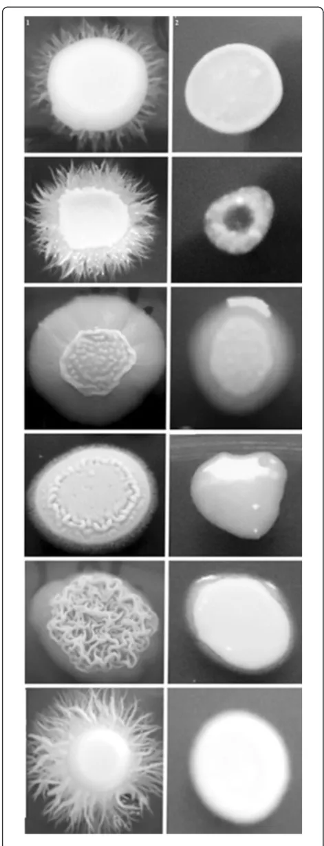
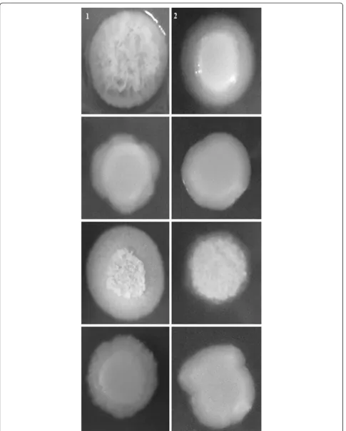
[image:9.595.55.291.85.701.2]We also evaluated a possible effect of the CE ofE. uniflora inC. albicanshypha formation on solid media by cultivat-ing all the 48 isolates and the two reference strains for seven days on YPD + 20% FBS agar, Spider medium and GlcNac agar medium at 30°C. The macromorphological aspect of the colonies was observed and the strains were markedly smoother than the respective control, grown in the absence of the CE, which showed wrinkled colonies (Table 3; Figures 3, 4 and 5). Despite the fact that YPD agar added 20% FBS was not an efficient inducer of fila-mentation for most of the isolates tested, for the strains
that showed a wrinkled phenotype grown in this condi-tion, smooth colonies were also observed for the respect-ive strain grown in the presence of the CE of E. uniflora (Figure 3).
Effect of CE ofEugenia uniflorain secretion of hydrolytic enzymes
Other important virulence attribute ofC. albicansis the ability to secrete hydrolytic enzymes. Therefore, we eval-uated phospholipase and proteinase activity of the 48 clinical isolates ofC. albicansand the references strains ATCC90028 and SC5314. When cells were grown on the surface of egg yolk emulsion agar (phospholipase induc-tion), the average phospholipase zone values for all the 50 strains was 0.54 ± 0.18, considered a moderate activity (Table 4). When the CE of E. uniflora was included in the phospholipase media, the activity was completely inhibited (Figure 6). The same trend was observed for cells grown on the surface of the proteinase induction medium without the CE of E. uniflora. All the strains were able to produce satisfactory levels of proteinase (meanPzvalue of 0.44 ± 0.21). A total inhibition of pro-teinase activity was observed for 48 out of 50 isolates of C. albicans, including the reference strains (Figure 7). In fact, only two strains (05 and 60; 4%) were still able to produce very low amounts of the hydrolytic enzyme (Pzof 0.88 ± 0.01 and 0.88 ± 0.00, respectively; Table 4).
Discussion
[image:11.595.57.289.117.727.2] [image:11.595.306.538.125.265.2]In this study we investigated a possible role of the CE of E. uniflorain some virulence factors ofC. albicans.In a previous study performed by Ferreira et al. [43], a screening of antifungal activity of medicinal plants from northeastern Brazil, including 30 different vegetal CEs, Table 4 Hydrolytic enzymes activity on solid media of
Candida albicansclinical isolates obtained from the oral cavity of kidney transplant recipients
Proteinase Activity (Pz†) Phospholipase Activity (Pz†)
Strain Absence of CE** vs. Presence of CE**
Absence of CE** vs. Presence of CE**
ATCC90028 0.47 ± 0.03 vs. 1 ± 0.00* 0.80 ± 0.11 vs. 1 ± 0.00*
SC5314 0.59 ± 0.10 vs. 1 ± 0.00* 0.59 ± 0.04 vs. 1 ± 0.00*
Strain01 0.36 ± 0.04 vs. 1 ± 0.00* 0.55 ± 0.08 vs. 1 ± 0.00*
Strain02 0.51 ± 0.02 vs. 1 ± 0.00* 0.68 ± 0.02 vs. 1 ± 0.00*
Strain03 1 ± 0.00 vs. 1 ± 0.00 1 ± 0.00 vs. 1 ± 0.00
Strain05 0.29 ± 0.01 vs. 0.88 ± 0.01* 0.64 ± 0.00 vs. 1 ± 0.00*
Strain06 0.72 ± 0.11 vs. 1 ± 0.00* 0.60 ± 0.00 vs. 1 ± 0.00*
Strain08 0.72 ± 0.02 vs. 1 ± 0.00* 0.57 ± 0.00 vs. 1 ± 0.00*
Strain10 1 ± 0.00 vs. 1 ± 0.00 1 ± 0.00 vs. 1 ± 0.00
Strain11 0.43 ± 0.01 vs. 1 ± 0.00* 0.63 ± 0.04 vs. 1 ± 0.00*
Strain12 0.31 ± 0.02 vs. 1 ± 0.00* 0.75 ± 0.00 vs. 1 ± 0.00*
Strain13 0.69 ± 0.02 vs. 1 ± 0.00* 0.59 ± 0.01 vs. 1 ± 0.00*
Strain17 0.74 ± 0.02 vs. 1 ± 0.00* 0.50 ± 0.03 vs. 1 ± 0.00*
Strain20 0.61 ± 0.11 vs. 1 ± 0.00* 0.55 ± 0.03 vs. 1 ± 0.00*
Strain21 0.63 ± 0.02 vs. 1 ± 0.00* 0.58 ± 0.07 vs. 1 ± 0.00*
Strain23 0.65 ± 0.05 vs. 1 ± 0.00* 0.67 ± 0.04 vs. 1 ± 0.00*
Strain24 0.67 ± 0.12 vs. 1 ± 0.00* 0.49 ± 0.02 vs. 1 ± 0.00*
Strain28 0.44 ± 0.02 vs. 1 ± 0.00* 0.55 ± 0.05 vs. 1 ± 0.00*
Strain30 0.27 ± 0.05 vs. 1 ± 0.00* 0.68 ± 0.04 vs. 1 ± 0.00*
Strain31 0.47 ± 0.02 vs. 1 ± 0.00* 0.52 ± 0.02 vs. 1 ± 0.00*
Strain32 0.42 ± 0.04 vs. 1 ± 0.00* 0.75 ± 0.06 vs. 1 ± 0.00*
Strain34 0.75 ± 0.00 vs. 1 ± 0.00* 0.73 ± 0.03 vs. 1 ± 0.00*
Strain37 0.60 ± 0.06 vs. 1 ± 0.00* 0.50 ± 0.03 vs. 1 ± 0.00*
Strain40 0.53 ± 0.06 vs. 1 ± 0.00* 0.58 ± 0.05 vs. 1 ± 0.00*
Strain41 0.76 ± 0.10 vs. 1 ± 0.00* 0.55 ± 0.02 vs. 1 ± 0.00*
Strain44 1 ± 0.00 vs. 1 ± 0.00 0.64 ± 0.03 vs. 1 ± 0.00*
Strain46 0.57 ± 0.04 vs. 1 ± 0.00* 0.65 ± 0.02 vs. 1 ± 0.00*
Strain50 0.54 ± 0.03 vs. 1 ± 0.00* 0.63 ± 0.11 vs. 1 ± 0.00*
Strain51 0.39 ± 0.03 vs. 1 ± 0.00* 0.53 ± 0.00 vs. 1 ± 0.00*
Strain53 0.36 ± 0.04 vs. 1 ± 0.00* 0.37 ± 0.05 vs. 1 ± 0.00*
Strain54 0.37 ± 0.06 vs. 1 ± 0.00* 0.57 ± 0.03 vs. 1 ± 0.00*
Strain60 0.28 ± 0.00 vs 0.88 ± 0.00* 0.55 ± 0.04 vs. 1 ± 0.00*
Strain61 1 ± 0.00 vs. 1 ± 0.00 0.5 ± 0.03 vs. 1 ± 0.00*
Strain70 0.39 ± 0.05 vs. 1 ± 0.00* 0.76 ± 0.08 vs. 1 ± 0.00*
Strain72 0.30 ± 0.01 vs. 1 ± 0.00* 0.62 ± 0.02 vs. 1 ± 0.00*
Strain82 0.38 ± 0.05 vs. 1 ± 0.00* 0.50 ± 0.06 vs. 1 ± 0.00*
Strain85 0.40 ± 0.03 vs. 1 ± 0.00* 1 ± 0.00 vs. 1 ± 0.00
Strain107 1 ± 0.00 vs. 1 ± 0.00 0.54 ± 0.01 vs. 1 ± 0.00*
Strain06A 0.28 ± 0.02 vs. 1 ± 0.00* 1 ± 0.00 vs. 1 ± 0.00
Strain06L 0.45 ± 0.03 vs. 1 ± 0.00* 0.56 ± 0.00 vs. 1 ± 0.00*
Table 4 Hydrolytic enzymes activity on solid media of
Candida albicansclinical isolates obtained from the oral cavity of kidney transplant recipients(Continued)
Strain10Li 0.59 ± 0.03 vs. 1 ± 0.00* 0.52 ± 0.02 vs. 1 ± 0.00*
Strain10S 0.31 ± 0.04 vs. 1 ± 0.00* 0.79 ± 0.04 vs. 1 ± 0.00*
Strain12A 0.72 ± 0.05 vs. 1 ± 0.00* 0.50 ± 0.00 vs. 1 ± 0.00*
Strain12L 0.56 ± 0.02 vs. 1 ± 0.00* 0.55 ± 0.02 vs. 1 ± 0.00*
Strain15A 0.75 ± 0.00 vs. 1 ± 0.00* 0.51 ± 0.00 vs. 1 ± 0.00*
Strain15G 0.44 ± 0.07 vs. 1 ± 0.00* 0.54 ± 0.02 vs. 1 ± 0.00*
Strain15L 0.42 ± 0.02 vs. 1 ± 0.00* 0.47 ± 0.03 vs. 1 ± 0.00*
Strain18A 0.4 ± 0.00 vs. 1 ± 0.00* 0.52 ± 0.03 vs. 1 ± 0.00*
Strain111L 0.29 ± 0.01 vs. 1 ± 0.00* 0.67 ± 0.04 vs. 1 ± 0.00*
Strain111R 0.42 ± 0.02 vs. 1 ± 0.00* 0.68 ± 0.02 vs. 1 ± 0.00*
*P < 0.05, **CE: Crude Extract ofEugenia uniflora(2000μg/mL);†
Pz = Precipitation zone.
The isolates were incubated in triplicates at 30°C for 72 h on proteinase or phospholipase agar in the absence (control experiments) or presence of 2000
revealed E. unifloraas the most active natural product, with minimal inhibitory concentration (MIC) values ran-ging from 31.25 to 62.5 μg/mL against C. albicans, as determined by broth microdilution methodology.
After the observation ofC. albicanscells growth inhib-ition with the CE of E. uniflora, we decided to investi-gate a possible action of this natural product in the impairment of the full expression of some virulence fac-tors inC. albicans. Therefore, we have used NGY broth to grow our strains in the presence of the CE previously to each attribute of virulence testedin vitro. We hypoth-esized that when growing our strains in the presence of this natural product,C. albicanscells would be impaired to fully express the pathogenicity factors investigated. Therefore a pilot experiment revealed that 2000 μg/ml of the E. uniflora CE was needed to inhibit approxi-mately 50% growth of cells incubated for 18 to 24 h in NGY broth under 200 rpm, 30°C, but not to kill all of them. Of note, this is a complete different condition compared to the MIC determination experiment per-formed by Ferreira et al. [43], where the cells were
incubated statically, in RPMI-1640, also in a different volume of culture medium. In our study, C. albicans cells were grown in NGY broth, 200 rpm. Incubation condition differences may explain discrepant MIC values within the two different methodologies.
When C. albicans cells were cultivated overnight in the presence of the natural product, bud cells demon-strated alterations in the cellular structure, suggesting a possible action of the CE of E. uniflora specifically onC. albicanscell wall.
It has been described that vegetal extracts can act on the cell wall ofC. albicans. By analyzing the mechanism of action of the vegetal extract of Stryphnodendron adstringens on C. albicans cells, Ishida et al. [35] ob-served that the integrity of the cell wall was damaged, with the presence of several deformations in buddy cells structure.
[image:12.595.58.540.89.233.2]It is important to emphasize that the recently devel-oped antifungal drugs named echinocandins target Can-dida and Aspergillus cell wall, by inhibiting the enzyme β-1,3-D-glucan synthase [46]. These chemical compounds Figure 6Phospholipase activity on solid media ofCandida albicansclinical isolates obtained from the oral cavity of kidney transplant recipients.The isolates were incubated in triplicates at 30°C for 72 h on phospholipase agar in the absence (control experiments;A)or presence of 2000μg/ml (test experiments;B)of the crude extract ofEugenia uniflora.
[image:12.595.58.540.563.695.2]are less toxic than polyenes, which can also bind to mam-malian cholesterol. Therefore, if E. uniflora compounds are truly acting on the cell wall, the natural product may be used for future development of antifungal drugs with lower side effects.
Bud-to-hyphae transition of C. albicans is a funda-mental step for the establishment of infection, contribut-ing to tissue invasion. We found that the ability to form true hyphae was reduced for the majority of isolates. Be-sides, all of the strains tested had a reduction in hyphal size in YPD + 20% FBS, when previously grown with the CE ofE. uniflora. Of note, shorter true hyphae were not observed whenC. albicanscells were incubated in liquid Spider medium and GlcNac liquid media, when previ-ously grown in the presence of the CE. This may be ex-plained because serum is the most potent inducer of hypha formation resulting in cells with higher MI [20]. Thus, due to the fact that the other two hyphal inducers already produced shorter true hyphae in C. albicans, a decrease in hyphal size could not be observed when cells were grown in the presence of the natural product.
Although others have found that natural products may impair morphogenesis [34,47,48], in our study we ana-lyzed a higher number of strains and generated quantita-tive data of decreased hypha formation, under different bud-to-hyphae transition inducing media.
When C. albicanscells suspension were plated on the surface of Spider medium, GlcNac agar and YPD + 20% FBS agar, several phenotypic filamentous colonies were observed. When the CE of E. uniflora was added in the composition of the three media, it was observed a no-torious reduction of filamentation of the colonies. Simi-lar results were observed by Tsang et al. [34], who investigated the role of purpurin, a compound derived fromRubia tinctorumL. roots, in the impairment of fila-mentation ofC. albicanson solid Spider medium.
Other important aspect of our findings is that the CE ofE. unifloraclearly affects bud-to-hypha transition, ob-served under three different conditions. It is well known that morphogenesis in C. albicans can be triggered by the stimulation of several pathways or by essentially a single pathway, which is fully activated [49].
The secretion of hydrolytic enzymes such as protein-ase and phospholipprotein-ase are important virulence factors of C. albicans,contributing for the establishment of infec-tion by damaging tissues and cellular membrane [25,50]. When the CE ofE. uniflorawas added to phospholipase and proteinase media, the zone of activity of the en-zymes was either highly reduced or completely inhibited. This is an important finding because a reduction of pro-duction and secretion of these enzymes are related with a decrease ofC. albicansvirulence [51].
To the best of our knowledge, this is the first report in literature where the action of a natural product obtained
from a plant is impairing the expression of both en-zymes. The effect of the CE of Dodonaea viscosa var. angustifolia leaves, an indigenous South African plant, on proteinase and phospholipase activity in C. albicans was investigated in 26 strains isolated from the oral cav-ity of 150 HIV patients. Nevertheless, the secretion of both enzymes was not affected by the addition of the plant extract [52].
Conclusions
This was the first study to deeply investigate the effect of the CE of E. uniflora in virulence factors of C. albicans clinical isolates collected in a specific clinical scenario (oral candidiasis in kidney transplant recipients). Our study has proved a satisfactory effect of this natural prod-uct in inhibiting three important virulence factors of C. albicans. We also verified that more than 80% of A549 cells (human alveolar basal epithelial cell line) remained viable even when exposed to 4 fold concen-tration of the CE of E. uniflora (8000 μg/ml; unpub-lished data), suggesting its possible safety if applied as an antifungal agent in the future. Our group is cur-rently working on the elucidation of the chemical compounds responsible for the inhibition of growth and specifically interactions with virulence factors.
Abbreviations
ATCC:American type culture collection; BSA: Bovine serum albumine; C.:Candida; CE: Crude extract; CO2: carbonic gas; °C: Grade celsius; DO: Optical density; et al.: Collaborators; FBS: Fetal bovine serum; g/l: Gram per liter; GlcNac: N-acetyl-D-glucosamine; h: Hour; MI: Morphological index; KCl: Sodium chloride; KH2PO4: Monobasic potassium phosphate; M: Molar; MIC: Minimal inhibitory concentration; min: Minute; MI: Morphology index; mL: Milliliter; mL/L: Milliliter per liter; mM: Milimolar; mm: Millimeter; MgCl2: Magnesium chloride; mg/mL: Milligram per milliliter;μg/ mL: Microgram per milliliter;μL: Microliter; N°: Number; NaCl: Sodium chloride; Na2HPO3: Sodium phosphate dibasic; NGY: Neopeptone–yeast extract - glucose; nm: Nanometer; pH: Hydrogen potential; Pz: Precipitation zone; ®: Trademark; Rpm: Revolutions per minute; Saps: Secreted aspartic proteinases; SDA: Sabouraud dextrose agar; spp.: Species; TM: Trade mark; UFRN: Universidade Federal of Rio Grande do Norte; YPD: Yeast peptone dextrose; YCB: Yeast carbon base.
Competing interests
The authors declare that they have no competing interests.
Authors’contributions
WP and GM analyzed data, performed research; wrote the paper. VL performed research, TI and EP, collected samples, MR and LA provided theE. unifloraextract. All authors read and approved the final manuscript.
Authors’information
GM, Professor of Basic and Clinical Mycology, Departamento de Análises Clínicas e Toxicológicas, Universidade Federal do Rio Grande do Norte, Brazil and Leader of the Laboratório de Micologia Médica e Molecular.
Acknowledgements
Author details 1
Laboratório de Micologia Médica e Molecular, Departamento de Análises Clínicas e Toxicológicas, Universidade Federal do Rio Grande do Norte, Natal, Rio Grande do Norte, Brazil.2Departamento de Farmácia, Universidade Federal de Pernambuco, Recife, Pernambuco, Brazil.3Departamento de Análises Clínicas, Universidade Estadual de Maringá, Maringá, Paraná, Brazil. 4Departamento de Infectologia, Universidade Federal do Rio Grande do
Norte, Natal, Rio Grande do Norte, Brazil.
Received: 20 October 2014 Accepted: 14 January 2015
References
1. Chaves GM, Santos FP, Colombo AL. The persistence of multifocal colonization by a single ABC genotype ofCandida albicansmay predict the transition from commensalism to infection. Mem Inst Oswaldo Cruz. 2012;107:198–204.
2. Gall F, Colella G, Di Onofrio V, Rossiello R, Angelillo IF, Liguori G.Candidaspp. in oral cancer and oral precancerous lesions. New Microbiol. 2013;36:283–8. 3. Muzyka BC, Epifanio RN. Update on oral fungal infections. Dent Clin North
Am. 2013;57:561–81.
4. Hebbar PB, Pai A, Sujatha D. Mycological and histological associations of Candidain oral mucosal lesions. J Oral Sci. 2013;55:157–60.
5. Runhke M. Skin and Mucous Membrane Infection. In: Calderone RA, editor. Candida and Candidiasis. Washington D.C: ASM Press; 2002. p. 14–27. 6. Chaves GM, Diniz MG, da Silva-Rocha WP, de Souza LB, Gondim LA, Ferreira
MA, et al. Species distribution and virulence factors of Candida spp. isolated from the oral cavity of kidney transplant recipients in Brazil. Mycopathologia. 2013;175:255–63.
7. Maurya V, Srivastava A, Mishra J, Gaind R, Marak RS, Tripathi AK, et al. Oropharyngeal candidiasis and Candida colonization in HIV positive patients in northern India. J Infect Dev Ctries. 2013;7:608–13.
8. Anwar KP, Malik A, Subhan KH. Profile of candidiasis in HIV infected patients. Iran J Microbiol. 2012;4:204–9.
9. de la Rosa-García E, Mondragón-Padilla A, Irigoyen-Camacho ME, Bustamante-Ramírez MA. Oral lesions in a group of kidney transplant patients. Med Oral Patol Oral Cir Bucal. 2005;10:196–204.
10. Tyrzyk S, Sadlak-Nowicka J, Kedzia A, Bochniak M, Szumska-Tyrzyk B, Rutkowski P. Clinical and mycological examinations of oral mucosa in cyclosporine A treated patients after renal transplantation. Przegl Lek. 2004;61:467–72.
11. Güleç AT, Demirbilek M, Seçkin D, Can F, Saray Y, Sarifakioglu E, et al. Superficial fungal infections in 102 renal transplant recipients: a case-control study. J Am Acad Dermatol. 2003;49:187–92.
12. Al-Mohaya MA, Darwazeh A, Al-Khudair W. Oral fungal colonization and oral candidiasis in renal transplant patients: the relationship to Miswak use. Oral Surg Oral Med Oral Pathol Oral Radiol Endod. 2002;93:455–60.
13. King GN, Healy CM, Glover MT, Kwan JT, Williams DM, Leigh IM, et al. Prevalence and risk factors associated with leukoplakia, hairy leukoplakia, erythematous candidiasis, and gingival hyperplasia in renal transplant recipients. Oral Surg Oral Med Oral Pathol. 1994;78:718–26.
14. Gow NA. A developmental program forCandidacommensalism. Nat Genet. 2001;45:967–8.
15. Menezes TO, Gillet LC, Menezes SA, Feitosa RN, Ishak MO, Ishak R, et al. Virulence factors ofCandida albicansisolates from the oral cavities of HIV-1-positive patients. Curr HIV Res. 2013;11:304–8.
16. Chaves GM, da Silva WP. Superoxide dismutases and glutaredoxins have a distinct role in the response ofCandida albicansto oxidative stress generated by the chemical compounds menadione and diamide. Mem Inst Oswaldo Cruz. 2012;107:998–1005.
17. Vediyappan G, Dumontet V, Pelissier F, D’Enfert C. Gymnemic acids inhibit hyphal growth and virulence inCandida albicans. PLoS One. 2013;8:e74189. 18. Adams DJ. Fungal cell wall chitinases and glucanases. Microbiology.
2004;150:2029–35.
19. Gow NA, van de Veerdonk FL, Brown AJ, Netea MG.Candida albicans morphogenesis and host defence: discriminating invasion from colonization. Nat Rev Microbiol. 2011;10:112–22.
20. Merson-Davies LA, Odds FC. A morphology index for characterization of cell shape inCandida albicans. J Gen Microbiol. 1989;135:3143–52.
21. Brown AJP. Expression of growth form-specific factors during morphogenesis inCandida albicans. In: Calderone RA, editor. Candida and Candidiasis. Washington D.C: ASM Press; 2002. p. 87–93.
22. Chaves GM, Bates S, Maccallum DM, Odds FC.Candida albicansGRX2, encoding a putative glutaredoxin, is required for virulence in a murine model. Genet Mol Res. 2007;6:1051–63.
23. Odds FC, Van Nuffel L, Gow NA. Survival in experimentalCandida albicans infections depends on inoculum growth conditions as well as animal host. Microbiology. 2000;146:1881–9.
24. Mattei AS, Alves SH, Severo CB, Guazzelli S, Oliveira M, Severo LC. Determination of germ tube, phospholipase, and proteinase production by bloodstream isolates ofCandida albicans. Rev Soc Bras Med Trop. 2013;46:340–2.
25. Ghannoum MA. Potential role of phospholipases in virulence and fungal pathogenesis. Clin Microbiol Rev. 2000;13:122–43.
26. Ran Y, Iwabuchi K, Yamazaki M, Tsuboi R, Ogawa H. Secreted aspartic proteinase fromCandida albicansacts as a chemoattractant for peripheral neutrophils. J Dermatol Sci. 2013;923:00216–8.
27. Dimopoulos G, Antonopoulou A, Armaganidis A, Vincent JL. How to select an antifungal agent in critically ill patients. J Crit Care. 2013;28:717–27. 28. Mora-Duarte J, Betts R, Rotstein C, Colombo AL, Thompson-Moya L,
Smietana J, et al. Comparison of caspofungin and amphotericin B for invasive candidiasis. N Engl J Med. 2002;19:2020–9.
29. Paquet MJ, Fournier I, Barwicz J, Tancrède P, Auger M. The effects of amphotericin B on pure and ergosterol- or cholesterol-containing dipalmi-toylphosphatidylcholine bilayers as viewed by 2H NMR. Chem Phys Lipids. 2002;119:1–11.
30. Hamill RJ. Amphotericin B, formulations: a comparative review of efficacy and toxicity. Drugs. 2013;73:919–34.
31. Wille MP, Guimarães T, Furtado GH, Colombo AL. Historical trends in the epidemiology of candidaemia: analysis of an 11-year period in a tertiary care hospital in Brazil. Mem Inst Oswaldo Cruz. 2013;3:108.
32. Perea S, López-Ribot JL, Kirkpatrick WR, McAtee RK, Santillán RA, Martínez M, et al. Prevalence of molecular mechanisms of resistance to azole antifungal agents in Candida albicans strains displaying high-level fluconazole resistance isolated from human immunodeficiency virus-infected patients. Antimicrob Agents Chemother. 2001;45:2676–84.
33. Hernandez S, López-Ribot JL, Najvar LK, McCarthy DI, Bocanegra R, Graybill JR. Caspofungin resistance inCandida albicans: correlating clinical outcome with laboratory susceptibility testing of three isogenic isolates serially obtained from a patient with progressiveCandidaesophagitis. Antimicrob Agents Chemother. 2004;48:1382–3.
34. Tsang PW, Bandara HM, Fong WP. Purpurin suppressesCandida albicans biofilm formation and hyphal development. PLoS One. 2012;7:e50866. 35. Ishida K, Mello JCP, Cortez DAG. Influence of tannins fromStryphnodendron
adstringenson growth and virulence factors ofCandida albicans. J Antimi Chemo. 2006;58:942–9.
36. Amorim AC, Lima CK, Hovell AM, Miranda AL, Rezende CM.
Antinociceptive and hypothermic evaluation of the leaf essential oil and isolated terpenoids fromEugenia unifloraL. (Brazilian Pitanga). Phytomedicine. 2009;16:923–8.
37. de Freitas Araújo MG, Pacífico M, Vilegas W, Dos Santos LC, Icely PA, Miró MS, et al. Evaluation ofSyngonanthus nitens(Bong.) Ruhl. extract as antifungal and in treatment of vulvovaginal candidiasis. Med Mycol. 2013;51:673–82.
38. Jothy SL, Zakariah Z, Chen Y, Sasidharan S. In vitro, in situ and in vivo studies on the anticandidal activity ofCassia fistulaseed extract. Molecules. 2012;17:6997–7009.
39. Liu Q, Luyten W, Pellens K, Wang Y, Wang W, Thevissen K, et al. Antifungal activity in plants from Chinese traditional and folk medicine. J Ethnopharmacol. 2012;143:772–8.
40. Brandão MG, Zanetti NN, Oliveira P, Grael CF, Santos AC, Monte-Mór RL. Brazilian medicinal plants described by 19th century European naturalists and in the Official Pharmacopoeia. J Ethnopharmacol. 2008;120:141–8. 41. Rodrigues KA, Amorim LV, de Oliveira JM, Dias CN, Moraes DF, Andrade EH,
et al. Eugenia uniflora L. Essential Oil as a Potential Anti-Leishmania Agent: Effects on Leishmania amazonensis and Possible Mechanisms of Action. Evid Based Complement Alternat Med. 2013;2013:279726.
42. Santos KK, Matias EF, Tintino SR, Souza CE, Braga MF, Guedes GM, et al. Enhancement of the antifungal activity of antimicrobial drugs byEugenia uniflora L. J Med Food. 2013;16:669–71.
44. Liu H, Kohler J, Fink GRN. Suppression of hyphal formation inCandida albicansby mutation of a STE12 homolog. Science. 1994;266:1723–6. 45. Price MF, Wilkinson ID, Gentry LO. Plate method for detection of
phospholipase activity inCandida albicans. Sabouraudia. 1982;20:7–14. 46. Douglas CM. Fungal beta(1,3)-D-glucan synthesis. Med Mycol. 2001;1:55–66. 47. Bernardes I, Felipe Rodrigues MP, Bacelli GK, Munin E, Alves LP, Costa MS.
Aloe veraextract reduces both growth and germ tube formation by Candida albicans. Mycoses. 2012;55:257–61.
48. Latha LY, Darah I, Jain K, Sasidharan S. Effects ofVernonia cinerealess methanol extract on growth and morphogenesis ofCandida albicans. Eur Rev Med Pharmacol Sci. 2011;15:543–9.
49. Ernst JF. Transcription factors inCandida albicans- environmental control of morphogenesis. Microbiology. 2000;146:1763–74.
50. Hube B, Naglik J.Candida albicansproteinases: resolving the mystery of a gene family. Microbiology. 2001;147:1997–2005.
51. Mohan V, Ballal M. Proteinase and phospholipase activity as virulence factors inCandidaspecies isolated from blood. Rev Iberoam Micol. 2008;25:208–10.
52. Patel M, Gulube Z, Dutton M. The effect ofDodonaea viscosavar. angustifoliaonCandida albicansproteinase and phospholipase production and adherence to oral epithelial cells. J Ethnopharmacol. 2009;124:562–5.
Submit your next manuscript to BioMed Central and take full advantage of:
• Convenient online submission
• Thorough peer review
• No space constraints or color figure charges
• Immediate publication on acceptance
• Inclusion in PubMed, CAS, Scopus and Google Scholar
• Research which is freely available for redistribution