Begus, Katarina and Southgate, Victoria and Gliga, Teodora (2015) Neural
mechanisms of infant learning: differences in frontal theta activity during
object exploration modulate subsequent object recognition. Biology Letters
11 (5), p. 20150041. ISSN 1744-9561.
Downloaded from:
Usage Guidelines:
Please refer to usage guidelines at
or alternatively
rsbl.royalsocietypublishing.org
Research
Cite this article:
Begus K, Southgate V, Gliga
T. 2015 Neural mechanisms of infant learning:
differences in frontal theta activity during
object exploration modulate subsequent
object recognition.
Biol. Lett.
11
: 20150041.
http://dx.doi.org/10.1098/rsbl.2015.0041
Received: 19 January 2015
Accepted: 30 April 2015
Subject Areas:
neuroscience, cognition, behaviour
Keywords:
theta oscillations, learning, infants, motivation
Author for correspondence:
Katarina Begus
e-mail: [email protected]
Electronic supplementary material is available
at http://dx.doi.org/10.1098/rsbl.2015.0041 or
via http://rsbl.royalsocietypublishing.org.
Animal behaviour
Neural mechanisms of infant learning:
differences in frontal theta activity during
object exploration modulate subsequent
object recognition
Katarina Begus, Victoria Southgate and Teodora Gliga
Centre for Brain and Cognitive Development, Birkbeck College, University of London, Malet Street, London, WC1E 7HX, UK
Investigating learning mechanisms in infancy relies largely on behavioural measures like visual attention, which often fail to predict whether stimuli would be encoded successfully. This study explored EEG activity in the theta frequency band, previously shown to predict successful learning in adults, to directly study infants’ cognitive engagement, beyond visual
atten-tion. We tested 11-month-old infants (N¼23) and demonstrated that
differences in frontal theta-band oscillations, recorded during infants’ object exploration, predicted differential subsequent recognition of these objects in a preferential-looking test. Given that theta activity is modulated by motiv-ation to learn in adults, these findings set the ground for future investigmotiv-ation into the drivers of infant learning.
1. Introduction
Investigating predictors of learning success in infancy has relied largely on behav-ioural measures like visual attention. While termination of visual attention might indicate successful encoding [1], longer visual attention to stimuli does not necess-arily predict better encoding or recognition at test [2]. This suggests that quality, rather than quantity, of attention may be more relevant for successful information processing. A promising means of elucidating how attentional quality supports learning in infancy is directly measuring the neural correlates, which have been shown to predict successful learning in adults.
A growing body of research is demonstrating that modulations in oscillatory activity in the theta frequency band (4–8 Hz in adults), believed to reflect pre-frontal–hippocampal information-processing loops, correlate with memory
performance at test. For example, Guderianet al.[3] demonstrated a linear
relation-ship between power of theta activity before item presentation and rate of recall for those items at test. A similar relationship was found between prestimulus frontal theta activity and memory accuracy [4], as well as between frontal theta activity during retention and the capacity of visual working memory [5].
In infants, an increase in theta oscillations has been reported in situations often associated with infant learning, such as during periods of sustained attention [6], when infants were involved in a social game and exploration of novel objects [7], when infants’ expectations were violated [8] and in response to infant-directed speech [9]. While some authors have interpreted theta oscillations as indexing implicit learning in infants [10], no study has so far directly explored whether theta oscillations in fact predict successful encoding in infants.
To address this, we recorded EEG activity while infants explored novel objects. Based on previous work demonstrating that object exploration induced the greatest modulation of theta oscillations over the frontal scalp location [7],
we ranked the explored objects for each infant based on the power of frontal theta-band oscillations during exploration. We then tested infants’ encoding of the objects’ features in a preferential-looking task. We predicted that infants would learn more about the objects that were associated with more frontal theta-band activity; thus, differences in the power of frontal theta oscillations during exploration should be reflected in differences in infants’ ability to discriminate the objects at test.
2. Material and methods
(a) Participants
Twenty-three 11-month-old infants (13 female) were included in the sample; 12 infants were excluded owing to fussiness (4), insufficient data (4), parental interference (2) or experimental error (2).
(b) Procedure
(i) Exploration phase
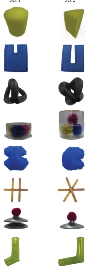
Materials:Infants were presented with one of two sets of eight novel objects (figure 1), approximately 1010 cm in size and easily grasped and manipulated by infants. Each object in Set 1 was partially matched to one object in Set 2. Paired objects were matched in colour, size and material, but differed in shape (infants of this age can readily detect changes in shape [11]). Infants’ behaviour was video recorded and their EEG was recorded at a sampling rate of 500 Hz using a 128-channel Geodesic Sensor Net (GSB; EGI Inc, Eugene, OR, USA).
[image:3.595.355.504.47.502.2]Procedure:Infants sat in a high chair with a tray attached, on which each of the eight objects was presented individually, in a random order, for 40 s each. Each trial started with the sentence: ‘This is for you to play with’, and was preceded by a period (approx. 20 s) of blowing bubbles. The parent and the exper-imenter did not interact with the infant or objects, unless the object was dropped, in which case it was returned to the table immediately.
(ii) Test phase
Materials:Photographs of object pairs were displayed on a 102 58 cm plasma screen. When presented at 150 cm distance from the infant, each image subtended approximately the same visual angle as the physical object would during exploration. Infants’ behaviour was video recorded.
Procedure:Infants were sat in a high chair or on their parent’s lap. The parent was instructed not to interact with the infant. Trials started with an audio –visual animation in the centre of the screen to attract the infant’s attention, followed by photo-graphs of the familiar object (explored during Exploration phase) and the matched shape-distorted object ( previously unseen), displayed side by side. Each trial lasted 12 s, with the side of presentation of the objects switching after 6 s.
(c) Data analysis
(i) Exploration phase
EEG analysis: Video recordings were coded frame by frame (25 fps) and time intervals during which the infant was visually attending to the object were extracted for EEG analysis. The raw EEG data were imported to EEGlab, Fieldtrip, and visually screened for motion and eye-blink artefacts. Epochs of 1 s were then extracted from periods of continuous artefact-free data (any remaining samples were discarded; artefact-free segments were not concatenated but segmented separately) and fast Fourier transformed (Hanning window, 50% overlap) to yield a
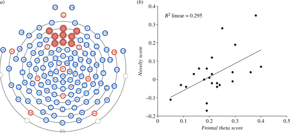
power spectrum between 1 and 50 Hz, in steps of 1 Hz. A mini-mum of 10 epochs of artefact-free data from a minimini-mum of four objects was required for an infant’s data to be analysed. Included objects were ranked according to the power of theta oscillations (3–5 Hz in infants [7]) measured at frontal central electrodes (figure 2a) during exploration. Two objects that elicited the high-est (high theta objects (HTO)) and two objects that elicited the lowest power of theta oscillations (low theta objects (LTO)) were identified and averaged together. Difference scores were then calculated for each participant’s EEG data (HTO2LTO/ HTOþLTO), creating the variable Frontal theta score.Variable Number of sampleswas created to account for possible differences in the amount of data analysed for each object.
Behavioural analysis: To control for any variation in how infants interacted with the objects, which could lead to differences in encoding, video recordings of HTO and LTO explorations were coded for each infant’s visual and manual exploration. Difference scores (HTO2LTO/HTOþLTO) were calculated to create variablesVisual exploration(total looking time at the object, regard-less of physical contact) andManual exploration (total time the infant handled the object, while visually attending to it).
Set 1 Set 2
Figure 1.
Novel objects. Infants explored all objects from one of the sets;
images of pairs of objects from both sets were used as stimuli during Test
phase. (Online version in colour.)
rsbl.r
oy
alsocietypublishing.org
Biol.
Lett.
11
:
20150041
(ii) Test phase
Infants’ looking behaviour was coded frame by frame (25 fps) to determine the magnitude of infants’ looking-time preference for the presented objects (Novel2Familiar/NovelþFamiliar). Trials in which the infant did not look for a minimum of 500 ms at each object presented on each side of the screen, were excluded from analysis. Looking-time difference scores were calculated for objects identified based on EEG data (HTO2LTO/HTOþLTO), creating the variable Novelty score. A score of 0 on this variable would mean the infant’s looking-time preference was identical when discriminating HTO and LTO objects; a positive value would indicate that infants exhib-ited a larger novelty preference for HTO than LTO objects, and vice versa for a negative score.
3. Results
To establish whether a relationship exists between theta acti-vity during exploration and infants’ later recognition of the explored objects, a stepwise linear regression was performed on the data. To account for any variation in infants’ exploration behaviour or amount of artefact-free data included in analyses,
Frontal theta score, Visual exploration, Manual exploration and
Number of sampleswere entered as predictors andNovelty score
as the dependent variable. A significant model emerged
(F1,21¼8.803,p¼0.007,R2¼0.295), explaining 29.5% of
var-iance of the dependent variable. The only significant predictor ofNovelty scorewasFrontal theta score (b¼0.543,t21¼2.967,
p¼0.007), whereasVisual exploration, Manual exploration and
Number of samples did not explain a significant amount of variance and were therefore dropped from the model (multiple
regression using Enter method produced the same results;
see the electronic supplementary material for details). This
relationship between Frontal theta score and Novelty score
means that when the power of theta activity recorded during exploration was similar for HTO and LTO objects, these objects were similarly well (or poorly) discriminated at test (resulting inNovelty score values just below and above 0 (figure 2b)). Conversely, when the difference in theta activity between HTO and LTO objects was large, it was also reflected in a
larger difference in infants’ preferential looking, showing a stronger looking-time preference for HTO compared with LTO objects.
To examine whether our effect was specific to oscillations in the theta frequency band, the data were also analysed by ranking the objects based on power of oscillations in delta (1– 3 Hz), alpha (6–8 Hz) and gamma (20 –40 Hz) frequency bands over the frontal central electrodes. No significant
relationship was found betweenNovelty score and power of
oscillations in any other frequency band over the frontal central electrodes. In addition, further analysis revealed that the power of theta oscillations recorded over other scalp locations (occipital and temporal sites; electronic supplemen-tary material, figure S1) did not significantly correlate with
Novelty score(see the electronic supplementary material). Note that while accounting for visual exploration, we could not control for potentially differential saccadic patterns during exploration. Altough this might be a caveat, previous findings showing within-trial modulations of saccadic ampli-tude in absence of modulation in the concurrently recorded frontal theta activity [12] suggest that an effect of saccades on our results is unlikely.
4. Discussion
This study is the first to demonstrate that modulations of frontal theta-band oscillations, recorded during infants’ object exploration, predict infants’ subsequent recognition of these objects. Specifically, the larger the difference between the power of theta activity recorded during exploration of two objects, the larger the difference in infants’ subsequent recognition of these objects. The relationship found was specific to theta-band oscillations (3–5 Hz) recorded over the frontal cortex and was not present in any other frequency band or scalp area. Importantly, this relationship was not mediated by the length of infants’ visual or manual exploration, suggesting that theta activity may provide a means of investigating infants’ learning processes that cannot be captured by behavioural measures like visual attention.
0.4 0.3 0.2 0.1 0 –0.1 –0.2 No velty scor e
0 0.1 0.2 0.3 0.4 0.5
Frontal theta score R2 linear = 0.295
127 21 14 8 125 1 2 3 123 118 5 12 20 6 112 111 110 103 105 106 80 87 93 98 97 102 101 107 91 85 86 79 78 72 84 90 89 94 88 82 81 S/N 74 73 95 99 100 RM 96 T6 92 P4 108 T4 104 C4 77 76 71 67 66 65 69 68 64 63 60 61 53 54 55 COM 62 Pz 52 P3 42 37 31 7 13 30 29 28 34 38 39 40 35 41 46 47 51 50 59 56 49 44 43 48 117 116 115 114 121 120 119 113 109 122 F8 124 F4 25 128 32 26 23 27 19
18 16 10
4 15
22
Fp1 Fp29
11 Fz 126 17 NAS 83 O2 75 Oz 70 O1 58 T5 57 LM 24 F3 REF Cz 33 F7 45 T3 36 C3
[image:4.595.60.543.44.264.2](a) (b)
Figure 2.
(
a
) EEG electrode map, with marked group of electrodes from which
Frontal theta score
data were extracted. (
b
) Relationship between
Frontal theta score
and
Novelty score
. (Online version in colour.)
While there is ample evidence that theta oscillations are involved in successful memory formation in adults, less is known about what drives the differences in the amount of theta activity for each individual. The timing and context dependency of theta activity in adult studies suggests that fluctuations in the power of theta are not random, but may reflect a strategic preparatory state for processing information [13]. Furthermore, it has been shown that theta activity can be modulated by expectancy of reward; only when participants were motivated to learn by monetary rewards did theta activity modulate recollection of words [4]. These findings are consistent with those of infant studies in which theta was recorded in situations where infants may expect to receive information, such as during infant-directed speech [9]; or be motivated to acquire new information, as in the case of violation of expectations [8].
Whether motivation modulates learning throughout life, including in infancy, remains largely unknown. Recent evi-dence that 16-month-olds use pointing to ask for information [14] and that information provided in response to pointing is better remembered [15] suggests the possibility that motivation drives learning even in infants. Theta activity, shown to be
modulated by motivation in adults and demonstrated to be involved in learning in both adults and infants, could provide an important measure for investigating early behaviours suggested to signal interest or motivation to learn in infants, such as babbling and pointing [11,15], as well as what drives differential learning in the absence of behavioural differences. Finally, future research should also clarify whether differences
in theta activitybetweenindividuals could explain individual
differences in exploration and learning.
Ethics.Informed consent was obtained from infants’ carers. The pro-cedure was approved by the ethics committee of the Department of Psychological Sciences, Birkbeck College, University of London. Authors’ Contributions.K.B., V.S. and T.G. conceived and designed the experiments. K.B. performed the experiments and analysed the data. K.B., V.S. and T.G. contributed to the writing of the manuscript. All authors approved the final version of the manuscript.
Competing Interests.We have no competing interests.
Funding.V.S. is supported by a Wellcome Trust Research Career Devel-opment Fellowship (088427/Z/09/Z) and T.G. is supported by MRC Programme grant (Nr. G0701484).
Acknowledgements.We thank Elena Orekhova, MEG Centre, Moscow State University, for help and advice on data processing of this study.
References
1. Houston-price C, Nakai S. 2004 Distinguishing
novelty and familiarity effects in infant preference
procedures.Infant Child Dev.13, 341 – 348. (doi:10.
1002/icd.364)
2. Colombo J, Richman WA, Shaddy DJ, Follmer
Greenhoot A, Maikranz JM. 2001 Heart rate-defined phases of attention, look duration, and infant performance in the paired-comparison paradigm.
Child Dev.72, 1605 – 1616. (doi:10.1111/1467-8624.00368)
3. Guderian S, Schott BH, Richardson-Klavehn A, Du¨zel
E. 2009 Medial temporal theta state before an event
predicts episodic encoding success in humans.Proc.
Natl Acad. Sci. USA106, 5365 – 5370. (doi:10.1073/ pnas.0900289106)
4. Gruber MJ, Watrous AJ, Ekstrom AD, Ranganath C,
Otten LJ. 2013 Expected reward modulates encoding-related theta activity before an event.
Neuroimage64, 68 – 74. (doi:10.1016/j.neuroimage. 2012.07.064)
5. Kawasaki M, Yamaguchi Y. 2013 Frontal theta
and beta synchronizations for monetary reward
increase visual working memory capacity.Soc.
Cogn. Affect. Neurosci.8, 523 – 530. (doi:10.1093/ scan/nss027)
6. Orekhova EV, Stroganova TA, Posikera IN. 1999
Theta synchronization during sustained anticipatory attention in infants over the second half of the first
year of life.Int. J. Psychophysiol.32, 151 – 172.
(doi:10.1016/S0167-8760(99)00011-2)
7. Orekhova EV, Stroganova TA, Posikera IN, Elam M.
2006 EEG theta rhythm in infants and preschool
children.Clin. Neurophysiol.117, 1047 – 1062.
(doi:10.1016/j.clinph.2005.12.027)
8. Berger A, Tzur G, Posner MI. 2006 Infant
brains detect arithmetic errors.Proc. Natl Acad.
Sci. USA103, 12649 – 12653. (doi:10.1073/pnas. 0605350103)
9. Zhang Y, Koerner T, Miller S, Grice-Patil Z, Svec A,
Akbari D, Tusler L, Carney E. 2011 Neural coding of formant-exaggerated speech in the infant brain.
Dev. Sci.14, 566 – 581. (doi:10.1111/j.1467-7687. 2010.01004.x)
10. Bosseler AN, Taulu S, Pihko E, Ma¨kela¨ JP, Imada T, Ahonen A, Kuhl PL. 2013 Theta brain rhythms index perceptual narrowing in infant speech perception.
Front. Psychol.4, 690. (doi:10.3389/fpsyg.2013. 00690)
11. Goldstein MH, Schwade J. 2010 Learning while babbling: prelinguistic object-directed vocalizations
indicate a readiness to learn.Infancy15, 362 – 391.
(doi:10.1111/j.1532-7078.2009.00020.x)
12. Fischer T, Graupner ST, Velichovsky BM, Pannasch S. 2013 Attentional dynamics during free picture viewing: evidence from oculomotor behavior and
electrocortical activity.Front. Syst. Neurosci.7, 17.
(doi:10.3389/fnsys.2013.00017)
13. Addante RJ, Watrous AJ, Yonelinas AP, Ekstrom AD, Ranganath C. 2011 Prestimulus theta activity
predicts correct source memory retrieval.Proc. Natl
Acad. Sci. USA108, 10702 – 10707. (doi:10.1073/ pnas.1014528108)
14. Begus K, Southgate V. 2012 Infant pointing serves
an interrogative function.Dev. Sci.15, 611 – 617.
(doi:10.1111/j.1467-7687.2012.01160.x) 15. Begus K, Gliga T, Southgate V. 2014 Infants learn
what they want to learn: responding to infant
pointing leads to superior learning.PLoS ONE9,
e108817. (doi:10.1371/journal.pone.0108817)