N A N O E X P R E S S
Open Access
Transient viscoelasticity study of tobacco mosaic
virus/Ba
2+
superlattice
Haoran Wang
1, Xinnan Wang
1*, Tao Li
2and Byeongdu Lee
2Abstract
Recently, we reported a new method to synthesize the rod-like tobacco mosaic virus (TMV) superlattice. To explore its potentials in nanolattice templating and tissue scaffolding, this work focused the viscoelasticity of the superlattice with a novel transient method via atomic force microscopy (AFM). For measuring viscoelasticity, in contrast to previous methods that assessed the oscillating response, the method proposed in this work enabled us to determine the transient response (creep or relaxation) of micro/nanobiomaterials. The mathematical model and numerical process were elaborated to extract the viscoelastic properties from the indentation data. The adhesion between the AFM tip and the sample was included in the indentation model. Through the functional equation method, the elastic solution for the indentation model was extended to the viscoelastic solution so that the time dependent forcevs.displacement relation could be attained. To simplify the solving of the differential equation, a standard solid model was modified to obtain the elastic and viscoelastic components of the sample. The viscoelastic responses with different mechanical stimuli and the dynamic properties were also investigated.
Keyword:Tobacco mosaic virus; Viscoelasticity; Atomic force microscopy; Nanoindentation
Background
The recognition of tobacco mosaic virus (TMV) since the end of nineteenth century [1] has sparked innumerable re-search towards its potential applications in biomedicine [2,3] and biotemplates for novel nanomaterial syntheses [4,5]. A TMV is composed of a single-strand RNA that is coated with 2,130 protein molecules, forming a special tubular structure with a length of 300 nm, an inner diam-eter of 4 nm, and an outer diamdiam-eter of 18 nm [6]. The TMVs observed under a microscope can reach several tens of microns in length due to its unique feature of head-to-tail self-assembly [7]. Practically useful properties of the TMVs include the ease of culture and broad range of thermal stability [8]. Biochemical studies have shown that the TMV mutant can function as extracellular matrix proteins, which guide the cell adhesion and spreading [8]. It has also been confirmed that stem cell differentiation can be enhanced by both native and chemically modified TMV through regulating the gene's expression [9-11]. Moreover, TMV can be electrospun with polyvinyl alcohol
(PVA) into continuous TMV/PVA composite nanofiber to form a biodegradable nonwoven fibrous mat as an extra-cellular matrix mimetic [12].
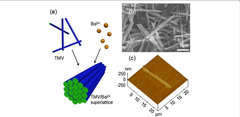
Very recently, we have reported that the newly synthe-sized hexagonally packed TMV/Ba2+ superlattice mater-ial can be formed in aqueous solution [13,14]. Figure 1 shows the schematic of the superlattice formation by hexagonal packing of TMVs, triggered by Ba ions, and the images observed from field emission scanning elec-tron microscopy (FESEM) and atomic force microscopy (AFM). The sample we used for this experiment was tens of microns in length, 2 ~ 3 microns in width (from FESEM), and several hundred nanometers in height (from AFM height image). It is known that the superlat-tice exhibits physical and mechanical properties that dif-fer significantly from its constituent materials [15-20]. The study on the viscoelastic properties of the TMV-derived nanostructured materials is still lacking despite the availability of the elastic property of the TMV and TMV-based nanotube composites [7]. The viscoelasticity of micro/nanobioarchitecture significantly affects the tis-sue regeneration [21] and repair [22], cell growth and aging [23], and human stem cell differentiation [24] as well as the appropriate biological functions of the
* Correspondence:xinnan.wang@ndsu.edu 1
Department of Mechanical Engineering, North Dakota State University, Fargo, ND 58108, USA
Full list of author information is available at the end of the article
membranes within a specific nanoenvironment [25]; in particular, the viscoelasticity of some viruses plays key roles in the capsid expansion for releasing nucleic acid and modifying protein cages for vaccine delivery pur-poses [26]. Specifically, for TMV superlattice, its nano-tube structure makes it a perfect biotemplate for synthesizing nanolattices that have been confirmed to possess extraordinary mechanical features with ultralow density [27,28]. Considering the biochemical functions of the TMV, its superlattice is an excellent candidate for bone scaffolding where the time-dependent mechanical properties become determinant [29], and research on scaffolding materials remains a hotspot [30]. Apart from contributing to the application of TMV superlattice, this work also pioneered in the viscoelasticity study of virus and virus-based materials. By far, most literature on viral viscoelasticity has been focused on the dynamic proper-ties of virus suspensions or solutions [31-34]. One of the rare viscoelasticity studies on individual virus particle is the qualitative characterization of the viscoelasticity of the cowpea chlorotic mottle virus [26] using quartz crystal microbalance with dissipation technique, which presents only the relative rigidity between two samples. To date, lit-tle literature is available on the quantitative study of the viscoelasticity of individual virus/virus-based particles. Considering the potential uses of TMV/Ba2+superlattice, its viscoelastic properties and responses under different mechanical stimuli need to be investigated.
A number of techniques for measuring the viscoelasticity of macro-scale materials have been used. A comprehensive
review of those methods can be found in the literature [35] that addresses the principles of viscoelasticity and experi-mental setup for time- and frequency-domain measure-ments. When the sample under investigation is in micro or even nanometer scale, however, the viscoelastic measure-ments become much more complicated. In dynamic methods, shear modulation spectroscopy [36] and mag-netic bead manipulation [37] are two common methodolo-gies to obtain the micro/nanoviscoelastic properties. To improve the measurement accuracy, efforts have been made to assess the viscoelasticity of micro/nanomaterials using contact-resonance AFM [38-41]. The adhesion be-tween the AFM probe tip and sample, however, is usually neglected. Furthermore, in order for the dynamic method to obtain a sinusoidal stress response, the applied strain amplitude must be kept reasonably small to avoid chaotic stress response and transient changes in material proper-ties [42]. In addition, the dynamic properproper-ties are frequency dependent, which is time consuming to map the viscoelas-ticity over a wide range of frequencies. An alternative way to measure the viscoelastic response of a material is the transient method. Transient indentation with an indenter was developed based on the functional equation methods [43], where the loading or traveling histories of the in-denter need to be precisely programmed.
[image:2.595.59.538.90.324.2]displacement. To realize the transient indentation in AFM, we introduced a novel experimental method. Viscoelas-tic nanoindentation theories were then developed based on the functional equation method [44]. The adhesion between the AFM tip and the sample, which signifi-cantly affected the determination of the viscoelastic properties [45], was included in the indentation model [20]. The viscoelastic responses of the sample with re-spect to different mechanical stimuli, including stress relaxation and strain creep, were further studied. The transition from transient properties to dynamic proper-ties was also addressed.
Methods
The TMV/Ba2+ superlattice solution was obtained from the mixture of the TMV and BaCl2solution (molar ratio of Ba2+/TMV = 9.2 × 10⁴:1) as stated in the reference [13]. It was further diluted with deionized water (volume ratio 1:1). A 10-μL drop of the diluted solution on a sili-con wafer was spun at 800 rpm for 10 s to form a mono-layer dispersion of the sample. The sample was dried for 30 min under ambient conditions (40% R.H., 21°C) for AFM (Dimension 3100, Bruker, Santa Barbara, CA, USA) observation and subsequent indentation tests.
The sample was observed with FESEM and AFM. The indentation was performed using the AFM nanoindenta-tion mode (AFM probe type: Tap150-G, NanoAndMore USA, Lady's Island, SC, USA). The geometry of the can-tilever was precisely measured using FESEM (S-4700, Hitachi, Troy, MI, USA), with a length of 125μm, width of 25μm, and thickness of 2.1 μm. To accurately meas-ure the tip radius, the tip was scanned on the standard AFM tip characterizer (SOCS/W2, Bruker) and the scanned data was curve fitted using PSI-Plot (Poly Software International, Orangetown, NY, USA). The tip radius calculated to be 12 nm. For a typical indentation test, the tip was pressed onto the top surface of the sample until a predefined force of ~100 nN. The cantilever end remained unchanged in position during the controlled delay time. A series of indentations of the same prede-fined indentation force and different delay times were performed to track the viscoelastic responses. A 10-min time interval of the two consecutive indentations was set for the sample to fully recover prior to the next in-dentation. The sample drift was minimized by turning off the light bulb in the AFM controller during scanning to keep the AFM chamber temperature constant and by shrinking the scan area gradually down to 1 nm × 1 nm on the top surface of the sample to rid the scanner piezo of the hysteresis effect.
Mathematical formulation
Derived from the functional equation method and the standard solid model (shown in the ‘Appendix’), the
differential equation governing the contact behavior of viscoelastic bodies can be obtained as
X2
i¼0 Ai ∂
i
∂ti
!
F tð Þ þ2πwR
½
¼4
ffiffiffi R
p
3
X2
i¼0 Bi ∂
i
∂ti
!
δ3 2ð Þt
ð1Þ
whereF(t) is the contact force history,δ(t) is the
inden-tation depth history, Ris the nominal radius of the two
contact spheres,wis the adhesive energy density,Aiand
Bi (i= 0, 1, 2) are the parameters determined by the
mechanical properties of two contact bodies, and the calculation of all these parameters can be found in the ‘Appendix.’
The elastic moduliE1andE2and viscosityηin Figure 2 are implicitly included in the above differential equation. To determine E1, E2, and η, besides experimental data for tandF, the function of the force historyF(t) is also required. The experimental data of t and F can be ob-tained as indicated in Figure 3. The force relaxation can be found in Figure 3a where the force decrease between the right ends of extension and retraction curves. By mapping the force decrease at different delay times as shown using the red asterisks in Figure 3b, the force re-laxation curve can be obtained, which decreases from 104 to 40 nN. The function ofF(t) can be obtained from Equation (1). Not only is Equation (1) applicable for the standard solid model in Figure 2(a) where it is derived from, but also it can be used for the modified standard solid model in Figure 2(b) where the elastic component of E1 is replaced by two elastic components in series. With this modification, the deflection of the cantilever can be incorporated into the deformation of the imagin-ary sample which is represented by the modified stand-ard solid model where the elastic component of E1c in Figure 2(b) denotes the cantilever and the rest compo-nents denote the TMV/Ba2+superlattice.
To be clearer, δ is substituted by D which represents the combined deformation. The relative approach, D, can be written as
Dð Þ ¼t D0H tð Þ ð2Þ
whereH(t) is the Heaviside unit step function and D0is
the relative approach between the substrate and the end of the cantilever.
Thus, Equation (1) can be rewritten as
X2
i¼0
Ai∂ i ∂ti
!
F tð Þ þ2πwR
ð Þ ¼ 4pffiffiffiR=3
D32
0
X2
i¼0
Bi∂ i ∂ti
! H tð Þ
ð3Þ
Applying Laplace transform, it yields
A0þA1sþA2s2
^
F sð Þ þ2πwR s
¼4pffiffiffiR=3D32
0 B0þB1sþB2s2
1
s ð4Þ
where a function with ‘∧’ denotes Laplace-transformed
function insdomain.
Performing inverse Laplace transform, the viscoelastic equation of AFM-based indentation becomes
F tð Þ ¼4 ffiffiffiffiffiffiffiffiffi
D3 0R q
3 Are
−αtþB
re−βtþCr
[image:4.595.59.539.88.315.2]
−2πwR ð5Þ Figure 2Standard solid model and modified standard solid model. (a)Schematic of the standard solid model for the TMV/Ba2+superlattice sample.(b/c)Modified standard solid model with the cantilever denoted by the blue spring and the sample denoted by the red springs and dashpot.
Figure 3Indentation force. (a)Indentation force decrease with delay time set as 100 ms, 200 ms, 500 ms, and 1,000 ms, respectively.(b)
[image:4.595.58.540.500.706.2]where
Ar¼ G
2 1 G1þG2;Br
¼ 27G21K21
3K1þ4G1
ð Þð3K1G1þ3K1G2þ4G1G2Þ
Cr¼
4G1G2
G1þG2 1−
3G1G2
3K1G1þ3K1G2þ4G1G2
;
α¼G1þηG2; β¼G2η þη 3K1G1
3K1þ4G1
ð Þ
Solution to AFM-based indentation equation
It is observed from Figure 3 that the initial indentation force at t= 0 was measured to be 104.21 nN, then the force started to decrease and then remained constant at 38 nN after ~5,000 ms. The force decrease shown as red asterisks in Figure 3b fits qualatitatively well with the ex-ponential function of Equation (5). E1,E2, and η, corre-sponding to the mechanical property parameters in Figure 2(a), can then be determined by fitting Equation (5) with the experimental data.
From the indentation data, D0 is obtained to be 78.457 nm. The pull-off force, 2πwR, calculated by aver-aging the pull-off forces of multiple indentations on the sample, is 16 nN. In comparison with the radius of the AFM tip, the surface of the sample can be treated as a flat plane. Hence, the nominal radiusR = Rtip=12 nm.
By invoking the force values at t =0, t=∞, and any intermediate point into Equation (5), the elasticity and viscosity components can be determined to be E1= 32.0 MPa,E2=21.3 MPa, and η=12.4 GPa ms. The co-efficient of determinationR2of the viscoelastic equation and the experimental data is ~0.9639.
Since the stress relaxation process is achieved by mod-eling a combination of the cantilever and the sample,
the viscoelasticity of the sample can be obtained by sub-tracting the component of the cantilever from the re-sults. The cantilever, acting as a spring, is in series with the sample, represented by a standard solid model. The schematic of the series organization is shown in Figure 2 (b). Thus the component of E1 comprises of E1s repre-senting the elastic part from the sample and E1c repre-senting the elastic part from the cantilever. To clarify the sources of the components in the modified standard solid model,E2,v2, andηin Figure 2(a) are now respect-ively denoted byE2s,v2s, andηsin Figure 2(b), where the subscript‘s’denotes the sample.
At the onset of indentation, only the spring with elastic modulus ofE1takes the instantaneous step load; therefore, the elastic modulus ofE1scan be determined from the ex-perimental data of zero-duration indentation. Applying the DMT model [46] with the force-displacement rela-tionship of the cantilever,
F¼kδcantilever ð6Þ
we can obtain the elastic equation of AFM-based indentation
δ¼F
kþ
Fþ2πwR EpffiffiffiR
2
3
ð7Þ
wherekis the spring constant of the cantilever, which is
5 nN/nm based on Sader's method [47] to calibrate k,
δcantilever is the cantilever deflection, and δ is recorded
directly as the Z-piezo displacement by AFM.
[image:5.595.56.541.90.233.2] [image:5.595.62.269.302.430.2]Results and discussion
Based on the solution obtained, the viscoelastic equation of AFM-based indentation for TMV/Ba2+superlattice is written as
F tð Þ ¼3:2098 0:007e−0:019312:4tþ0:0136e−0:016312:4tþ0:0168
−16
ð8Þ
The force decrease curve is shown in Figure 3b with the experimental data.
Specifically, for the TMV/Ba2+ superlattice whose viscoelastic behavior is simulated by a standard solid model, the differential equation governs its stress-strain behavior and becomes
_
σþE2s
ηs
σ ¼E1sE2s
ηs
εþðE1sþE2sÞε_ ð9Þ
whereE1s=3 GPa,E2s= 21.3 MPa, andηs=12.4GPa ms.
In the standard solid model, the initial experimental data point is determined by the instantaneous elastic modulus E1s. For the indentation that is held for over 5,000 ms, the indentation force becomes steady at ~38 nN, when the force exerts on the two springs in series. In contrast to E1s, E2s is much smaller, as can be seen from the significant force decrease of from ~104 to ~38 nN. The tip traveled down 13.2 nm from the beginning
of indentation. It is noted that for our indentation test, the ratio of the maximum indentation depth to the sam-ple diameter is less than 10% [48,49]; the substrate effect to the elastic modulus calculation is neglected.
From the determined viscoelastic model, the mechanical response of the superlattice under a variety of mechanical loads can be predicted. Several simulation results were included as follows.
When the TMV/Ba2+ superlattice sample undergoes a uniformly constant tensile/compressive strain, the stress relaxation can be obtained from the standard solid model as below
σð Þ ¼t ε0 E1sþE2se−E2st=η
ð10Þ
whereε0is the constantly applied strain.
When the sample undergoes a uniformly constant tensile/compressive stress, the strain creep can then be obtained as
εð Þ ¼t σ0
1
E1sþ
1
E1sþE2s−
1
E
e−E1sE2st=ηsðE1sþE2sÞ
ð11Þ
whereσ0is the constantly applied stress.
The stress relaxationvs. applied strains and the strain creep vs. applied stresses are shown in Figure 6a,b, re-spectively. In Figure 6a, the stress reduces to a steady state after ~2 s when the applied strain is ~10%. In Figure 7b, strain increases to a steady value after ~5 s when the applied stress is ~ 1 GPa.
When the sample is indented with a spherical in-denter, the indentation depth history can be analytically obtained when a step force is applied. Similar to the procedures above where the force history of Equation (5) is obtained, a step force function is used as input, and the creep indentation depth history function can be derived as
d tð Þ ¼
3ðF0þ2πwRÞ
4pffiffiffiR
G1sþG2s
2G1sð1þ6K1sÞ
A0þB 0
ηe
−G2st
ηs þC
0 G1
sþG2s
ð Þ
ηs e
−ðG1sþG2sÞt
ηs
23
[image:6.595.58.291.90.256.2]ð12Þ where F0is the step force,A0¼G14sGþ1Gs2sþ3GK21ss
Figure 5Indentation force data as a function of Z-piezo displacement, a comparison of experimental measurement and fitted results.
B0¼4G1sG 2
2sηsð1−G1s−G2sÞ−3K1sηsG1sG2sðG1sþ2G2sþ1Þ þ 3K1sηs G22s−G21s−G32s
G2sðG1sþG2sÞðG1sþ2G2sÞ
C0¼4G1sG2sηs−3K1sηsG1s−3K1sηsG 2 2s
G1sþG2s
ð Þ2
G1sþ2G2s
ð Þ −
4G1sG2sηs
G1sþ2G2s
ð ÞðG1sþG2sÞ−
3K1sηsG1s
G1sþG2s
ð Þ2þ
3K1sηsðG2s−G1sÞ
G2sðG1sþG2sÞðG1sþ2G2sÞþ
3K1sηsG1s
G2sðG1sþG2sÞ2
G1s¼2 1E1þs
v1s
ð Þ;K1s¼3 1E1−2s
v1s
The indentation force history has been obtained in Equation (5), where the elastic shear modulus G1 as a combined elastic response of two springs shown in Figure 2(b) should be replaced byG1sof one spring only. Then, the simulated curves for the two situations can be found in Figures 6c,d. It is concluded that the creep depth variation under different forces gets larger through creep while the indentation force variation under differ-ent depths gets smaller through relaxation. Particularly, in Figure 6d, the force finally decreases to negative values, which represent attractive forces. The attraction cannot be found when G1sandG2s are very small. This phenomenon can be interpreted by the conformability of materials determined by the elastic modulus. When
G1s and G2s get smaller, the materials are more con-formable. Accordingly, in the final equilibrium state, the materials around the indenter tend to be more de-formable to enclose the spherical indenter. This will re-sult in a smaller attraction.
[image:7.595.58.540.88.421.2] [image:7.595.57.292.479.715.2]In addition, the example of shear dynamic experiment is simulated to obtain the storage and loss moduli of Figure 6Stress relaxation, strain creep, and indention depth creep and force relaxation. (a)Stress relaxation of TMV/Ba2+superlattice under uniform tensile/compressive strains.(b)strain creep under uniform tensile/compressive stresses.(c)Indentation depth creep with a rigid spherical indenter (R= 12 nm) under constant forces.(d)Indentation force relaxation with a rigid spherical indenter (R= 12 nm) under constant indentation depths.
TMV/Ba2+superlattice. The storage and loss shear mod-uli are calculated by [42]
G0ð Þ ¼ω ω Z∞
0
Gsð Þt sinωtdt ð13Þ
G}ð Þ ¼ω ω Z∞
0
Gsð Þt cosωtdt ð14Þ
whereG′and G″are storage and loss moduli,
respect-ively, ω is the angular velocity which is related to
the frequency of the dynamic system, and Gsð Þ ¼t
G1sþG2se−G2st=η is the shear stress relaxation
modu-lus, determined by the ratio of shear stress and con-stant shear strain.
Based on the relation between the transient and dy-namic viscoelastic parameters in Equations (13) and (14), the storage and loss shear moduli are finally deter-mined to be
G0ð Þ ¼ω ω 2G2
sη2s
G22sþω2η2s
ð15Þ
G″ð Þ ¼ω G 2 2sωηs
G22sþω2η2s
ð16Þ
whereG2s=E2s/ 2(1 +v2s).
Figure 7 shows the curves of storage and loss shear moduli vs. the angular velocity. The storage shear modulus,G′, increases with the increase of angular vel-ocity, while the increasing rate ofG′decreases and the angular velocity of ~2 rad/s is where the increasing rate changes most drastically. However, the loss shear modulus,G″, first increases and then decreases reach-ing the maximum value, ~3.9 MPa, at the angular vel-ocity of ~0.7 rad/s. The storage and loss moduli in other cases as uniform tensile, compressive, and inden-tation experiments can also be obtained.
Conclusions
This paper presented a novel method to characterize the viscoelasticity of TMV/Ba2+ superlattice with the AFM-based transient indentation. In comparison with previ-ous AFM-based dynamic methods for viscoelasticity measurement, the proposed experimental protocol is able to extract the viscosity and elasticity of the sample. Furthermore, the adhesion effect between the AFM tip and the sample was included in the indentation model. The elastic moduli and viscosity of TMV superlattice were determined to be E1s=2.14 GPa, E2s= 21.3 MPa, and ηs=12.4 GPa∙ms. From the characterized viscoelas-tic parameters, it can be concluded that the TMV/Ba2+ superlattice was quite rigid at the initial contact and
then experienced a large deformation under a constant pressure. Finally, the simulation of the mechanical be-havior of TMV/Ba2+ superlattice under various loading cases, including uniform tension/compression and nano-indentation, were conducted to predict the mechanical response of sample under different loadings. The storage and loss shear moduli were also demonstrated to extend the applicability of the proposed method. With the char-acterized viscoelastic properties of TMV superlattice, we are now able to predict the process of tissue regener-ation around the superlattice where the time-dependent mechanical properties of scaffold interact with the growth of tissue.
Appendix
Modeling of adhesive contact of viscoelastic bodies The functional equation method was employed to de-velop a contact mechanics model for indenting a visco-elastic material with adhesion. A modified standard solid model was used to extract the viscous and elastic param-eters of the sample.
Several adhesive contact models are available, such as Johnson-Kendall-Roberts (JKR) model [50], Derjaguin-Muller-Toporov (DMT) model [46], etc. [51-53]. De-tailed comparisons can be found in reference [54]. As the DMT model results in a simpler differential equa-tion, it was used in this study for the simulation to solve the indentation on an elastic body with adhesion.
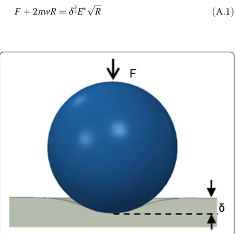
[image:8.595.305.540.474.707.2]For the DMT model [46], the relation between the in-dentation force F and relative approach δ, shown in Figure 8, can be expressed as
Fþ2πwR¼δ32EpffiffiffiR ðA:1Þ
whereRis the nominal radius of the two contact spheres
of R1 and R2, given by R=R1R2/(R1+R2); the adhesive
energy density w is obtained from the pull-off force Fc,
where Fc= 3πwR/2; and the reduced elastic modulus E*
is obtained from the elastic modulus Es and Poisson's
ratio νs of the sample by E¼4Es= 3 1−v2s
with the assumption that the elastic modulus of the tip is much larger than that of the sample.
In Equation (A.1), E*, which governs the contact de-formation behavior, is decided by the sample's mechan-ical properties. In the functional equation method [43],
E* needs to be replaced by its equivalence in the visco-elastic system, so that the contact deformation behavior can be governed by the viscoelastic properties. To achieve it, the elastic/viscoelastic constitutive equations are needed.
As a premise of the functional equation method, quasi-static condition is assumed so that the inertial forces of deformation can be neglected [43,44]. The gen-eral constitutive equations for a linear viscoelastic/elastic system in Cartesian coordinate configuration can be written as
Pdsij¼Qdeij ðA:2Þ
Pmσkk ¼Qmεkk ðA:3Þ
wheresij, eij,σkk, andεkk are the deviatoric stress, strain,
mean stress, and strain, respectively. The linear
opera-torsPd,Qd,Pm, andQmcan be expressed in the form of
Pd¼XN
1
i¼0 pd
i
∂i
∂ti; Qd¼
XN2
i¼0 qd
i ∂i
∂ti ðA:4aÞ
Pm¼X
N3
i¼0 pmi
∂i
∂ti; Qm¼
XN4
i¼0 qmi
∂i
∂ti ðA:4bÞ
where i (i =0, 1, 2,…) is determined by the
viscoelas-tic model to be selected, t is time, and pd
i, qdi, pmi ,
and qm
i are the components related to the materials
property constants, such as elastic modulus and
Pois-son's ratio etc.
For a pure elastic system, the four linear operators are reduced to
Pd¼pd0; Qd¼qd0; Pm¼pm0; Qm¼qm0 ðA:5Þ
which, according to the elastic stress-strain relations, are correlated as
qd
0 pd
0
¼2G¼Q
d
Pd; qm
0 pm
0
¼3K¼Q
m
Pm ðA:6Þ
where Gand K are the shear modulus and bulk
modu-lus, respectively.
Combining Equation (A.6) with
G¼ E
2 1ð þvÞ;K ¼
E
3 1ð −2vÞ ðA:7Þ
the reduced elastic modulus can be expressed by the elastic linear operators as
E¼4 q
d
0pm0qd0þ2pd0qm0qd0
3 2qd
0pm0pd0þpd0pd0qm0
¼4 QdPmQdþ2PdQmQd
3 2 QdPmPdþPdPdQm
ðA:8Þ
Hence, Equation (A.1) becomes
2QdPmPdþPdPdQm
F tð Þ þ2πwR
½ ¼4 ffiffiffi R p 3 Q
dPmQdþ2PdQmQd
δ3
2ð Þt ðA:9Þ
To evolve the elastic solution into a viscoelastic solu-tion, the linear operators in the viscoelastic system need to be determined. To this end, the standard solid model, shown in Figure 2(a), was used to simulate the viscoelas-tic behavior of the sample, since both the instantaneous and retarded elastic responses can be reflected in this model, which well describes the mechanical response of most viscoelastic bodies.
It is customary to assume that the volumetric response under the hydrostatic stress is elastic deformation; thus, it is uniquely determined by the spring in series [55]. Hence, the four linear operators for the standard solid model can be expressed as
Pd¼1þpd1
∂ ∂t;Q
d¼qd
0þqd1
∂ ∂t; Pm¼1;Qm ¼3K1
ðA:10Þ
where pd
1¼G1þηG2; qd0¼G2G1þ1GG22; q
d 1¼
2G1η
G1þG2; G1¼
E1
2 1ðþv1Þ; G2¼ E2
2 1ðþv2Þ; K1¼
E1
3 1ð−2v1Þ,E1,E2,v1, andv2are the elas-tic modulus and Poisson's ratio of the two elaselas-tic
com-ponents, respectively, shown in Figure2.
Plugging Equation (A.10) into Equation (A.9), the rela-tion between F(t) and δ(t) can be found. The functional differential equation that extends the elastic solution of indentation to viscoelastic system is obtained
X2
i¼0 Ai ∂
i
∂ti
!
F tð Þ þ2πwR
½ ¼4 ffiffiffi R p 3 X2
i¼0 Bi ∂
i
∂ti
!
δ3 2ð Þt
ðA:11Þ
where A0= 2q0+ 3K1, A1=p1(3K1+ 2q0) + (3p1K1+ 2q1),
A2=p1(3p1K1+ 2q1), B0=q0(1 + 6K1), B1=q0(p1+ 6K1p1) +
Abbreviations
AFM:atomic force microscopy; DMT: Derjaguin-Muller-Toporov; FESEM: field emission scanning electron microscopy; JKR: Johnson-Kendall-Roberts; PVA: polyvinyl alcohol; TMV: tobacco mosaic virus.
Competing interests
The authors declare that they have no competing interests.
Authors' contributions
HW carried out the experiment and drafted the manuscript. XW supervised and guided the overall project and involved in drafting the manuscript. TL and BL provided the FESEM analysis on the sample. All authors read and approved the final manuscript.
Acknowledgements
Funding support is provided by ND NASA EPSCoR FAR0017788. Use of the Advanced Photon Source, Electron Microscopy Center, and Center of Nanoscale Materials, an Office of Science User Facilities operated for the U. S. Department of Energy (DOE) Office of Science by Argonne National Laboratory, was supported by the U.S. DOE under Contract No. DE-AC02-06CH11357.
Author details
1
Department of Mechanical Engineering, North Dakota State University, Fargo, ND 58108, USA.2X-ray Science Division, Advanced Photon Source of
Argonne National Laboratory, 9700 S. Cass Avenue, Argonne, IL 60439, USA.
Received: 7 May 2014 Accepted: 6 June 2014 Published: 13 June 2014
References
1. Zaitlin M:Discoveries in Plant Biology, ed S D K a S F Yang.HongKong: World Publishing Co., Ltd; 1998:105–110.
2. Hou CX, Luo Q, Liu JL, Miao L, Zhang CQ, Gao YZ, Zhang XY, Xu JY, Dong ZY, Liu JQ:Construction of GPx active centers on natural protein nanodisk/ nanotube: a new way to develop artificial nanoenzyme.ACS Nano2012, 6:8692–8701.
3. Hefferon KL:Plant virus expression vectors set the stage as production platforms for biopharmaceutical proteins.Virology2012,433:1–6. 4. Atanasova P, Rothenstein D, Schneider JJ, Hoffmann RC, Dilfer S, Eiben S,
Wege C, Jeske H, Bill J:Virus-templated synthesis of ZnO nanostructures and formation of field-effect transistors.Adv Mater2011,23:4918–4922. 5. Balci S, Bittner AM, Hahn K, Scheu C, Knez M, Kadri A, Wege C, Jeske H, Kern
K:Copper nanowires within the central channel of tobacco mosaic virus particles.Electrochim Acta2006,51:6251–6257.
6. Klug A:The tobacco mosaic virus particle: structure and assembly.Philos Trans Biol Sci1999,354:531–535.
7. Wang XN, Niu ZW, Li SQ, Wang Q, Li XD:Nanomechanical characterization of polyaniline coated tobacco mosaic virus nanotubes.J Biomed Mater Res A2008,87A:8–14.
8. Lee LA, Nguyen QL, Wu LY, Horyath G, Nelson RS, Wang Q:Mutant plant viruses with cell binding motifs provide differential adhesion strengths and morphologies.Biomacromolecules2012,13:422–431.
9. Petrie TA, Raynor JE, Dumbauld DW, Lee TT, Jagtap S, Templeman KL, Collard DM, Garcia AJ:Multivalent integrin-specific ligands enhance tissue healing and biomaterial integration.Sci Transl Med2010,2:1–6. 10. Kaur G, Wang C, Sun J, Wang Q:The synergistic effects of multivalent
ligand display and nanotopography on osteogenic differentiation of rat bone marrow stem cells.Biomaterials2010,31:5813–5824.
11. Kaur G, Valarmathi MT, Potts JD, Jabbari E, Sabo-Attwood T, Wang Q: Regulation of osteogenic differentiation of rat bone marrow stromal cells on 2D nanorod substrates.Biomaterials2010,31:1732–1741. 12. Wu LY, Zang JF, Lee LA, Niu ZW, Horvatha GC, Braxtona V, Wibowo AC,
Bruckman MA, Ghoshroy S, zur Loye HC, Li XD, Wang Q:Electrospinning fabrication, structural and mechanical characterization of rod-like virus-based composite nanofibers.J Mater Chem2011,21:8550–8557. 13. Li T, Winans RE, Lee B:Superlattice of rodlike virus particles formed in
aqueous solution through like-charge attraction.Langmuir2011, 27:10929–10937.
14. Li T, Zan X, Winans RE, Wang Q, Lee B:Biomolecular assembly of thermoresponsive superlattices of the tobacco mosaic virus with large tunable interparticle distances.Angew Chem Int Ed2013,52:6638–6642. 15. Agrawal BK, Pathak A:Oscillatory metallic behaviour of carbon nanotube
superlattices - an ab initio study.Nanotechnology2008,19:135706–135706. 16. Hultman L, Engstrom C, Oden M:Mechanical and thermal stability of TiN/
NbN superlattice thin films.Surface Coatings Technol2000,133:227–233. 17. Jaskolski W, Pelc M:Carbon nanotube superlattices in a magnetic field.
Int J Quantum Chem2008,108:2261–2266.
18. Wu MJ, Wen HC, Wu SC, Yang PF, Lai YS, Hsu WK, Wu WF, Chou CP: Nanomechanical characteristics of annealed Si/SiGe superlattices.
Appl Surf Sci2011,257:8887–8893.
19. Xu JH, Li GY, Gu MY:The microstructure and mechanical properties of TaN/TiN and TaWN/TiN superlattice films.Thin Solid Films2000,370:45–49. 20. Wang HR, Wang XN, Li T, Lee B:Nanomechanical characterization of
rod-like superlattice assembled from tobacco mosaic viruses.J Appl Phys
2013,113(024308):1–6.
21. Belfiore LA, Floren ML, Paulino AT, Belfiore CJ:Stress-sensitive tissue regeneration in viscoelastic biomaterials subjected to modulated tensile strain.Biophys Chem2011,158:1–8.
22. Coulombe PA, Wong P:Cytoplasmic intermediate filaments revealed as dynamic and multipurpose scaffolds.Nat Cell Biol2004,6:699–706. 23. Drozdov AD:Viscoelastic Structures: Mechanics of Growth and Aging.San
Diego, CA, the United States: Academic Press; 1998.
24. Tan SCW, Pan WX, Ma G, Cai N, Leong KW, Liao K:Viscoelastic behaviour of human mesenchymal stem cells.BMC Cell Biol2008,9:40–40. 25. Rico F, Picas L, Colom A, Buzhynskyy N, Scheuring S:The mechanics of
membrane proteins is a signature of biological function.: Soft: Matter; 2013. 26. Rayaprolu V, Manning BM, Douglas T, Bothner B:Virus particles as active
nanomaterials that can rapidly change their viscoelastic properties in response to dilute solutions.Soft Matter2010,6:5286–5288.
27. Jang D, Meza LR, Greer F, Greer JR:Fabrication and deformation of three-dimensional hollow ceramic nanostructures.Nat Mater2013,12:893–898. 28. Schaedler TA, Jacobsen AJ, Torrents A, Sorensen AE, Lian J, Greer JR,
Valdevit L, Carter WB:Ultralight metallic microlattices.Science2011, 334:962–965.
29. Bawolin NK, Chen XB, Zhang WJ:A method for modeling time-dependant mechanical properties of tissue scaffolds. In2007 IEEE International Conference on Mechatronics and Automation, Vols I-V, IEEE Conference Proceedings, Harbin, Heilongjiang, China; 2007:1423–1427.
30. Leung LH, Naguib HE:Characterization of the viscoelastic properties of poly(epsilon-caprolactone)-hydroxyapatite microcomposite and nanocomposite scaffolds.Polym Eng Sci2012,52:1649–1660. 31. Nemoto N, Schrag JL, Ferry JD, Fulton RW:Infinite-dilution viscoelastic
properties of tobacco mosaic-virus.Biopolymers1975,14:409–417. 32. Graf C, Kramer H, Deggelmann M, Hagenbuchle M, Johner C, Martin C,
Weber R:Rheological properties of suspensions of interacting rodlike Fd-virus particles.J Chem Phys1993,98:4920–4928.
33. Huang F, Rotstein R, Fraden S, Kasza KE, Flynn NT:Phase behavior and rheology of attractive rod-like particles.Soft Matter2009,5:2766–2771. 34. Schmidt FG, Hinner B, Sackmann E, Tang JX:Viscoelastic properties of
semiflexible filamentous bacteriophage fd.Phys Rev E2000,62:5509–5517. 35. Lakes RS:Viscoelastic measurement techniques.Rev Sci Instrum2004,
75:797–810.
36. Wahl KJ, Stepnowski SV, Unertl WN:Viscoelastic effects in nanometer-scale contacts under shear.Tribol Lett1998,5:103–107.
37. MacKintosh FC, Schmidt CF:Microrheology.Curr Opin Colloid Interface Sci
1999,4:300–307.
38. Mahaffy RE, Shih CK, MacKintosh FC, Kas J:Scanning probe-based frequency-dependent microrheology of polymer gels and biological cells.Phys Rev Lett2000,85:880–883.
39. Yuya PA, Hurley DC, Turner JA:Contact-resonance atomic force microscopy for viscoelasticity.J Appl Phys2008,104:074916-1–7. 40. Yablon DG, Gannepalli A, Proksch R, Killgore J, Hurley DC, Grabowski J, Tsou
AH:Quantitative viscoelastic mapping of polyolefin blends with contact resonance atomic force microscopy.Macromolecules2012,45:4363–4370. 41. Herbert EG, Oliver WC, Pharr GM:Nanoindentation and the dynamic
characterization of viscoelastic solids.J Phys D Appl Phys2008, 41:074021-1–9.
43. Radok JRM:Visco-elastic stress analysis.Quart Appl Math1957,15:198–202. 44. Lee EH:Stress analysis in visco-elastic bodies.Quart Appl Math1955,
13:183–190.
45. Gupta S, Carrillo F, Li C, Pruitt L, Puttlitz C:Adhesive forces significantly affect elastic modulus determination of soft polymeric materials in nanoindentation.Mater Lett2007,61:448–451.
46. Derjaguin BV, Muller VM, Toporov YP:Effect of contact deformations on adhesion of particles.J Colloid Interface Sci1975,53:314–326. 47. Sader JE, Larson I, Mulvaney P, White LR:Method for the calibration of
atomic force microscope cantilevers.Rev Sci Instrum1995,66:3789–3798. 48. Gamonpilas C, Busso EP:On the effect of substrate properties on the
indentation behaviour of coated systems.Mater Sci Eng A Struct Mater Properties Microstruct Process2004,380:52–61.
49. Tsui TY, Pharr GM:Substrate effects on nanoindentation mechanical property measurement of soft films on hard substrates.J Mater Res1999, 14:292–301.
50. Johnson KL, Kendall K, Roberts AD:Surface energy and contact of elastic solids.Proc Royal Soc Lond A Math Phys Sci1971,324:301–313.
51. Maugis D:Extension of the Johnson-Kendall-Roberts theory of the elastic contact of spheres to large contact radii.Langmuir1995,11:679–682. 52. Maugis D:Adhesion of spheres - the jkr-dmt transition using a Dugdale
model.J Colloid Interface Sci1992,150:243–269.
53. Sneddon IN:The relation between load and penetration in the axisymmetric Boussinesq problem for a punch of arbitrary profile.Int J Engng Sci1965, 3:47–57.
54. Johnson KL, Greenwood JA:An adhesion map for the contact of elastic spheres.J Colloid Interface Sci1997,192:326–333.
55. Malvern LE:Introduction to the mechanics of a continuous medium.
Englewood Cliffs, New Jersey: Prentice-Hall, Inc; 1969.
doi:10.1186/1556-276X-9-300
Cite this article as:Wanget al.:Transient viscoelasticity study of tobacco mosaic virus/Ba2+superlattice.Nanoscale Research Letters
20149:300.
Submit your manuscript to a
journal and benefi t from:
7 Convenient online submission
7 Rigorous peer review
7 Immediate publication on acceptance
7 Open access: articles freely available online
7 High visibility within the fi eld
7 Retaining the copyright to your article