REVIEW
Spindle orientation: a question of complex positioning
Dan T. Bergstralh1,*, Nicole S. Dawney1and Daniel St Johnston2ABSTRACT
The direction in which a cell divides is determined by the orientation of its mitotic spindle at metaphase. Spindle orientation is therefore important for a wide range of developmental processes, ranging from germline stem cell division to epithelial tissue homeostasis and regeneration. In multiple cell types in multiple animals, spindle orientation is controlled by a conserved biological machine that mediates a pulling force on astral microtubules. Restricting the localization of this machine to only specific regions of the cortex can thus determine how the mitotic spindle is oriented. As we review here, recent findings based on studies in tunicate, worm, fly and vertebrate cells have revealed that the mechanisms for mediating this restriction are surprisingly diverse.
KEY WORDS: Asymmetric cell division, Mitotic spindle, Spindle orientation
Introduction
Spindle orientation plays a crucial role in animal development. In particular, its importance for asymmetric cell division (ACD) has long been recognized. The term ACD is typically used to mean that the two products of cell division have different fates. This can be a cell-intrinsic process that relies on the asymmetric distribution of cell fate factors, such that only one daughter cell inherits proteins or RNAs that determine its identity (Fig. 1). Multiple cell types use oriented divisions to direct this distribution. For example, in the one-cell stage Caenorhabditis elegans embryo, cell fate factors are differentially localized at either the anterior or posterior of the cell, and the orientation of the spindle, along with its position along the anterior-posterior (A-P) axis, ensures that these factors are partitioned unequally (reviewed by Gönczy and Rose, 2005). A similar mechanism for asymmetric segregation of fate determinants has been observed in Drosophila neuroblasts and sensory organ precursor ( pI) cells, and more recent work suggests that it also functions inDrosophilaintestinal stem cells (reviewed by Knoblich, 2010; Morin and Bellaïche, 2011; Siller and Doe, 2009; see also Goulas et al., 2012; Guo and Ohlstein, 2015). In the mollusc Ilyanassa obsoleta, cell fate-determining mRNAs associate with one of the two centrosomes, and are thus differentially inherited (Lambert and Nagy, 2002). Cell intrinsic division asymmetry has also been observed in vertebrate systems, namely in T lymphocytes and embryonic stem cells (Chang et al., 2007; Habib et al., 2013). In other instances, cell fate determination is cell extrinsic (Fig. 1). In these cases, division orientation controls cell fate not through differential distribution of factors, but rather by dictating the position in which each daughter cell is born within the tissue. This position,
meaning the microenvironment in which the cell comes to find itself, makes the cell more or less accessible to signaling factors that control differentiation. Extrinsic control is often seen in the case of germline stem cells that, in a number of organisms, reside within a specialized niche, where they are contacted by supporting somatic cells. Oriented divisions cause one daughter cell to leave the niche, going on to become a gamete, while the other remains (reviewed by Inaba and Yamashita, 2012; Spradling et al., 2011). Cell-extrinsic mechanisms also determine fate asymmetry in the developing vertebrate brain and in stratifying embryonic mammalian skin (reviewed by Paridaen and Huttner, 2014; Williams and Fuchs, 2013).
The importance of spindle orientation for symmetrically dividing cells is less well understood. It is known that cells within an epithelial monolayer tend to divide symmetrically (Fig. 1), orienting their divisions such that both daughters are born within the layer. In this way, the tissue expands as a two-dimensional sheet. It might therefore be predicted that the loss of spindle orientation control in the apical-basal (A-B) polarity axis of the tissue, which is perpendicular to the sheet, would generate tissue disorganization by causing daughter cells to be born outside the sheet. Studies that have addressed this possibility in monolayered epithelial tissues have instead found that spindle misorientation along the A-B axis is not disruptive. In most of the tissues examined so far, the misplaced cell simply reintegrates into the layer in a manner that depends on adhesion between neighbouring cells (Bergstralh et al., 2015; Strzyz et al., 2015). Another mechanism that protects tissues from the consequence of spindle misorientation is at work in theDrosophilaimaginal wing disc, where cells that are misplaced by misoriented divisions do not reintegrate but are instead extruded and die (Nakajima et al., 2013).
Although these findings suggest that spindle orientation in the A-B axis is inessential, additional work in the fruit fly shows that spindle orientation can help to determine the direction of tissue expansion, which is governed by mechanical tension across the tissue (Baena-López et al., 2005; Bosveld et al., 2016; Mao et al., 2011). This is consistent with earlier work implicating spindle orientation in determining the shape of epithelial tubes in the lung (Tang et al., 2011). Furthermore, a recent study has highlighted the broad extent to which directed spindle orientation in expanding epithelia is relevant across the animal kingdom. In this study, it was shown that cells in the developing epidermis of the tunicate C. intestinalis orient their spindles in an unusual way; a membrane invagination appears to capture the interphase centriole, anchoring it at one side of the cell until the spindle has formed and thereby ensuring that cells divide along the A-P axis of the organism (Negishi et al., 2016). Although the nature of this structure remains to be elucidated, it is likely to intersect with a well-studied molecular mechanism that controls spindle orientation in a variety of organisms and cell types.
Work in multiple tissues and organisms has identified a core cortical machinery that orients mitotic spindles at metaphase by exerting a pulling force on astral microtubules, essentially‘reeling’ them in until the spindle is aligned (Fig. 2A). This machinery consists of three conserved proteins: the first isDrosophila Mud (Mushroom body defective), C. elegans LIN-5 (abnormal cell
1Department of Biology, University of Rochester, Rochester, NY 14627, USA.2The
Gurdon Institute and the Department of Genetics, University of Cambridge, Tennis Court Road, Cambridge CB2 1QN, UK.
*Author for correspondence (dan.bergstralh@rochester.edu)
D.T.B., 0000-0003-1689-3715
DEVEL
O
lineage 5) and vertebrate NuMA (nuclear mitotic apparatus); the second is Drosophila Pins, C. elegans GPR1/2 (G-protein regulators 1 and 2) or vertebrate LGN (leucine-glycine-asparagine); the third isDrosophilaand vertebrate Gαi, which has two redundantC. elegansorthologues called GOA-1 and GPA-16 (Table 1). Together, these proteins are believed to secure a complex consisting of dynein/dynactin motor at the cell cortex, and thus localise the astral-microtubule pulling force. A model that describes their interaction is well established in the literature (reviewed by di Pietro et al., 2016). Briefly, Gαi, which attaches to the plasma membrane, anchors Pins/LGN by binding to its C-terminus (Du and Macara, 2004; Gotta and Ahringer, 2001; Kaushik et al., 2003; Nipper et al., 2007; Schaefer et al., 2000; Yu et al., 2003). The tetricopeptide repeats (TPRs) at the other end of Pins/LGN bind to Mud/NuMA, which itself binds to dynein (Bowman et al., 2006; Du et al., 2001; Izumi et al., 2006; Pecreaux et al., 2006; Siller et al., 2005; Yu et al., 2000). The importance of this machinery is highlighted by studies concerning its dysregulation, particularly with regard to cancer phenotypes. Indeed, loss of spindle orientation in the central nervous system has been associated with hyperplasia in both the fly and chick (de Belle and Heisenberg, 1996; Morin et al., 2007; Yu et al., 2006). In the fly imaginal wing disc, expression of a baculoviral anti-apoptotic protein in misplaced epithelial cells promotes tumour-like overgrowth (Guilgur et al., 2012; Nakajima et al., 2013).
In this Review, we discuss key aspects of the canonical spindle-orienting machinery and its function. First, we describe recent studies that raise questions about the regulation of astral microtubule ends at the cortex. Second, we describe a body of work addressed at defining how the spindle-orienting machinery is restricted to only certain regions of an epithelial cell cortex.
Astral microtubule plus tips: a consideration for spindle orientation
A recent study identified two proteins – small kinetochore associated protein (SKAP) and Astrin–at astral microtubule plus ends, and implicated these proteins in spindle orientation (Kern et al., 2016). SKAP and Astrin form a complex that is known to play a role at kinetochores, which mediate the attachment between
spindle microtubules and chromosomes (Dunsch et al., 2011; Schmidt et al., 2010). Their astral microtubule localization is probably determined directly by SKAP, which has an end-binding protein 1 (EB-1) motif that is required for plus-end localization (Kern et al., 2016). Intriguingly, in single HeLa cells, mutation of the EB-1-binding motif (yielding SKAPΔEB-1) causes spindle mispositioning (Kern et al., 2016). This effect is not caused by astral microtubule shortening; unlike disruption of EB-1 itself, deletion of the SKAP EB-1 motif does not affect astral microtubule length (Kern et al., 2016; Toyoshima and Nishida, 2007). Another potential explanation for mispositioning, namely that plus tips carry spindle-orienting factors to the cortex, is ruled out by the observation that cortical dynein is still observed in SKAPΔEB-1 cells (Kern et al., 2016). However, an additional possibility to consider is that the Astrin/SKAP complex mediates the interaction between astral microtubules and the cortical machinery, analogous to the role played by the complex at kinetochores. This model is supported by recent evidence identifying Astrin as a binding partner for the core machinery protein NuMA, although this study focused on the main spindle body (Chu et al., 2016).
The functional importance of an association between astral microtubule plus tips and the core machinery is not yet clear, but may relate to the mechanism of force generation. An isolated region of NuMA (called NuMA-TIP) shows microtubule tip-binding activity, remaining associated even during microtubule depolymerization (Seldin et al., 2016). The affinity of NuMA-TIP for microtubule tips is increased by gentle microtubule disruption but decreased by microtubule stabilization, indicating that it binds preferentially to shrinking microtubules (Seldin et al., 2016). Notably, in addition to dynein-mediated reeling, pulling can also be achieved through the regulation of microtubule dynamics. In this model, the microtubules hit the cortex end-on and are subsequently brought into alignment by controlled depolymerisation, i.e. the microtubules shorten until they are taut. Elegantin vitrowork has provided proof of principle for this mechanism and demonstrated that it can be mediated by dynein (Laan et al., 2012). In combination with the results described above, these findings raise the possibility that the spindle-orienting machinery regulates shrinkage rather than pulling. Additional functional analyses will be needed to clarify this notion.
Asymmetric Symmetric
Cell intrinsic Cell extrinsic
Differentiating ganglion mother cell
Self-renewing
cell
Neuroblast
Apical
Basal Follicular epithelium
Differentiating gonialblast
Self-renewing cell Male germline stem cell
Apical
Basal Cell fate
factors
[image:2.612.51.433.59.285.2]+
Fig. 1. Asymmetric and symmetric cell division.Directed cell division influences cell and tissue
development. This is illustrated using examples fromDrosophila
melanogaster. The neuroblast (left) uses a cell-intrinsic mechanism for asymmetric cell division (ACD), relying on spindle orientation for unequal distribution of cell fate factors (orange). In the male germline stem cell (centre), ACD is cell-extrinsic, using directed division to ensure that, when germline stem cells divide, only one daughter cell remains in proximity to the somatic hub cells (light green) that comprise the niche, while the other daughter cell differentiates into a gonialblast. By contrast, cell division in the follicular epithelium (right) is symmetric. Cells in this tissue divide to simply expand the monolayer. Green, microtubules; red, spindle poles; blue, DNA.
DEVEL
O
The cycling of core machinery components
Although the list of factors involved in spindle orientation is ever expanding, a long-standing question still remains: how is the spindle-orientation machinery restricted to specific regions of the cortex? Historically, this problem has been considered primarily with regard to cortical polarity-based cues. However, work performed in isolated cultured cells, which generally lack these cues, and in Drosophila neuroblasts has illuminated the role played by the mitotic apparatus (meaning the spindle and chromosomes) in regulating the spindle-orienting machinery (Kiyomitsu and Cheeseman, 2012; Siegrist and Doe, 2005). This activity means that the spindle can influence its own position in the cell (in single cells, this position is usually examined relative to the cell centre, rather than along a polarity axis). Proximity of the spindle-orienting machinery to the mitotic apparatus diminishes cortical pulling, resulting in a bipolar balance of forces. This is accomplished by signalling from the chromosomes and/or the spindle poles that reduces cortical localization and activity of the spindle-orienting machinery when the apparatus is close and/or the chromosomes are misaligned (Kiyomitsu and Cheeseman, 2012; Tame et al., 2016).
The mechanism of dynamic cortical localization of the spindle-orientation machinery in these cells may be clarified by evidence that LGN and Gαi are not glued to the membrane during mitosis, but rather cycle back and forth between the cortex and the spindle poles (Zheng et al., 2013). Their transit from the cortex to the poles relies on dynein and astral microtubules (Zheng et al., 2013). This indicates that dynein not only acts to mediate a pulling force for spindle orientation, but also transports Gαi-bound LGN as cargo. Redelivery to the cortex also appears to require astral microtubules, although the mechanism of transport is unknown (Tame et al., 2014; Zheng et al., 2013). A possible regulatory step for this movement is suggested by work in human cells showing that NuMA is regulated by the mitotic kinase Aurora A, which phosphorylates NuMA directly at Ser1969 and thereby promotes its mobility from the spindle poles to the cortex (Gallini et al., 2016; Kettenbach et al., 2011; Toughiri et al., 2013). However, two lines of evidence suggest that this mechanism may not be important outside mammals. First, the S1969 residue is not obviously conserved. Second, knockdown of the Aurora A homologue AIR-1 promotes, rather than diminishes, cortical
pulling on microtubules inC. elegans(Kotak et al., 2016). This is opposite of what would be expected if AIR-1-mediated phosphorylation of the core machinery protein LIN-5 caused it to move from the spindle poles to the cortex.
The phosphorylation of LIN-5 on other residues is crucial for its function in mediating spindle orientation. For example, CDK1 phosphorylates LIN-5 at its N-terminus to promote interaction with dynein (Portegijs et al., 2016), while sequential phosphorylation at the C-terminus by glycogen synthase kinase 3 (GSK-3) and casein kinase 1 (CK-1) mediates association with GPR1/2 (Portegijs et al., 2016). Another C-terminal phosphorylation, by the aPKC homologue PKC-3, has an inhibitory effect on cortical pulling (Galli et al., 2011). As is the case for AIR-1, some of these regulatory modifications may not be conserved outside of worms. Indeed, although CDK1 phosphorylates NuMA at the C-terminus in vertebrate cells, this step is not thought to affect interaction with dynein. Rather, phosphorylation acts as a temporal switch for activity; at anaphase, dephosphorylation allows NuMA to bind the membrane directly, where it associates with dynein to anchor the spindle poles at opposite sides as the cell elongates and divides (Kiyomitsu and Cheeseman, 2013; Kotak et al., 2013; Seldin et al., 2013; Zheng et al., 2014). In Drosophila, the kinase Warts is reported to phosphorylate Mud to promote association with Pins (Dewey et al., 2015). Roles for GSK-3 and CK-1 in the regulation of Mud/NuMA have not yet been identified, but several studies suggest a role for aPKC in the regulation of spindle orientation. In the zebrafish retinal neuroepithelium, morpholino-mediated disruption of aPKCλ/ζ causes division misorientation (Cui et al., 2007; Strzyz et al., 2015). This is presumably caused by influence on the spindle, although spindle misorientation has not been measured directly in this system. One possibility to consider is that aPKC does not regulate NuMA in this tissue, but rather LGN. In support of this, aPKC in MDCK cell cysts is thought to regulate spindle orientation by phosphorylating LGN at a conserved serine residue (401 in mammals, 436 inDrosophila), and thereby excluding it from the apical cortex (Hao et al., 2010). However, a role for aPKC in spindle orientation is not universal, as disruption of aPKC in the chick neuroepithelium, Drosophila follicular epithelium andDrosophilaimaginal wing disc does not affect spindle orientation (Bergstralh et al., 2016, 2013; Peyre et al., 2011).
?
Canoe/Afadin
Dynein
Khc73
Discs large
NudE
14-3-3 Discs large
Astral microtubule
Astral microtubule
A Basic model B Updated model
Towards spindle pole
Dynein
Astral microtubule
Cortical actin
Pins/LGN Pins/LGN
Mud/NuMA Mud/NuMA
G
[image:3.612.48.434.57.266.2]αi-GDP Gαi-GDP
Fig. 2. A model for spindle orientation.
(A) The canonical machinery comprising Mud/NuMA (blue), Pins/LGN (dark green) and Gαi (yellow) links the membrane with dynein (light green), which pulls spindles into alignment by‘walking’towards astral microtubule minus ends at the spindle poles. (B) Recent studies of additional proposed spindle-orientation factors (shown in various shades of grey) suggest an updated model for spindle orientation in epithelial cells. In this model, Discs large and Canoe (dark grey) provide positional information for the canonical machinery, while Discs large, in combination with other factors (e.g. Khc73, 14-3-3, NudE and unknown factors; shown in light grey), helps connect the dynein-mediated pulling force with a Khc73-mediated pushing/ capturing activity.
DEVEL
O
Is Gαi sufficient to anchor the core machinery?
Although Gαi is long-established as a spindle-orientation factor, the finding that it–like LGN–cycles between the cortex and spindle poles raises questions about its role in this process. Gαi, which is regulated by the guanine nucleotide exchange factor Ric-8, interacts in its GDP-bound state with the C-terminal GoLoco motifs of Pins/ LGN (Afshar et al., 2004, 2005; Couwenbergs et al., 2004; David et al., 2005; Hampoelz et al., 2005; Wang et al., 2005). These motifs allow Pins/LGN to act as a guanine dissociation inhibitor (GDI), thereby promoting the release of GDP from Gαi (Jia et al., 2012; McCudden et al., 2005). Although GDI activity has been shown to negatively regulate G-protein-coupled receptor signalling, preventing heterotrimer formation by holding Gαi uncoupled from Gβγ, this GDI activity of Pins/LGN is thought to positively regulate spindle orientation (Siderovski and Willard, 2005).
One proposed function for Gαi, described above, is as a membrane anchor for the cortical pulling force that acts on astral microtubules. In support of this role, it has been shown that Gα proteins can undergo covalent lipid modification, either palmitoylation or myristoylation, and thereby associate with the plasma membrane directly (reviewed by Chen and Manning, 2001). Thus, lipid modified Gαi may bind Pins/LGN to position it in proximity to the membrane. However, Gαi also serves another activating function. Pins/LGN is held in an auto-inhibited conformation by interaction between its TPR and GoLoco domains, and biochemical assays demonstrate that this inhibition is relieved by the binding of Gαi to the GoLoco domains (Du and Macara, 2004; Nipper et al., 2007; Pan et al., 2013; Smith and Prehoda, 2011). Once in its open conformation, Pins/LGN becomes a high-affinity binding partner for Mud/NuMA (Du and Macara, 2004; Nipper et al., 2007), and the subsequently assembled complex can then act to orient the spindle through dynein.
A straightforward interpretation of these findings is that Gαi performs two roles: it activates Pins/LGN (by relieving autoinhibition) and provides a membrane anchor. However, its ability to cycle to the centrosomes shows that Gαi is not simply stuck to the membrane (Zheng et al., 2013). Furthermore, Gαi localization is more uniform around the cortex in chick neuroepithelial cells and HeLa cells than localization of either LGN or NuMA, although this observation does not account for
different nucleotide states (Kiyomitsu and Cheeseman, 2012; Peyre et al., 2011). These findings indicate that another cortical factor is required to provide positional information and potentially anchoring for the spindle-orienting machinery, either in cooperation with Gαi or by regulating nucleotide binding. Several such positional factors have been identified. In C. elegans, LET-99 serves to regulate positioning of the complex by acting as a negative regulator of GPR1/2 (Krueger et al., 2010; Tsou et al., 2002). Conservation of this function in vertebrates, which have homologous proteins, has not been demonstrated, and LET-99 does not have an orthologue in flies. This means that Drosophila at least must use a different mechanism to localize the pulling force.
Canoe/Afadin: linking adhesion to spindle orientation
As mentioned above, epithelial cells tend to orient their divisions such that both daughter cells are born within the plane of the tissue. To achieve this orientation, the cortical pulling force should originate from the lateral cortex, but be excluded from the apical and/or basal cortex. Consistent with this, Pins/LGN is observed at the lateral cortex during mitosis in MDCK cell cysts, Drosophila follicle cells and chick neuroepithelial cells (Bergstralh et al., 2013; Hao et al., 2010; Peyre et al., 2011; Zheng et al., 2010). The mechanism by which it achieves this localization is unclear, although recent studies are beginning to provide some insight (Fig. 2B).
[image:4.612.47.565.68.231.2]One candidate for localizing the cortical pulling force is Drosophila Canoe, which is called Afadin in vertebrates. This protein is well-studied for its role at adherens junctions, where it links the adhesion protein Echinoid inDrosophila(an orthologue of the vertebrate Nectin family) to the actin cytoskeleton (reviewed by Niessen and Gottardi, 2008). Canoe/Afadin also interacts directly with N-terminal Pins/LGN TPRs (Carminati et al., 2016; Wee et al., 2011). Canoe/Afadin has been implicated in spindle orientation in several systems, but evidence for a single function has not emerged. In theDrosophilaembryonic neuroepithelium, Canoe exhibits its characteristic adherens junction localization, while in delaminated neuroblasts (which derive from the neuroepithelium) it localizes to the apical cortex, where it acts to control spindle orientation (Speicher et al., 2008). Work in this system and in cultured DrosophilaS2 cells suggests that Canoe acts as an accessory factor that facilitates interaction between Pins and Mud (Speicher et al., Table 1. Key factors involved in spindle orientation
Model organism Tissue Division orientation Canonical factors Additional factors
Caenorhabditis elegans
One-cell stage embryo A-P LIN-5, GPR-1/2, GOA-1, GPA-16
and Dynein
AFD-1 and DLG-1
Drosophila melanogaster
Neuroblasts A-B Mud, Pins, Gαi and Dynein Canoe, Discs large and Khc73
pI cells A-P and A-B (tilted)
S2 cultured cells Induced (artificial) polarity Follicular epithelium Within the tissue plane
(symmetric) Imaginal wing disc/pupal
notum
Vertebrates Epidermis Both A-B (asymmetric) and
symmetric
NuMa, LGN, Gαi and dynein Afadin, SAP97 (Dlg1), Dlg2, Dlg3, PSD-95 (Dlg4), GAKIN or KIF13B
Neuroepithelium Both A-B (asymmetric/tilted) and symmetric
HeLa cells Symmetric
MDCK cells
Cultured keratinocytes
The core factors governing spindle orientation are highly conserved, whereas a number of diverse additional factors have been identified in each specific system. Listed in this table are some prominent model systems for studying spindle orientation, along with the respective names of their spindle-orienting orthologues. A-B, apical-basal; A-P, anterior-posterior; AFD-1, afadin homolog 1; DLG/Dlg, Discs large; GAKIN, guanylate kinase-associated kinesin; GOA-1, Go alpha subunit; GPA-16, guanine nucleotide-binding protein alpha-16 subunit; GPR1/2, G-protein regulators 1 and 2; LIN-5, abnormal cell lineage 5; LGN, leucine-glycine-asparagine; MDCK, Madin-Darby canine kidney; Mud, Mushroom body defective; NuMa, nuclear mitotic apparatus; SAP97, synapse-associated protein 97.
DEVEL
O
2008; Wee et al., 2011). In these studies, Canoe is thought to function at a step downstream from Pins localization. By contrast, work in HeLa cells shows that the cortical localization of LGN depends on Afadin, and that Afadin competes with NuMA for LGN binding (Carminati et al., 2016). It has therefore been proposed that rather than acting to recruit NuMA, as does Drosophila Canoe, Afadin helps to localize LGN by connecting it to cortical actin. This is consistent with earlier studies showing that cortical actin is required for the pulling force in epithelial cells (Busson et al., 1998; Kaushik et al., 2003; Machicoane et al., 2014; Nakajima et al., 2013). Additional roles for actin in spindle orientation have also recently been proposed (as reviewed by di Pietro et al., 2016).
The influence of cortical actin may also explain a third proposed function for Canoe/Afadin, downstream of the planar polarity protein Dishevelled (Dsh), inDrosophilasensory organ precursor ( pI) cells. These cells, which are found in the pupal notum, divide asymmetrically to produce distinct daughter cells–pIIa and pIIb (Gho and Schweisguth, 1998; Rhyu et al., 1994). To accomplish this, the spindle is carefully oriented along the A-P axis of the cell and tilted in the A-B axis. Orientation is controlled by the Wnt receptor Frizzled and its intracellular mediator Dsh, which both localize to the posterior cortex (Bellaïche et al., 2001; David et al., 2005). From there, Dsh is thought to determine the localization of Mud, and thus the pulling force, by interacting with it directly (Ségalen et al., 2010). Work in cultured cells has shown that Dsh can simultaneously recruit Canoe, which is thought to enhance cortical actin through Rho signalling and thereby promote spindle orientation (Johnston et al., 2013). Thus, the evidence from flies does not support a role for Canoe in localizing the pulling force. Instead, it suggests that Canoe functions as a downstream mediator of Dsh in the pI cell. It should, however, be noted that Afadin does appear to contribute to the cortical positioning of LGN in HeLa cells, although the relevance of this observation to polarized tissues, in which Afadin localization is restricted to adherens junctions, is uncertain (Carminati et al., 2016).
A very recent report (Gloerich et al., 2017) demonstrates that another adherens junction protein, E-cadherin, can also bind directly to LGN. Using a clever cultured cell system, this study shows that E-cadherin recruits LGN to cell-cell contacts and can serve as an instructive cue for spindle orientation. As is the case for Afadin, LGN cannot be bound to both E-cadherin and NuMA simultaneously, which suggests that LGN uses E-cadherin as an initial positional cue, but is anchored by Gαi during mitosis. Intriguingly, these results suggest that vertebrate and insect epithelia use different systems for localizing the spindle-orientation machinery, as spindles are not oriented by adherens junctions in Drosophila(Bergstralh et al., 2013; Bosveld et al., 2016).
Diverse roles for Discs large
Discs large (Dlg) is another candidate spindle-orientation factor in epithelial cells. Like other membrane-associated guanylate kinase (MAGUK) family proteins, Dlg contains N-terminal PDZ domains, an SH3 domain and a catalytically inactive guanylate kinase (GK or GUK) domain at its C-terminus that binds phosphorylated partner proteins. In theDrosophilafollicular epithelium, Dlg is important for spindle orientation at metaphase; spindles are misoriented in cells homozygous for the mutant alleledlg1P20, which encodes a truncated form of Dlg missing approximately one-third of the GUK domain (Bellaïche et al., 2001; Bergstralh et al., 2013). This role is likely to be conserved, as RNAi-mediated depletion of Dlg in the chick neuroepithelium causes spindle misorientation at metaphase and, less severely, at anaphase (Saadaoui et al., 2014). However, and
as we discuss below, precisely how Dlg might function during spindle orientation is unclear.
Dlg can provide a cortical cue for Pins
Long studied for its role as a determinant of lateral cortical identity, Dlg is an obvious candidate to provide a spatial cue for the pulling force during spindle orientation (Bilder et al., 2000; St Johnston and Ahringer, 2010).In vitroexperiments show that the GUK domain of Dlg and its mammalian homologues binds Pins/LGN when it is phosphorylated at a conserved serine residue in its unstructured ‘linker’region (Hao et al., 2010; Johnston et al., 2012; Sans et al., 2005; Zhu et al., 2011). Accordingly, in GUK domain-disrupted dlg1P20mutant follicle cells, Pins is not restricted to the lateral cortex but rather spreads around the entire cortex (Bergstralh et al., 2013). Likewise, work in MDCK cells shows that the phosphorylation site (serine 401) that facilitates binding between Dlg and LGN/Pins is required to exclude cortical LGN from the apical cortex; when S401 is converted to alanine, LGN is not strictly lateral but spreads around the entire cortex (Hao et al., 2010). Similarly, though not identically, conversion of S401 to alanine prevents a truncated LGN (lacking the TPRs) from becoming completely cortical in chick neuroepithelial cells; the truncated protein is instead partially cytoplasmic, suggesting that Dlg works in cooperation with another localization factor, probably Gαi (Saadaoui et al., 2014). Cumulatively, these results suggest that the interaction between phosphorylated Pins and the GUK domain of Dlg helps to localize Pins to the lateral cortex of epithelial cells during mitosis (Fig. 2B) and that this localization is required for spindle orientation. Moreover, recent findings have suggested that the lateral polarity factor Lethal (2) giant larvae (Lgl) is involved in a regulatory step that controls this interaction. Like Pins, Lgl can bind to the GUK domain of Dlgin vitro(Zhu et al., 2014).In theDrosophilafollicular epithelium and imaginal wing disc, Lgl is cortical during interphase but becomes cytoplasmic at mitosis, a switch that is triggered by its phosphorylation by Aurora kinases (Bell et al., 2015; Carvalho et al., 2015). It has therefore been proposed that Lgl controls Pins localization by physically preventing Pins from binding Dlg at the lateral cortex until mitosis, when Lgl is released (Bell et al., 2015; Carvalho et al., 2015); however, direct evidence for this model has not yet been provided.
The studies discussed above concern the behaviour of Pins and Dlg in epithelia but both proteins are also implicated in the control of spindle orientation in asymmetrically-dividing cells. For example, Drosophila neuroblasts orient their spindles along the A-B axis, perpendicular to the plane of the tissue (Knoblich et al., 1995), and this orientation relies on Pins and Gαi, which are recruited by a protein called Inscuteable to form a crescent at the apical cortex (Kraut et al., 1996; Yu et al., 2000). Dlg is also apically enriched in mitotic neuroblasts, but in the presence of Inscuteable it is not needed to provide positional information for Pins; genetic removal of Dlg from neuroblasts disrupts cell polarity but does not affect either Pins crescent formation or spindle orientation (Albertson and Doe, 2003; Ohshiro et al., 2000; Peng et al., 2000; Yu et al., 2000). However, more recent studies have shown that Dlg becomes more important when Inscuteable is removed: in inscuteable-null mutant neuroblasts, Pins and Dlg both still localize to a cortical crescent at mitosis, although it may not be apical, and this crescent is lost in most dlg1P20, inscuteable double mutant neuroblasts (Siegrist and Doe, 2005). Dlg is also likely to participate in spindle orientation inDrosophilapI cells. Indeed, whereas Dsh controls spindle orientation along the tissue plane in these cells (as discussed above), spindle orientation in the z-axis requires Pins, which is enriched along with Dlg at the anterior/lateral cortex during
DEVEL
O
mitosis (Bellaïche et al., 2001; David et al., 2005). This enrichment is severely impaired in dlg1P20 mutant pI cells (Bellaïche et al., 2001). Together, these observations are consistent with the notion that Dlg can provide a positional cue for Pins at the cortex in asymmetrically dividing cells, as it does in epithelial cells.
Dlg links up with Khc73 to regulate microtubule capture
As highlighted above, Dlg appears to play a central role in positioning Pins at the cortex. But what, then, controls the localization of Dlg? In inscuteable mutant neuroblasts and in pI cells, the cortical enrichment of Dlg and Pins is mutually dependent (Bellaïche et al., 2001; Siegrist and Doe, 2005). This is not the case in the follicular epithelium, where Dlg is observed at the lateral cortex even in the absence of Pins (Bergstralh et al., 2013). A potential explanation for this difference is hinted at by experiments showing that the cortical crescent of Dlg in inscuteable mutant neuroblasts relies on astral microtubules (Siegrist and Doe, 2005), suggesting that Dlg is transported along these microtubules. One candidate for transporting Dlg and Pins along astral microtubules to the cortex is the plus end-directed microtubule motor protein kinesin heavy chain 73 (Khc73), which co-immunoprecipitates with Dlg from Drosophila embryonic lysates (Siegrist and Doe, 2005). In neuroblasts lacking Inscuteable, Khc73 knockdown prevents the formation of the cortical crescent of Dlg and Pins (Siegrist and Doe, 2005). Furthermore, guanylate kinase-associated kinesin (GAKIN), which is the mammalian orthologue of Khc73, was initially identified in T-lymphocyte-derived cells as a binding partner of SAP97, which is one of four mammalian Dlg homologues (Hanada et al., 2000). Subsequent biochemical work suggests that GAKIN is held inactive by an intramolecular inhibition that is relieved by binding to the C-terminus of SAP97, and that this allows the two molecules to travel together along microtubules (Asaba et al., 2003; Yamada et al., 2007). Full-length SAP97 does not allow for this activity, which implies that another factor is also involved (Yamada et al., 2007). It is tempting to speculate that LGN/Pins is that factor. This model is complicated, however, by evidence showing that binding between SAP97 and GAKIN is mediated by the guanylate kinase domain of GAKIN, which also mediates binding between SAP97/Dlg and LGN/Pins (Asaba et al., 2003; Hanada et al., 2000; Zhu et al., 2016). It is therefore not clear how all three factors could traffic together. One explanation is that Dlg could function as an oligomer, with individual units binding either Pins or Khc73.
It should also be noted that Dlg has not been shown to bind the membrane directly. Analogous to its human homologue PSD-95, the cortical localization of Dlg is therefore likely to be determined by an interaction between its PDZ domains and at least one membrane-associated protein (Kim and Sheng, 2004). Thus, Dlg can only provide positional information for Pins/LGN by acting as an adaptor for something else. The identity of this anchoring factor and the mechanism by which it is localized remain to be determined. A further possibility to consider is that Dlg is not just a cortical cue, but can also act as an adaptor protein in a complex that links
cortical Pins to Khc73, thereby allowing Pins to exert a second effect on astral microtubules via Khc73 (Fig. 2B). Again, as Dlg cannot bind both Khc73 and Pins simultaneously, this model calls for multiple Dlg subunits within the complex (Zhu et al., 2016). Consistent with this idea, it was shown that Khc73 knockdown has a mild effect on spindle orientation in neuroblasts (Siegrist and Doe, 2005). Likewise, in a culturedDrosophilacell system with induced cell polarity, Khc73 and Dlg can cooperate with the GUK-binding Pins linker domain to partially orient spindles (Johnston et al., 2009). This activity relies on 14-3-3, which binds phosphorylated residues, and NudE, a binding partner of dynein (Lu and Prehoda, 2013). Together, Pins, Dlg, 14-3-3 and NudE are thought to form a bridge between Khc73 on one astral microtubule and dynein on another (Fig. 2B). This allows the spindle-orienting machinery to not only exert a pulling force on astral microtubules (through Mud/ dynein), but also to push or more likely capture microtubules (through Dlg/Khc73) (Johnston et al., 2009).
In summary, although it is clear that Dlg helps to orient spindles in epithelial tissues, in asymmetrically dividing cells and in cultured cells, a unifying model for understanding its involvement has not emerged and may in fact not exist. As is the case for Canoe/Afadin, the role of Dlg appears to vary depending on cell type. This variability is underlined by the fact that Dlg is not essential in every tissue; it is dispensable for Inscuteable-mediated spindle orientation, and recent work shows that although Mud-mediated pulling is needed to orient spindles in theDrosophilalarval wing and pupal notum, neither Pins nor Dlg is required (Bergstralh et al., 2016; Bosveld et al., 2016).
Mud/NuMA: translating cell shape into spindle orientation The Drosophila wing and notum, which derive from the same imaginal disc, have proved exceptionally useful for investigating the influence of cortical tension on cell division and spindle orientation. Previous work has demonstrated that the direction of cell division can be predicted by local tension at the tissue level (Baena-López et al., 2005; Mao et al., 2013; Wyatt et al., 2015). This is not unanticipated; a cell under tension should be expected to elongate, and Hertwig’s rule states that a cell with a longer axis will divide across that axis. However, this is inconsistent with the observation that epithelial cells tend to round up during mitosis. How, then, is interphase cell shape translated through mitosis? The spindle-orienting machinery, or at least part of it, provides the answer.
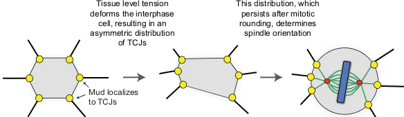
In theDrosophilapupal notum and larval wing, Mud localizes to tricellular junctions (TCJs), specialized structures at the intersection of more than two cells (Bosveld et al., 2016). The molecular link between TCJs, which have very few known components, and Mud is currently unknown. However, because the shape of the cell is influenced by tissue tension, so too is the distribution of TCJs, and as these structures persist throughout the cell cycle, their distribution provides a mechanism for ensuring that interphase cell shape, which is lost upon mitotic rounding, determines planar division angle (Fig. 3) (Bosveld et al., 2016).
Tissue level tension deforms the interphase
cell, resulting in an asymmetric distribution
of TCJs
This distribution, which persists after mitotic rounding, determines spindle orientation
[image:6.612.46.338.649.734.2]Mud localizes to TCJs
Fig. 3. The role of tricellular junctions during spindle orientation.In theDrosophilawing disc and pupal notum, tissue tension governs spindle orientation. Mud (yellow) is enriched at tricellular junctions (TCJs) throughout the cell cycle. The distribution of TCJs, and thus of Mud/dynein, translates interphase cell shape into division orientation by producing an asymmetric pulling force on astral microtubules. Green red and blue structures?
DEVEL
O
Intriguingly, the localization of Mud to TCJs is independent of Pins in the larval wing and pupal notum (Bosveld et al., 2016). Furthermore, Pins is not required to orient spindles in the A-B axis (Bergstralh et al., 2016; Bosveld et al., 2016; David et al., 2005). The developing wing and notum are therefore the first epithelial tissues identified in which spindle orientation is Pins independent, although other examples of Pins-independent mechanisms have emerged. In the pI cell (as discussed above), Mud can be anchored by interaction with Dsh (David et al., 2005; Ségalen et al., 2010). Intriguingly, LGN is not required for the development of the murine hair follicle, suggesting the possibility that a similar mechanism is at work (Byrd et al., 2016).
It is tempting to speculate that epithelial tissues, or at least those in the wing and notum, may have evolved a distinct system for spindle orientation. However, the extent to which the TCJ mechanism is used in other tissues is still unclear. As described above, spindle orientation in the Drosophila follicular epithelium and the chick neuroepithelium, as well as in epithelia-derived cells grown in culture, does require Pins/LGN (Bergstralh et al., 2013; Du et al., 2001; Peyre et al., 2011). How the direction of expansion is controlled in these tissues is unknown. The mitotic follicular epithelium at least is characterized by‘immature’septate junctions; markers such as Dlg are not tightly focused to one region of the lateral cortex and TCJs are absent (Goode and Perrimon, 1997). Furthermore, TCJ-mediated distribution of Mud/NuMA is not the only mechanism for translating interphase shape into division orientation in rounded cells. In HeLa cells, this is thought to be accomplished by caveolin 1-dependent recruitment of the spindle-orienting machinery to the retracting edge of a rounding cell (Matsumura et al., 2016), although the precise mechanism underlying this remains to be elucidated.
Together, these studies show that spindle orientation in epithelial cells does not occur through a generalized mechanism, as might have been predicted by earlier work showing that the canonical complex is conserved across organisms and tissue types. Instead, distinct cell types, even those in epithelial tissues within the same organism (e.g. Drosophila), have different requirements for localizing the cortical force.
Conclusions
Although the core components of the spindle-orientation machinery have been known for well over a decade, a number of questions concerning how this complex works still remain. The pulling force has generally been thought to rely on minus-end-directed dynein walking, but new findings suggest that regulated depolymerization of microtubules might also be involved. Questions about the localization of the machinery persist too. Work inDrosophilaand in isolated cells shows that the spindle can help to determine its own position, probably by regulating the delivery of spindle-orienting machinery proteins to and from the spindle poles. This shows that the location of the machinery within the cell is more dynamic than anticipated. However, as both LGN and Gαi cycle between the cortex and the spindle poles, these findings also suggest a requirement for cortical polarity factors in anchoring. It is not yet clear that the candidates identified already, such as Canoe/Afadin, E-cadherin and Discs large, can fully explain how spindles are anchored to the cortex in some epithelial cell types, and it is now apparent that different types of epithelia use distinct mechanisms to position their mitotic spindles. A more complete understanding of how spindles are oriented will therefore require more extensive research, e.g. using CRISPR-based strategies to interrogate the proposed interactionsin vivo, into how the spindle-orienting machinery is localized and regulated in multiple systems.
Competing interests
The authors declare no competing or financial interests.
Funding
D.S.J. is supported by The Wellcome Trust.
References
Afshar, K., Willard, F. S., Colombo, K., Johnston, C. A., Mccudden, C. R., Siderovski, D. P. and Gönczy, P. (2004). RIC-8 is required for GPR-1/2-dependent Galpha function during asymmetric division of C. elegans embryos.
Cell119, 219-230.
Afshar, K., Willard, F. S., Colombo, K., Siderovski, D. P. and Gonczy, P.(2005). Cortical localization of the Galpha Protein GPA-16 requires RIC-8 function during
C. elegans asymmetric cell division.Development132, 4449-4459.
Albertson, R. and Doe, C. Q.(2003). Dlg, Scrib and Lgl regulate neuroblast cell size
and mitotic spindle asymmetry.Nat. Cell Biol.5, 166-170.
Asaba, N., Hanada, T., Takeuchi, A. and Chishti, A. H.(2003). Direct interaction with a kinesin-related motor mediates transport of mammalian discs large tumor
suppressor homologue in epithelial cells.J. Biol. Chem.278, 8395-8400.
Baena-López, L. A., Baonza, A. and Garcı́a-Bellido, A.(2005). The orientation of
cell divisions determines the shape of Drosophila organs. Curr. Biol. 15,
1640-1644.
Bell, G. P., Fletcher, G. C., Brain, R. and Thompson, B. J.(2015). Aurora kinases phosphorylate Lgl to induce mitotic spindle orientation in Drosophila epithelia.
Curr. Biol.25, 61-68.
Bellaïche, Y., Radovic, A., Woods, D. F., Hough, C. D., Parmentier, M.-L., O’Kane, C. J., Bryant, P. J. and Schweisguth, F. (2001). The partner of inscuteable/discs-large complex is required to establish planar polarity during
asymmetric cell division in Drosophila.Cell106, 355-366.
Bergstralh, D. T., Lovegrove, H. E. and St Johnston, D.(2013). Discs large links
spindle orientation to apical-basal polarity in Drosophila epithelia.Curr. Biol.23,
1707-1712.
Bergstralh, D. T., Lovegrove, H. E. and St Johnston, D.(2015). Lateral adhesion
drives reintegration of misplaced cells into epithelial monolayers.Nat. Cell Biol.17,
1497-1503.
Bergstralh, D. T., Lovegrove, H. E., Kujawiak, I., Dawney, N. S., Zhu, J., Cooper, S., Zhang, R. and St Johnston, D.(2016). Pins is not required for spindle
orientation in the Drosophila wing disc.Development143, 2573-2581.
Bilder, D., Li, M. and Perrimon, N.(2000). Cooperative regulation of cell polarity
and growth by drosophila tumor suppressors.Science289, 113-116.
Bosveld, F., Markova, O., Guirao, B., Martin, C., Wang, Z., Pierre, A., Balakireva, M., Gaugue, I., Ainslie, A., Christophorou, N. et al.(2016). Epithelial tricellular
junctions act as interphase cell shape sensors to orient mitosis.Nature530,
495-498.
Bowman, S. K., Neumüller, R. A., Novatchkova, M., Du, Q. and Knoblich, J. A.
(2006). The Drosophila NuMA homolog mud regulates spindle orientation in
asymmetric cell division.Dev. Cell10, 731-742.
Busson, S., Dujardin, D., Moreau, A., Dompierre, J. and Mey, J. R. D.(1998). Dynein and dynactin are localized to astral microtubules and at cortical sites in
mitotic epithelial cells.Curr. Biol.8, 541-544.
Byrd, K. M., Lough, K. J., Patel, J. H., Descovich, C. P., Curtis, T. A. and Williams, S. E.(2016). LGN plays distinct roles in oral epithelial stratification,
filiform papilla morphogenesis and hair follicle development.Development143,
2803-2817.
Carminati, M., Gallini, S., Pirovano, L., Alfieri, A., Bisi, S. and Mapelli, M.(2016). Concomitant binding of afadin to LGN and F-actin directs planar spindle
orientation.Nat. Struct. Mol. Biol.23, 155-163.
Carvalho, C. A., Moreira, S., Ventura, G., Sunkel, C. E. and Morais-de-Sá, E.
(2015). Aurora a triggers Lgl cortical release during symmetric division to control
planar spindle orientation.Curr. Biol.25, 53-60.
Chang, J. T., Palanivel, V. R., Kinjyo, I., Schambach, F., Intlekofer, A. M., Banerjee, A., Longworth, S. A., Vinup, K. E., Mrass, P., Oliaro, J. et al.(2007). Asymmetric T lymphocyte division in the initiation of adaptive immune responses.
Science315, 1687-1691.
Chen, C. A. and Manning, D. R.(2001). Regulation of G proteins by covalent
modification.Oncogene20, 1643-1652.
Chu, X., Chen, X., Wan, Q., Zheng, Z. and Du, Q.(2016). Nuclear Mitotic Apparatus
(NuMA) interacts with and regulates astrin at the mitotic spindle.J. Biol. Chem.
291, 20055-20067.
Couwenbergs, C., Spilker, A. C. and Gotta, M.(2004). Control of embryonic
spindle positioning and galpha activity by C. elegans RIC-8.Curr. Biol.14,
1871-1876.
Cui, S., Otten, C., Rohr, S., Abdelilah-Seyfried, S. and Link, B. A.(2007). Analysis of aPKClambda and aPKCzeta reveals multiple and redundant functions
during vertebrate retinogenesis.Mol. Cell. Neurosci.34, 431-444.
David, N. B., Martin, C. A., Segalen, M., Rosenfeld, F., Schweisguth, F. and Bellaïche, Y.(2005). Drosophila Ric-8 regulates Galphai cortical localization to
DEVEL
O
promote Galphai-dependent planar orientation of the mitotic spindle during
asymmetric cell division.Nat. Cell Biol.7, 1083-1090.
de Belle, J. S. and Heisenberg, M.(1996). Expression of Drosophila mushroom body mutations in alternative genetic backgrounds: a case study of the Mushroom
Body Miniature Gene (Mbm).Proc. Natl. Acad. Sci. USA.93, 9875-9880.
Dewey, E. B., Sanchez, D. and Johnston, C. A.(2015). Warts phosphorylates mud to promote pins-mediated mitotic spindle orientation in Drosophila, independent of
Yorkie.Curr. Biol.25, 2751-2762.
di Pietro, F., Echard, A. and Morin, X.(2016). Regulation of mitotic spindle
orientation: an integrated view.EMBO Rep.17, 1106-1130.
Du, Q. and Macara, I. G.(2004). Mammalian pins is a conformational switch that
links NuMA to heterotrimeric G proteins.Cell119, 503-516.
Du, Q., Stukenberg, P. T. and Macara, I. G.(2001). A mammalian partner of
inscuteable binds NuMA and regulates mitotic spindle organization.Nat. Cell Biol.
3, 1069-1075.
Dunsch, A. K., Linnane, E., Barr, F. A. and Gruneberg, U.(2011). The Astrin-Kinastrin/SKAP complex localizes to microtubule plus ends and facilitates
chromosome alignment.J. Cell Biol.192, 959-968.
Galli, M., Muñoz, J., Portegijs, V., Boxem, M., Grill, S. W., Heck, A. J. R. and Van Den Heuvel, S.(2011). aPKC phosphorylates NuMA-related LIN-5 to position the
mitotic spindle during asymmetric division.Nat. Cell Biol.13, 1132-1138.
Gallini, S., Carminati, M., De Mattia, F., Pirovano, L., Martini, E., Oldani, A., Asteriti, I. A., Guarguaglini, G. and Mapelli, M.(2016). NuMA phosphorylation
by aurora-a orchestrates spindle orientation.Curr. Biol.26, 458-469.
Gho, M. and Schweisguth, F.(1998). Frizzled signalling controls orientation of
asymmetric sense organ precursor cell divisions in Drosophila.Nature 393,
178-181.
Gloerich, M., Bianchini, J. M., Siemers, K. A., Cohen, D. J. and Nelson, W. J.
(2017). Cell division orientation is coupled to cell-cell adhesion by the E-cadherin/
LGN complex.Nat. Commun.8, 13996.
Gönczy, P. and Rose, L. S.(2005). Asymmetric cell division and axis formation in
the embryo.WormBook, ed. TheC. elegansResearch Community, WormBook,
http://www.wormbook.org.
Goode, S. and Perrimon, N.(1997). Inhibition of patterned cell shape change and
cell invasion by discs large during Drosophila oogenesis. Genes Dev. 11,
2532-2544.
Gotta, M. and Ahringer, J.(2001). Distinct roles for Galpha and Gbetagamma in regulating spindle position and orientation in Caenorhabditis elegans embryos.
Nat. Cell Biol.3, 297-300.
Goulas, S., Conder, R. and Knoblich, J. A.(2012). The Par complex and integrins
direct asymmetric cell division in adult intestinal stem cells.Cell Stem Cell11,
529-540.
Guilgur, L. G., Prudencio, P., Ferreira, T., Pimenta-Marques, A. R. and Martinho, R. G.(2012). Drosophila aPKC is required for mitotic spindle orientation during
symmetric division of epithelial cells.Development139, 503-513.
Guo, Z. and Ohlstein, B.(2015). Bidirectional notch signaling regulates Drosophila
intestinal stem cell multipotency.Science350, aab0988.
Habib, S. J., Chen, B.-C., Tsai, F.-C., Anastassiadis, K., Meyer, T., Betzig, E. and Nusse, R.(2013). A localized Wnt signal orients asymmetric stem cell division in
vitro.Science339, 1445-1448.
Hampoelz, B., Hoeller, O., Bowman, S. K., Dunican, D. and Knoblich, J. A.
(2005). Drosophila Ric-8 is essential for plasma-membrane localization of
heterotrimeric G proteins.Nat. Cell Biol.7, 1099-1105.
Hanada, T., Lin, L., Tibaldi, E. V., Reinherz, E. L. and Chishti, A. H.(2000). GAKIN, a novel kinesin-like protein associates with the human homologue of the
Drosophila discs large tumor suppressor in T lymphocytes.J. Biol. Chem.275,
28774-28784.
Hao, Y., Du, Q., Chen, X., Zheng, Z., Balsbaugh, J. L., Maitra, S., Shabanowitz, J., Hunt, D. F. and Macara, I. G. (2010). Par3 controls epithelial spindle
orientation by aPKC-mediated phosphorylation of apical pins. Curr. Biol. 20,
1809-1818.
Inaba, M. and Yamashita, Y. M.(2012). Asymmetric stem cell division: precision for
robustness.Cell Stem Cell11, 461-469.
Izumi, Y., Ohta, N., Hisata, K., Raabe, T. and Matsuzaki, F.(2006). Drosophila pins-binding protein mud regulates spindle-polarity coupling and centrosome
organization.Nat. Cell Biol.8, 586-593.
Jia, M., Li, J., Zhu, J., Wen, W., Zhang, M. and Wang, W.(2012). Crystal structures of the scaffolding protein LGN reveal the general mechanism by which GoLoco
binding motifs inhibit the release of GDP from Gαi. J. Biol. Chem. 287,
36766-36776.
Johnston, C. A., Hirono, K., Prehoda, K. E. and Doe, C. Q.(2009). Identification of an aurora-a/PinsLINKER/Dlg spindle orientation pathway using induced cell
polarity in S2 cells.Cell138, 1150-1163.
Johnston, C. A., Doe, C. Q. and Prehoda, K. E.(2012). Structure of an
enzyme-derived phosphoprotein recognition domain.PLoS ONE7, e36014.
Johnston, C. A., Manning, L., Lu, M. S., Golub, O., Doe, C. Q. and Prehoda, K. E.
(2013). Formin-mediated actin polymerization cooperates with Mushroom Body
Defect (Mud)-dynein during frizzled-dishevelled spindle orientation.J. Cell Sci.
126, 4436-4444.
Kaushik, R., Yu, F., Chia, W., Yang, X. and Bahri, S. (2003). Subcellular localization of LGN during mitosis: evidence for its cortical localization in mitotic
cell culture systems and its requirement for normal cell cycle progression.Mol.
Biol. Cell14, 3144-3155.
Kern, D. M., Nicholls, P. K., Page, D. C. and Cheeseman, I. M.(2016). A mitotic
SKAP isoform regulates spindle positioning at astral microtubule plus ends.J. Cell
Biol.213, 315-328.
Kettenbach, A. N., Schweppe, D. K., Faherty, B. K., Pechenick, D., Pletnev, A. A. and Gerber, S. A.(2011). Quantitative phosphoproteomics identifies substrates and functional modules of aurora and polo-like kinase activities in mitotic cells.
Sci. Signal.4, rs5.
Kim, E. and Sheng, M.(2004). PDZ domain proteins of synapses. Nat. Rev. Neurosci.5, 771-781.
Kiyomitsu, T. and Cheeseman, I. M.(2012). Chromosome- and
spindle-pole-derived signals generate an intrinsic code for spindle position and orientation.Nat.
Cell Biol.14, 311-317.
Kiyomitsu, T. and Cheeseman, I. M.(2013). Cortical dynein and asymmetric
membrane elongation coordinately position the spindle in anaphase.Cell154,
391-402.
Knoblich, J. A.(2010). Asymmetric cell division: recent developments and their
implications for tumour biology.Nat. Rev. Mol. Cell Biol.11, 849-860.
Knoblich, J. A., Jan, L. Y. and Jan, Y. N.(1995). Asymmetric segregation of numb
and prospero during cell division.Nature377, 624-627.
Kotak, S., Busso, C. and Gönczy, P.(2013). NuMA phosphorylation by CDK1
couples mitotic progression with cortical dynein function.EMBO J.32, 2517-2529.
Kotak, S., Afshar, K., Busso, C. and Gönczy, P.(2016). Aurora a kinase regulates
proper spindle positioning in C. elegans and in human cells.J. Cell Sci.129,
3015-3025.
Kraut, R., Chia, W., Jan, L. Y., Jan, Y. N. and Knoblich, J. A.(1996). Role of
inscuteable in orienting asymmetric cell divisions in Drosophila.Nature383,
50-55.
Krueger, L. E., Wu, J.-C., Tsou, M.-F. B. and Rose, L. S.(2010). LET-99 inhibits lateral posterior pulling forces during asymmetric spindle elongation in C. elegans
embryos.J. Cell Biol.189, 481-495.
Laan, L., Pavin, N., Husson, J., Romet-Lemonne, G., Van Duijn, M., López, M. P., Vale, R. D., Jülicher, F., Reck-Peterson, S. L. and Dogterom, M.(2012). Cortical dynein controls microtubule dynamics to generate pulling forces that
position microtubule asters.Cell148, 502-514.
Lambert, J. D. and Nagy, L. M.(2002). Asymmetric inheritance of centrosomally
localized mRNAs during embryonic cleavages.Nature420, 682-686.
Lu, M. S. and Prehoda, K. E.(2013). A NudE/14-3-3 pathway coordinates dynein
and the kinesin Khc73 to position the mitotic spindle.Dev. Cell26, 369-380.
Machicoane, M., De Frutos, C. A., Fink, J., Rocancourt, M., Lombardi, Y., Garel, S., Piel, M. and Echard, A.(2014). SLK-dependent activation of ERMs controls
LGN-NuMA localization and spindle orientation.J. Cell Biol.205, 791-799.
Mao, Y., Tournier, A. L., Bates, P. A., Gale, J. E., Tapon, N. and Thompson, B. J.
(2011). Planar polarization of the atypical myosin dachs orients cell divisions in
Drosophila.Genes Dev.25, 131-136.
Mao, Y., Tournier, A. L., Hoppe, A., Kester, L., Thompson, B. J. and Tapon, N.
(2013). Differential proliferation rates generate patterns of mechanical tension that
orient tissue growth.EMBO J.32, 2790-2803.
Matsumura, S., Kojidani, T., Kamioka, Y., Uchida, S., Haraguchi, T., Kimura, A. and Toyoshima, F.(2016). Interphase adhesion geometry is transmitted to an
internal regulator for spindle orientation via Caveolin-1. Nat. Commun. 7,
ncomms11858.
Mccudden, C. R., Willard, F. S., Kimple, R. J., Johnston, C. A., Hains, M. D., Jones, M. B. and Siderovski, D. P.(2005). G Alpha selectivity and inhibitor
function of the multiple GoLoco motif protein GPSM2/LGN.Biochim. Biophys.
Acta1745, 254-264.
Morin, X. and Bellaïche, Y.(2011). Mitotic spindle orientation in asymmetric and
symmetric cell divisions during animal development.Dev. Cell21, 102-119.
Morin, X., Jaouen, F. and Durbec, P.(2007). Control of planar divisions by the
G-protein regulator LGN maintains progenitors in the chick neuroepithelium.Nat.
Neurosci.10, 1440-1448.
Nakajima, Y., Meyer, E. J., Kroesen, A., Mckinney, S. A. and Gibson, M. C.
(2013). Epithelial junctions maintain tissue architecture by directing planar spindle
orientation.Nature500, 359-362.
Negishi, T., Miyazaki, N., Murata, K., Yasuo, H. and Ueno, N.(2016). Physical association between a novel plasma-membrane structure and centrosome orients
cell division.Elife5, e16550.
Niessen, C. M. and Gottardi, C. J.(2008). Molecular components of the adherens
junction.Biochim. Biophys. Acta1778, 562-571.
Nipper, R. W., Siller, K. H., Smith, N. R., Doe, C. Q. and Prehoda, K. E.(2007). Galphai generates multiple pins activation states to link cortical polarity and
spindle orientation in Drosophila neuroblasts.104, 14306-14311.
Ohshiro, T., Yagami, T., Zhang, C. and Matsuzaki, F.(2000). Role of cortical tumour-suppressor proteins in asymmetric division of Drosophila neuroblast.
Nature408, 593-596.
DEVEL
O
Pan, Z., Zhu, J., Shang, Y., Wei, Z., Jia, M., Xia, C., Wen, W., Wang, W. and Zhang,
M.(2013). An autoinhibited conformation of LGN reveals a distinct interaction
mode between GoLoco motifs and TPR motifs.Structure21, 1007-1017.
Paridaen, J. T. M. L. and Huttner, W. B.(2014). Neurogenesis during development
of the vertebrate central nervous system.EMBO Rep.15, 351-364.
Pecreaux, J., Röper, J.-C., Kruse, K., Jülicher, F., Hyman, A. A., Grill, S. W. and Howard, J.(2006). Spindle oscillations during asymmetric cell division require a
threshold number of active cortical force generators.Curr. Biol.16, 2111-2122.
Peng, C.-Y., Manning, L., Albertson, R. and Doe, C. Q.(2000). The tumour-suppressor genes Lgl and Dlg regulate basal protein targeting in Drosophila
neuroblasts.Nature408, 596-600.
Peyre, E., Jaouen, F., Saadaoui, M., Haren, L., Merdes, A., Durbec, P. and Morin,
X. (2011). A lateral belt of cortical LGN and NuMA guides mitotic spindle
movements and planar division in neuroepithelial cells.J. Cell Biol.193, 141-154.
Portegijs, V., Fielmich, L.-E., Galli, M., Schmidt, R., Muñoz, J., van Mourik, T., Akhmanova, A., Heck, A. J. R., Boxem, M. and van den Heuvel, S.(2016). Multisite phosphorylation of NuMA-related LIN-5 controls mitotic spindle
positioning in C. elegans.PLoS Genet.12, e1006291.
Rhyu, M. S., Jan, L. Y. and Jan, Y. N.(1994). Asymmetric distribution of numb protein during division of the sensory organ precursor cell confers distinct fates to
daughter cells.Cell76, 477-491.
Saadaoui, M., Machicoane, M., Di Pietro, F., Etoc, F., Echard, A. and Morin, X.
(2014). Dlg1 controls planar spindle orientation in the neuroepithelium through
direct interaction with LGN.J. Cell Biol.206, 707-717.
Sans, N., Wang, P. Y., Du, Q., Petralia, R. S., Wang, Y.-X., Nakka, S., Blumer, J. B., Macara, I. G. and Wenthold, R. J.(2005). mPins modulates PSD-95 and
SAP102 trafficking and influences NMDA receptor surface expression.Nat. Cell
Biol.7, 1179-1190.
Schaefer, M., Shevchenko, A. and Knoblich, J. A. and Knoblich, J. A.(2000). A protein complex containing inscuteable and the Galpha-binding protein pins
orients asymmetric cell divisions in Drosophila.Curr. Biol.10, 353-362.
Schmidt, J. C., Kiyomitsu, T., Hori, T., Backer, C. B., Fukagawa, T. and Cheeseman, I. M.(2010). Aurora B kinase controls the targeting of the
Astrin-SKAP complex to bioriented kinetochores.J. Cell Biol.191, 269-280.
Ségalen, M., Johnston, C. A., Martin, C. A., Dumortier, J. G., Prehoda, K. E., David, N. B., Doe, C. Q. and Bellaïche, Y.(2010). The Fz-Dsh planar cell polarity pathway induces oriented cell division via Mud/NuMA in Drosophila and zebrafish.
Dev. Cell19, 740-752.
Seldin, L., Poulson, N. D., Foote, H. P. and Lechler, T.(2013). NuMA localization,
stability, and function in spindle orientation involve 4.1 and Cdk1 interactions.Mol.
Biol. Cell24, 3651-3662.
Seldin, L., Muroyama, A. and Lechler, T.(2016). NuMA-microtubule interactions are critical for spindle orientation and the morphogenesis of diverse epidermal
structures.Elife5, e12504.
Siderovski, D. P. and Willard, F. S.(2005). The GAPs, GEFs, and GDIs of
heterotrimeric G-Protein Alpha subunits.Int. J. Biol. Sci.1, 51-66.
Siegrist, S. E. and Doe, C. Q.(2005). Microtubule-induced pins/Galphai cortical
polarity in Drosophila neuroblasts.Cell123, 1323-1335.
Siller, K. H. and Doe, C. Q.(2009). Spindle orientation during asymmetric cell
division.Nat. Cell Biol.11, 365-374.
Siller, K. H., Serr, M., Steward, R., Hays, T. S. and Doe, C. Q.(2005). Live imaging of Drosophila brain neuroblasts reveals a role for Lis1/Dynactin in spindle
assembly and mitotic checkpoint control.Mol. Biol. Cell16, 5127-5140.
Smith, N. R. and Prehoda, K. E.(2011). Robust spindle alignment in Drosophila
neuroblasts by ultrasensitive activation of pins.Mol. Cell43, 540-549.
Speicher, S., Fischer, A., Knoblich, J. and Carmena, A.(2008). The PDZ protein canoe regulates the asymmetric division of Drosophila neuroblasts and muscle
progenitors.Curr. Biol.18, 831-837.
Spradling, A., Fuller, M. T., Braun, R. E. and Yoshida, S.(2011). Germline stem
cells.Cold Spring Harb. Perspect. Biol.3, a002642.
St Johnston, D. and Ahringer, J.(2010). Cell polarity in eggs and epithelia:
parallels and diversity.Cell141, 757-774.
Strzyz, P. J., Lee, H. O., Sidhaye, J., Weber, I. P., Leung, L. C. and Norden, C.
(2015). Interkinetic nuclear migration is centrosome independent and ensures
apical cell division to maintain tissue integrity.Dev. Cell32, 203-219.
Tame, M., Raaijmakers, J., Van Den Broek, B., Lindqvist, A., Jalink, K. and Medema, R. H.(2014). Astral microtubules control redistribution of dynein at the
cell cortex to facilitate spindle positioning.Cell Cycle13, 1162-1170.
Tame, M. A., Raaijmakers, J. A., Afanasyev, P. and Medema, R. H.(2016).
Chromosome misalignments induce spindle-positioning defects.EMBO Rep.17,
317-325.
Tang, N., Marshall, W. F., Mcmahon, M., Metzger, R. J. and Martin, G. R.(2011). Control of mitotic spindle angle by the RAS-regulated ERK1/2 pathway
determines lung tube shape.Science333, 342-345.
Toughiri, R., Li, X., Du, Q. and Bieberich, C. J.(2013).Phosphorylation of NuMA by Aurora-a Kinase in PC-3 prostate cancer cells affects proliferation, survival, and interphase NuMA localization.J. Cell. Biochem.114, 823-830.
Toyoshima, F. and Nishida, E.(2007). Integrin-mediated adhesion orients the spindle parallel to the substratum in an EB1- and myosin X-dependent manner.
EMBO J.26, 1487-1498.
Tsou, M.-F. B., Hayashi, A., DeBella, L. R., McGrath, G. and Rose, L. S.(2002). LET-99 determines spindle position and is asymmetrically enriched in response to
PAR polarity cues in C. elegans embryos.Development129, 4469-4481.
Wang, H., Ng, K. H., Qian, H., Siderovski, D. P., Chia, W. and Yu, F.(2005). Ric-8 controls Drosophila neural progenitor asymmetric division by regulating
heterotrimeric G proteins.Nat. Cell Biol.7, 1091-1098.
Wee, B., Johnston, C. A., Prehoda, K. E. and Doe, C. Q.(2011). Canoe binds
RanGTP to promote Pins(TPR)/Mud-mediated spindle orientation.J. Cell Biol.
195, 369-376.
Williams, S. E. and Fuchs, E.(2013). Oriented divisions, fate decisions.Curr. Opin. Cell Biol.25, 749-758.
Wyatt, T. P. J., Harris, A. R., Lam, M., Cheng, Q., Bellis, J., Dimitracopoulos, A., Kabla, A. J., Charras, G. T. and Baum, B.(2015). Emergence of homeostatic epithelial packing and stress dissipation through divisions oriented along the long
cell axis.Proc. Natl. Acad. Sci. USA112, 5726-5731.
Yamada, K. H., Hanada, T. and Chishti, A. H.(2007). The effector domain of human Dlg tumor suppressor acts as a switch that relieves autoinhibition of
kinesin-3 motor GAKIN/KIF13B.Biochemistry46, 10039-10045.
Yu, F., Morin, X., Cai, Y., Yang, X. and Chia, W.(2000). Analysis of partner of inscuteable, a novel player of Drosophila asymmetric divisions, reveals two
distinct steps in inscuteable apical localization.Cell100, 399-409.
Yu, F., Cai, Y., Kaushik, R., Yang, X. and Chia, W.(2003). Distinct roles of Galphai and Gbeta13F subunits of the heterotrimeric G protein complex in the mediation of
Drosophila neuroblast asymmetric divisions.J. Cell Biol.162, 623-633.
Yu, J. X., Guan, Z. and Nash, H. A.(2006). The mushroom body defect gene product is an essential component of the meiosis II spindle apparatus in
Drosophila oocytes.Genetics173, 243-253.
Zheng, Z., Zhu, H., Wan, Q., Liu, J., Xiao, Z., Siderovski, D. P. and Du, Q.(2010).
LGN regulates mitotic spindle orientation during epithelial morphogenesis.J. Cell
Biol.189, 275-288.
Zheng, Z., Wan, Q., Liu, J., Zhu, H., Chu, X. and Du, Q.(2013). Evidence for dynein
and astral microtubule-mediated cortical release and transport of Gαi/LGN/NuMA
complex in mitotic cells.Mol. Biol. Cell24, 901-913.
Zheng, Z., Wan, Q., Meixiong, G. and Du, Q. (2014). Cell cycle-regulated membrane binding of NuMA contributes to efficient anaphase chromosome
separation.Mol. Biol. Cell25, 606-619.
Zhu, J., Shang, Y., Xia, C., Wang, W., Wen, W. and Zhang, M.(2011). Guanylate kinase domains of the MAGUK family scaffold proteins as specific
phospho-protein-binding modules.EMBO J.30, 4986-4997.
Zhu, J., Shang, Y., Wan, Q., Xia, Y., Chen, J., Du, Q. and Zhang, M.(2014). Phosphorylation-dependent interaction between tumor suppressors Dlg and Lgl.
Cell Res.24, 451-463.
Zhu, J., Shang, Y., Xia, Y., Zhang, R. and Zhang, M.(2016). An atypical MAGUK GK target recognition mode revealed by the interaction between DLG and
KIF13B.Structure24, 1876-1885.