N A N O E X P R E S S
Open Access
Structure of Biocompatible Coatings
Produced from Hydroxyapatite
Nanoparticles by Detonation Spraying
Valentyna Nosenko
1*, Nataliia Strutynska
2, Igor Vorona
1, Igor Zatovsky
2, Volodymyr Dzhagan
1, Sergiy Lemishko
1,3,
Matthias Epple
4, Oleg Prymak
4, Nikolai Baran
1, Stanislav Ishchenko
1, Nikolai Slobodyanik
2, Yuriy Prylutskyy
2,
Nickolai Klyui
1and Volodymyr Temchenko
1Abstract
Detonation-produced hydroxyapatite coatings were studied by scanning electron microscopy (SEM), X-ray powder diffraction (XRD), Raman spectroscopy, and electron paramagnetic resonance (EPR) spectroscopy. The source material for detonation spraying was a B-type carbonated hydroxyapatite powder. The coatings consisted of tetracalcium phosphate and apatite. The ratio depended slightly on the degree of crystallinity of the initial powder and processing parameters of the coating preparation. The tetracalcium phosphate phase was homogeneous; the apatite phase contained defects localized on the sixfold axis and consisted of hydroxyapatite and oxyapatite. Technological factors contributing to the transformation of hydroxyapatite powder structure during coating formation by detonation spraying are discussed.
Keywords:Calcium phosphate, Hydroxyapatite coating, EPR, Raman, XRD, SEM, Detonation spraying
Background
Titanium is the most widely used implant material in orthopedic surgery and stomatology due to its durabil-ity under high load, as well as the applicabildurabil-ity in com-plex mechanical systems, such as knee and elbow joints. However, there are some challenges in titanium implant applications, such as possible degradation under the corrosive action of biological tissues in com-bination with continuous and/or cyclic loads, harmful action of electrochemical products of corrosion, and metal sensitivity of the human body [1, 2]. To over-come these disadvantages, orthopedic and stomatologi-cal titanium implants are coated with ceramics which provides good biocompatibility with living tissues. The materials commonly used for producing such ceramics are various calcium phosphates, especially
hydroxyapa-tite (HAP), Ca10(PO4)6(OH)2 [3–6]. Titanium-based
implants coated with HAP facilitate quick bone adapta-tion and allow to firmly fasten the implant to the bone
and to significantly reduce the time of healing. The good biocompatibility of such coatings is caused by the fact that HAP is the mineral phase of bone (~60 %) and teeth (~85 %) [3, 7]. To further improve the biocom-patibility, the coating properties must be as close to natural tissue characteristics as possible. Taking into ac-count that the biological HAP contains at least 4–6 wt % of carbonate impurity, the aim of the present study was to obtain a carbonate-containing HAP (CO32−-HAP) coating.
Among various methods to obtain HAP coatings, we have chosen the detonation spraying method [8]. Its ad-vantages are the ability to coat surfaces of arbitrary shapes and/or large-scale surfaces, and the method pro-vides a good adhesion to the substrate. However, this coating technique can change the phase composition, structure, and other properties of the initial material.
The changes can occur during either explosion
(temperature-induced changes) or deposition on the titanium substrate (impact-induced changes). We re-port on the characterization of these coatings and analyze how the properties of the source material and technological parameters influence on the properties of the coatings. This knowledge is very important for * Correspondence:[email protected]
1
V. Lashkaryov Institute of Semiconductor Physics, National Academy of Sciences of Ukraine, 45, Pr. Nauky, Kyiv 03028, Ukraine
Full list of author information is available at the end of the article
control of biological performances of the coatings used as implants.
Methods
Sample Preparation
The source material for coating fabrication was carbonate-containing hydroxyapatite. It was synthe-sized by wet precipitation method using Ca(NO3)2· 4
H2O, Na2CO3, and Na3PO4· 12 H2O as the initial
components. The solution with a mixture of sodium carbonate and sodium phosphate was pumped into a reactor containing a calcium nitrate solution (0.1 M). The molar ratio of Ca/P was 1.67:1, corresponding to stoichiometric hydroxyapatite. The molar ratio of CO32 −/PO43−
was 1:1. The obtained amorphous precipitates were filtered and washed several times with water to eliminate any residual alkali ions. The samples were prepared at different conditions at 25 or 80 °C (here-after denoted as powder P1 and powder P2, respect-ively). Then, the samples were dried at 80 °С. The well-ground powders were used to coat Ti plates.
A detonation spraying setup was used for deposition. The coating was generated by igniting an explosive mixture of oxygen and combustible gas (propane-bu-tane) in the explosion chamber. The wave propagated along the gun tube and caught up a portion of the HAP powder injected into the gun. Particles of the material were accelerated up to 5 M of speed (M is the Mach number ~340 m/s) and bombarded the substrate, form-ing a continuous coatform-ing due to physical and chemical interactions with the substrate material. The prog-rammable displacement of the detonation gun or the substrate provided coating of a large area or multi-surface substrates. The coating of plates was done at two different distances from the gun to the titanium plate. Table 1 gives a short description of the studied samples.
Methods
X-ray powder diffraction (XRD) was used to analyze the phase composition of the samples. Diffractometers type Shimadzu XRD-6000 and Bruker D8 ADVANCE with Cu Kαradiation were used. Data were collected over the 2θ range 5.0°–90.0°, with the steps 0.02 and 0.01°.
Identification of the phases was achieved by comparing the diffraction patterns of the samples with ICDD stan-dards. Scanning electron microscopy (SEM) (FEI Quanta 400 ESEM instrument in a high vacuum after sputtering with Au:Pd) was applied to observe the morphological changes of the coatings. Raman spectroscopy was used to identify the chemical composition and molecular structure. Raman spectra of the initial powders and de-posited coatings were investigated at room temperature using a DFS-24 double monochromator and a photon-counting system for recording. The radiation of a CW argon ion laser (514.5 nm, 50 mW) was used for ex-citation. Electron paramagnetic resonance (EPR) spec-troscopy was applied to analyze structural changes on the atomic level. EPR spectra were measured with a Radiopan X-band EPR spectrometer (~9.5 GHz). EPR signals were recorded at 2 mW microwave power using 100 kHz modulation of magnetic field with 0.05 mT amp-litude (spectra of the samples were recorded together with the spectrum of MgO:Cr3+ reference sample (g= 1.9800) that allowed to compare the intensities of the EPR spectra of different samples). The estimated accuracy of the g-factor determined for the EPR lines observed was ±2 · 10−4.
Results and Discussion SEM and XRD Studies
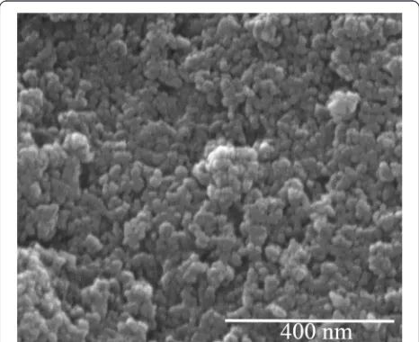
[image:2.595.305.538.513.703.2]The morphology of the initial powders was studied by scanning electron microscopy. For both powders (P1 and P2), SEM showed similar structures: the powders consist of irregular spherical particles with the sizes about 20–30 nm. The SEM image of P1 sample is shown as an example (Fig. 1).
Table 1Description of the studied samples
Sample Description
P1 (powder) Synthesis at 25 °С
P2 (powder) Synthesis at 80 °С
T1 (coating) Spraying of P2; distance to the Ti plate—150 mm
T2 (coating) Spraying of P2; distance to the Ti plate—200 mm
T3 (coating) Spraying of P1; distance to the Ti plate—150 mm
T4 (coating) Spraying of P1; distance to the Ti plate—200 mm
[image:2.595.56.291.637.733.2]The X-ray diffraction patterns of the powders P1 and P2 are shown in Fig. 2, together with the reported peak positions for HAP (ICDD pattern #00-089-6495). They corresponded to a crystalline HAP phase with a hex-agonal unit cell and lattice parameters a= 9.432 Å and c= 6.881 Å. It is seen that the XRD patterns of P1 and P2 powders are typical patterns of an HAP structure (Fig. 2). All reflexes are significantly broadened which is due to the small particle size (Fig. 1) and to the poor crystallinity of the HAP. The XRD patterns of P1 and P2 samples are similar; however, for P1 all reflexes are slightly broadened, suggesting that crystallinity of P2 powder is better than that of P1.
Figure 3 shows the X-ray diffraction patterns of car-bonated apatite coatings on Ti plates (T1 and T3 sam-ples). The narrower reflections as compared to the XRD patterns in Fig. 2 indicate the increase of crystallinity. XRD patterns of all coatings show both the expected HAP reflexes and some additional peaks that are most evidently observed in the range 27°–31°. These peaks can be attributed to tetracalcium phosphate (TTCP) phase, Ca4(PO4)2O; the positions of the corresponding reflections are shown in the upper part of Fig. 3. The ex-ample of morphology of an obtained coating is demon-strated in Fig. 4.
Figure 3 shows that the T1 spectrum in comparison with T3 demonstrates narrower reflexes and better reso-lution. This indicates that the T1 coating has a higher crystallinity.
Lattice parameters for both phases of all coatings were calculated from the XRD data. The TTCP lattice param-eters were the same for all samples (monoclinic system, space group P21, а= 7.023 Å, b= 11.986 Å, and c= 9.473 Å,β= 90.9°), whereas small changes were observed for the HAP phase (see Table 2). The parameterаfor all
coatings was considerably smaller than the value of a= 9.432 Å of B-type HAP. Since the value а for the stoichiometric (without carbonate) HAP is 9.4176 Å [7], it can be assumed that, first of all, the variation ofа is re-lated to the partial escape of water and carbonate during the process of coating deposition. Furthermore, as it is seen from Table 2, the calculated values of the lattice par-ameter a for all samples are slightly smaller than a= 9.4176 Å; this decrease can be caused by the formation of some structural defects such as vacancies. Note that a value ofasmaller than 9.4 Å is often observed in B-type carbonate-containing apatite annealed at different tem-peratures (see, for example, [9]). Previously in [19], it was shown that the high-temperature annealing (above 700 °С) of carbonate-containing HAP causes partial es-cape of carbonate and structural-bound water. As a re-sult, HAP transformed partially into TTCP. During the detonation spraying, this process apparently takes place in the surface layers of the powder grains, which are subjected to maximum temperature action.
Raman Spectroscopy
The values of the frequencies of PO43− in water obtained from Raman scattering measurements areν1= 936.6 cm−1, ν2= 415 cm−1, ν3= 1010 cm−1, andν4= 558 cm−1[10]. In the case when the PO43− tetrahedron is a part of symmet-rical lattice HAP and/or TTCP, the crystal field induces distortions in PO43− tetrahedron which change the intra-tetrahedral bond lengths and angles, and as a result, the normal modes of PO43−are shifted and split.
Fig. 2X-ray diffraction patterns of carbonated apatite powders P1 and P2. The calculated positions of main HAP reflections are shown byvertical lines(from ICDD (#00-089-6495))
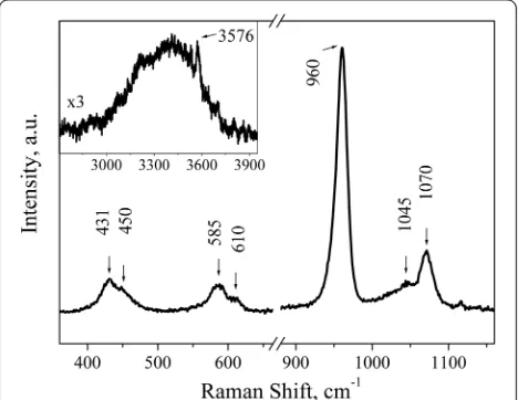
[image:3.595.305.539.89.314.2] [image:3.595.56.291.526.693.2]Figure 5 shows the Raman spectrum of P2 sample that is similar to the one of P1 sample. The main lines are observed in 360–1160 cm−1spectral range. Source ma-terial demonstrates typical HAP vibrations. The phos-phate ν2 vibrations (431 and 450 cm−1), phosphate ν4 vibrations (585 and 610 cm−1), and phosphate ν1 PO43− vibrations (960 cm−1) are seen. The structure observed in the 1020–1080-cm−1 range can be ascribed to the stretching ν1 mode of carbonate in B position, i.e., 1070 cm−1[11–14], and the bending mode of carbonate at 1045 cm−1 [11] which overlaps with the wide back-ground of the phosphate ν3 vibrations [14]. The broad band of low intensity in the range 3000–3750 cm−1(see the inset in Fig. 5) can be attributed to the traces of water that is incorporated into the structure. A sharp peak at 3576 cm−1 is associated with the stretching vi-bration of the structural OH−group in HAP [15–17].
Raman spectra of the coatings have a number of differ-ences from the spectra of source powders, namely, there is no band at 1070 cm−1that indicates the escape of car-bonate from B position of apatite lattice; the broad band 3000–3750 cm−1disappears after coating deposition, in-dicating that water was removed from the crystal lattice. The similar carbonate and water escape during anneal-ing of apatite was observed by other characterization methods [18, 19].
The main differences in the Raman spectra of differ-ent coatings are the changes of position, intensity, and bandwidth of ν1 PO43− vibrations, which allows
analyz-ing the composition and structural features of the coat-ings obtained. Fig. 6 shows the Raman features in the
frequency range corresponding to ν1 for source HAP
powder and coatings. It was found that ν1 component
in both P1 and P2 powders is well fitted by single line at 960 cm−1. The best fitting for coatings was obtained using three fitting components with varied intensities but fixed positions and line widths. The first
compo-nent at 960 cm−1 is caused by PO43− mode in HAP
phase. To describe the low-frequency shoulder, the sec-ond fitting component at 945 cm−1was used. Usually, the band at 945 cm−1is assigned to amorphous calcium phosphate [20] that indicates of a highly disordered
structure, although not necessarily a completely
amorphous one. On the other hand, the position of this band corresponds to one of theν1components of PO43− vibrations of TTCP [21]. Since the X-ray diffraction patterns (Figs. 2 and 3) showed that the coatings have better crystallinity than the initial powders, therefore, the band at 945 cm−1 is more likely related to one of
the ν1 components of PO43− vibrations of the TTCP
phase. Moreover, the presence of TTCP phase was ob-served by XRD.
[image:4.595.58.541.91.216.2]The third component is used to fit the high-frequency part of the spectrum. This component is placed at Fig. 4Scanning electron micrograph of the coating T1
Table 2The calculated cell parameters of HAP phase in the coatings
Sample Lattice parameters
a(Å) c(Å)
T1 9.405(2) 6.902(2)
T2 9.400(3) 6.903(2)
T3 9.413(4) 6.905(4)
[image:4.595.305.539.532.713.2] [image:4.595.56.291.651.733.2]970 cm−1and can be assigned to ν1PO43− of oxyapatite [22]. The presence of oxyapatite phase is clearly ob-served in Raman spectra, while XRD data do not allow distinguishing of HAP and oxyapatite phases. This find-ing correlates with the generally accepted fact that dehydrated HAP contains various intermediate phases, particularly oxyapatite [17]. As it is seen from Fig. 6, the more amorphous is the source powder, the larger is the content of oxyapatite in the ceramics, while the ratio between TTCP and HAP content is almost con-stant. The change of the distance to the substrate in the investigated range has less influence on the phase composition of the coating, although a slight increase in the amount of the oxyapatite phase is observed with increasing of the distance to the substrate.
Electron Paramagnetic Resonance (EPR)
EPR was used to investigate the local structure changes of HAP after the coating deposition. For the initial pow-ders, the EPR signals were not observed. EPR spectra of
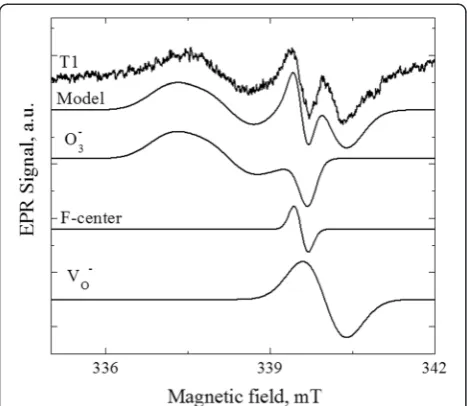
the coatings are shown in Fig. 7. The difference in the EPR lineshape of coatings indicates that the EPR spectra have composite nature and are formed by contributions of at least two or three (as in the case of T1 sample) dif-ferent types of paramagnetic centers.
Pretty well, description of the experimental EPR spec-tra of T2, T3, and T4 coatings can be achieved taking into account the contributions from two signals. The first fitting component is characterized by the parame-ters gx= 2.0022, gy= 2.0169, and gz= 2.0108. These
pa-rameters are close to the values gx= 2.0029, gy= 2.0176,
and gz= 2.0105 observed for the center in HAP that is
attributed to О3- located between two vacant hydroxyl sites [23]. The small difference in the parameters indi-cates that it is the same О3− center with slightly differed nearest surrounding. The second fitting component is the isotropic line with giso= 1.9990. The same signal was previously observed in modified HAP powders and at-tributed to VO- center—an electron localized on an oxygen vacancy [24]. To describe the spectrum of T1 sample, one more line with giso= 2.0026 is needed. Isotropic lines in the range of 2.002–2.003 have been re-peatedly observed in the annealed or mechanically treated HAP (g= 2.0019 (F-centers—electron local-ized on hydroxyl group vacancy) [25], g= 2.002 (un-known center) [26],g= 2.0029 (unknown center) [25], and g= 2.0032 (unknown center) [27]). The absence of hyper-fine interaction features complicates the unambiguous identification of isotropic lines. Perhaps, the authors of all these works have dealt with the same center with slightly different nearest surrounding. In our opinion, the most suitable candidate for this center is the F-center. In Fig. 8, we show an example of modeling of the EPR spectrum of T1 coating by three fitting compo-nents described above.
Fig. 6The experimental Raman spectra of initial HAP powder and coatings, their simulation, and fitting components:black curves—experimental Raman spectra of initial HAP powder and coatings;red curves—model spectra; green curves—fitting component at 960 cm−1;magenta curves
—fitting component at 945 cm−1;blue curves
—fitting component at 970 cm−1
[image:5.595.57.291.89.410.2] [image:5.595.305.540.531.715.2]Mechanism of Paramagnetic Defects Formation
In a carbonate-containing HAP material, PO43- phos-phate groups are partly replaced by carbonate CO32-. It is obvious that the substitution CO32-→PO43-is not isova-lent, and charge compensation is necessary for the exist-ence of a stable structure. At present, the generally accepted mechanism for charge compensation at noniso-valent replacement CO32-→PO43- in HAP is the forma-tion of vacancies of calcium and hydroxyl groups in neighboring lattice sites (see, e.g., [28]). The presence of
VOH in carbonated HAP was confirmed experimentally
[29]. Carbonate replacement of B-type in the apatite structure leads to the formation of structural defects lo-cated on the sixfold axis (vacancy VOH at OH site) and near this axis (vacancy VCa at Ca site); as a result, CO32 −-HAP becomes less stable as compared to
stoichiomet-ric HAP. Another source of hydroxyl group vacancies and additional structure changes is the influence of high temperatures. Loss of carbonate and water that are seen in the Raman spectra changes the lattice constant of HAP. Presumably, additional changes of the material structure occur when the heated HAP particles hit the substrate. This can lead to the displacement of atoms and redistribution of electrons. In particular, the charged paramagnetic defects are formed:
VOHþе→VOH−ðF‐centerÞ;
VOþе→VO−:
At the same time, extended defects (nanopores) are formed along the sixfold axis; these defects can capture some molecules from the atmosphere, in particular, О2.
This molecular О2is most likely bonded to atomic oxy-gen (the residual of OH groups on the sixfold axis); as a result, theО3structural unit is formed. Presumably, this process is similar to the A-type СО32--HAP formation (high-temperature annealing of HAP inСО2flow) [30]:
2ОH−þСО2→СО32−þН2О↑−A‐typeСО32−‐HAP formation;
2ОH−þО2→О3−þН2О↑þ e−О3−structural unit formation:
Conclusions
Coatings obtained by detonation spraying of B-type carbonate-containing HAP are a mixture of two com-pounds: apatite and tetracalcium phosphate. The relative contributions of these phases depend on technological conditions of the coating production. The analysis of the Raman spectra demonstrated the appearance of oxyapa-tite phase in the ceramics caused by partial dehydration of HAP. The more amorphous is the initial powder, the more effective is the dehydrogenation. Transformations of the HAP structure occur predominantly along the six-fold axis and in its nearest surroundings. As a result, paramagnetic defects such as VO-, F-center, andО3- center are formed. The above changes are caused by the sequen-tial influence of various factors, the dominating one being the thermal heating of carbonate-containing HAP powder.
Abbreviations
HAP:hydroxyapatite; SEM: scanning electron microscopy; TTCP: tetracalcium phosphate; XRD: X-ray diffraction.
Competing interests
The authors declare that they have no competing interests.
Authors’Contributions
VN and IV analyzed and discussed the result and wrote the final version of the paper. VN, IP, NSt, IZ, ME, VD, and SI organized and performed the experiments, analyzed and discussed the result, and wrote the drafted version of the manuscript. NS and YP synthesized HAP powders. NK and VT deposited coatings. NS, ME, and OP carried out SEM and XRD studies. VD carried out Raman measurements. VN, NB, and IV carried out EPR measurements. All authors analyzed and discussed the result. All authors read and approved the final manuscript.
Acknowledgements
This project is financially supported by Branch target preparation Taras Shevchenko National University of Kyiv and National Academy of Sciences of Ukraine (Grant no. 2–03, D114U003876). Dr. N. Strutynska thanks the Ministry of Education and Science of Ukraine for the financial support.
Author details
1V. Lashkaryov Institute of Semiconductor Physics, National Academy of
Sciences of Ukraine, 45, Pr. Nauky, Kyiv 03028, Ukraine.2Taras Shevchenko National University of Kyiv, Volodymyrska Str., 64/13, 01601 Kyiv, Ukraine.
3
National Technical University of Ukraine“KPI”, 03056 Kyiv, Ukraine.4Inorganic Chemistry and Center for Nanointegration Duisburg-Essen (CeNIDE), University of Duisburg-Essen, Universitaetsstrasse, 5-7, 45117 Essen, Germany.
Received: 12 October 2015 Accepted: 17 November 2015
Reference
1. Long M, Rack HJ (1998) Titanium alloys in total joint replacement—a materials science perspective. Biomaterials 19:1621–1639
[image:6.595.57.291.88.291.2]2. Brown SA, Farnsworth L, Merrit K, Crowe TD (1988) In vitro and in vivo metal ion release. J Biomed Mater Res 22:321–338
3. Ong JL, Chan DCN (1999) Hydroxyapatite and their use as coatings in dental implants: a review. Critical Reviews™in Biomed Eng 28:1–41 4. Denissen HW, Walk W, Veldhuis AAH, Van der Hooff A (1989) Eleven-year
study of hydroxyapatite implants. J Prosthet Dent 61:708–712
5. Dorozhkin S (2012) Calcium orthophosphate coatings, films and layers. Prog Biomater 1:1–40
6. Dorozhkin SV (2010) Nanosized and nanocrystalline calcium orthophosphates. Acta Biomater 6:715–734
7. Ellion JC (1994) Structure and chemistry of the apatites and other calcium orthophosphates. Elsevier, Amsterdam
8. Singh L, Chawla V, Grewa JS (2012) A review on detonation gun sprayed coatings. JMMCE 11:243–265
9. Brik AB, Danilchenko SN, Radchuk VV, Karbovsky VL, Kalinichenko AM, Bagmut NN (2007) Thermally activated changes in the properties of biogenic and synthetic carbonate-containing apatite by XRD and EPR data. Mineral Journ (Ukraine) 29:32–47
10. Rudolph WW, Irmer G (2007) Raman and infrared spectroscopic investigations on aqueous alkali metal phosphate solutions and density functional theory calculations of phosphate–water clusters. Appl Spectroscopy 61:1312–1327
11. Berger SB, Soares LES, Martin AA, Ambrosano GMB, Tabchoury CPM, Giannini M (2014) Effects of various hydrogen peroxide bleaching concentrations and number of applications on enamel. Braz J Oral Sci 13:22–27
12. Awonusi A, Morris MD, Tecklenburg MMJ (2007) Carbonate assignment and calibration in the Raman spectrum of apatite. Calcif Tissue Int 81:46–52 13. Penel G, Cau E, Delfosse C, Rey C, Hardouin P, Jeanfils J, Delecourt C,
Lemaitre J, Leroy G (2003) Raman microspectrometry studies of calcified tissues and related biomaterials. Raman studies of calcium phosphate biomaterials. Dent Med Probl 40:37–43
14. Penel G, Leroy G, Rey C, Bres E (1998) MicroRaman spectral study of the PO4
and CO3vibrational modes in synthetic and biological apatites. Calcif Tissue
Int 63:475–481
15. Iqbal Z, Tomaselli V, Fahrenfeld O, Miller KD, Ruszala FA, Kostiner E (1977) Polarized Raman scattering and low frequency infrared study of hydroxyapatite. J Phys Chem Solids 38:923–927
16. Saber-Samandari S, Alamara K, Saber-Samandari S, Gross KA (2013) Micro-Raman spectroscopy shows how the coating process affects the characteristics of hydroxylapatite. Acta Biomater 9:9538–9546 17. Yu H, Zhang H, Wang X, Gu Z, Li X, Deng F (2007) Local structure of
hydroxy–peroxy apatite: a combined XRD, FT-IR, Raman, SEM, and solid-state NMR study. J Phys Chem Solids 68:1863–1871
18. Holcomb DW, Young RA (1980) Thermal decomposition of human tooth enamel. Calcif Tissue Int 31:189–201
19. Strutynska N, Zatovsky I, Slobodyanik N, Malyshenko A, Prylutskyy Yu, Prymak O, Vorona I, Ishchenko S, Baran N, Byeda A, Mischanchuk A (2015) Preparation, characterization, and thermal transformation of poorly crystalline sodium- and carbonate-substituted calcium phosphate. Eur J Inorg Chem 2015:622–629.
20. Sauer GR, Zunic WB, Durig JR, Wuthier RE (1994) Fourier transform Raman spectroscopy of synthetic and biological calcium phosphates. Calcif Tissue Int 54:414–420
21. Jillavenkatesa A, Condrate RA (1997) The infrared and Raman spectra of tetracalcium phosphate (Ca4P2O9). Spectroscopy Letters 30:1561–1570
22. Fowler BO, Markovic M, Brown WE (1993) Octacalcium phosphate. 3. Infrared and Raman vibrational spectra. Chem Mater 5:1417–1423 23. Van Doorslaer S, Moens P, Cailens F, Matthys P, Verbeeck R (1996)31P and
1H powder ENDOR study of ozonide radicals in carbonated apatites,
synthesized from aqueous solutions. Appl Magn Reson 10:87–102 24. Fanovich MA, Castro MS, Porto Lopez JM (2001) Structural analysis of
modified hydroxyapatite. Mater Res Bull 36:487–496
25. Pietak AM, Reid JW, Sayer M (2005) Electron spin resonance in silicon substituted apatite and tricalcium phosphate. Biomaterials 26:3819–3830 26. Fattibene P, Callens F (2010) EPR dosimetry with tooth enamel: a review.
Appl Radiat Isot 68:2033–2116
27. Aragno D, Fattibene P, Onori S (2001) Mechanically induced EPR signals in tooth enamel. Appl Radiat Isot 55:375–382
28. Gilinskaya LG, Zanin YN (1998) Factors stabilizing the paramagnetic radicals CO2−, CO3−and CO33−in natural carbonated apatites. J Struct
Chem 39:671–686
29. Ishchenko S, Vorona I, Okulov S (1999) ENDOR study of irradiated tooth enamel. SPQEO 2:84–92
30. Schramm DU, Terra J, Rossi AM, Ellis DE (2000) Configuration of CO2−radicals
inγ-irradiated A-type carbonated apatites: theory and experimental EPR and ENDOR studies. Phys Rev B 63:024107 (14 pages)
Submit your manuscript to a
journal and benefi t from:
7 Convenient online submission
7 Rigorous peer review
7 Immediate publication on acceptance
7 Open access: articles freely available online
7 High visibility within the fi eld
7 Retaining the copyright to your article