D
E
V
E
LO
P
M
E
N
T
INTRODUCTION
In many animal embryos, cells destined to the germline fate incorporate a region containing a distinct cytoplasm, termed the germ plasm (reviewed by Houston and King, 2000). The germ plasm contains many maternally inherited RNAs and proteins, which play crucial roles in germ cell formation. These germ plasm components often form electron-dense granulofibrillar structures, called germinal granules. In later embryogenesis, germinal granules are reorganized to form perinuclear granules, or nuage, a hallmark of germ cells in a wide range of animal species. Thus, the identification and characterization of the germ plasm and the germinal granules are crucial for understanding the mechanism of germline formation.
Ascidian embryos are well known to develop in a typical mosaic fashion (Conklin, 1905), and they undergo rapid cell-fate determinations, which are regulated by maternal RNAs and proteins localized to specific regions of unfertilized eggs and early embryos (reviewed by Nishida, 2005). As several lines of evidence have indicated that the somatic cell-fate determination is completed by the 110-cell stage (Conklin, 1905; Nishida, 1987), it is believed that germline cells are also specified by this stage.
Within ascidian embryos, a specific cytoplasm, called the postplasm, is sequestered to a region at the posterior pole (Sasakura et al., 1998; Sasakura et al., 2000), where many maternal RNAs and proteins are highly concentrated (Yoshida et al., 1996; Satou, 1999; Imai et al., 2004; Yamada et al., 2005) (reviewed by Sardet et al., 2005). The postplasm contains a specialized cytoplasmic structure,
called the centrosome-attracting body (CAB). The CAB is composed of electron-dense masses (EDM), which are morphologically very similar to germinal granules, and the cortical endoplasmic reticulum (cER) domains (Hibino et al., 1998; Iseto and Nishida, 1999). Recent observations have shown that in Ciona intestinalis, the RNA and protein of CiVH, a homolog of the Drosophila germline-specific gene vasa,are highly concentrated in the postplasm, presumably in the CAB (Fujimura and Takamura, 2000; Takamura et al., 2002). The CiVH-positive cells are later incorporated into the primitive gonads of juveniles to form germ cells. Vasa is an integral component of Drosophilagerminal granules (polar granules and nuage), and its homologs have also been identified as germline-specific genes in a wide range of animals (reviewed by Raz, 2000). The postplasm-containing blastomeres are also likely to be transcriptionally inactive, as the zygotic transcriptions of several genes, including the housekeeping gene elongation factor-1␣, are detected at a high level in all blastomeres, except for the postplasm-containing cells (Tomioka et al., 2002) (M.S.-K. and A.N., unpublished). Furthermore, it has been proposed that, following the cleavage stage, a pair of the postplasm-containing blastomeres at the posterior pole of the embryo, the B7.6 cells, are mitotically inactive during the subsequent embryogenesis. This transcriptional quiescence and cell division arrest are also seen in the primordial germ cells (PGCs) in Drosophilaand C. elegansduring early embryogenesis (reviewed by Leatherman and Jongens, 2003). Based on these observations, it has been proposed that in ascidians, the postplasm acts as the germ plasm, and that the B7.6 cells develop into PGCs.
Although the postplasm is thought to act as the germ plasm, the CAB and several postplasmic components are also known to play crucial roles in unequal cleavages and somatic cell differentiation. First, the CAB functions in promoting unequal cleavages during early embryogenesis (Hibino et al., 1998; Iseto and Nishida, 1999; Nishikata et al., 1999). Second, posterior end mark(PEM) RNA, which was the first molecule identified as a postplasmic component
Dynamic redistribution of
vasa
homolog and exclusion of
somatic cell determinants during germ cell specification in
Ciona intestinalis
Maki Shirae-Kurabayashi1,*, Takahito Nishikata2, Katsumi Takamura3, Kimio J. Tanaka4, Chiaki Nakamoto1and
Akira Nakamura1,*
Ascidian embryos sequester a specific cytoplasm, called the postplasm,at the posterior pole, where many maternal RNAs and proteins accumulate. Although the postplasm is thought to act as the germ plasm, it is also highly enriched in several factors essential for somatic cell development, and how the postplasm components regulate both germ and somatic cell differentiation remains elusive. Using a vasahomolog, CiVH, and other postplasmic components as markers, we found that the postplasm-containing blastomeres, the B7.6 cells, undergo an asymmetric cell division during gastrulation to produce two distinct daughter cells: B8.11 and B8.12. Most of the postplasmic components segregate only into the B8.11 cells, which never coalesce into the gonad. By contrast, the maternal CiVHRNA and protein are specifically distributed into the B8.12 cells, which divide further and are incorporated into the gonad in juveniles. In the B8.12 cells, CiVH production is upregulated from the maternal RNA source, resulting in the formation of perinuclear CiVH granules, which may be the nuage, a hallmark of germ cells in many animal species. We propose that the redistribution of specific maternal molecules into the B8.12 cells is essential for germ-cell specification in ascidians.
KEY WORDS: Ciona intestinalis, vasa, Germ cell, Germ plasm, Nuage, Asymmetric cell division
Development 133, 2683-2693 (2006) doi:10.1242/dev.02446
1Laboratory for Germline Development, RIKEN Center for Developmental Biology,
Kobe, Hyogo 650-0047, Japan. 2Department of Biology, Konan University, Kobe,
Hyogo 658-8501, Japan. 3Department of Marine Biotechnology, Fukuyama
University, Fukuyama, Hiroshima 729-0292, Japan. 4Laboratory of Cellular
Biochemistry, RIKEN, Wako, Saitama 351-0198, Japan.
*Authors for correspondence (e-mail: [email protected]; [email protected])
D
E
V
E
LO
P
M
E
N
T
(Yoshida et al., 1996), appears to control the positioning of the cleavage planes, promoting unequal cleavages (Nishida, 2005). Third, macho-1 RNA, which encodes a transcription factor, is tightly associated with the CAB and acts as a muscle determinant in Halocynthiaand Ciona(Nishida and Sawada, 2001; Satou et al., 2002a; Kobayashi et al., 2003). Maternal macho-1activity in the postplasm-containing blastomeres also promotes the posterior adjacent blastomeres to differentiate into mesenchyme during early cleavage stages (Kondoh et al., 2003; Kobayashi et al., 2003). Fourth, CiYB1 protein has been identified as a component of the postplasm in C. intestinalisembryos (Tanaka et al., 2004). CiYB1 is a member of the Y-box protein family, which includes RNA-binding proteins that control the translation of specific mRNAs (reviewed by Matsumoto and Wolffe, 1998; Sommerville, 1999). Consistent with this function, CiYB1 binds Ci-PEMand Ci-macho-1 RNAs in vitro, and represses their translation in the rabbit reticulocyte lysate system, suggesting that CiYB1 regulates the translation of postplasmic/PEM RNAs (Tanaka et al., 2004). Previous studies have indicated that these postplasmic/PEM RNAs and proteins, which are involved in somatic cell differentiation during the cleavage stages, can be detected in the presumed B7.6 cells in the tailbud embryo. Therefore, it is thought that postplasmic components with known functions in somatic cell development may have additional roles in germ cell formation. However, how the postplasm components regulate germ cell specification and somatic cell differentiation remains elusive.
Here, we show that B7.6 cells undergo an asymmetric cell division during gastrulation to produce two distinct daughter cells: CAB-containing anterior cells, named B8.11, and CAB-negative posterior cells, the B8.12 cells. Most of the postplasmic components remain associated with the CAB and are partitioned only into the B8.11 cells, which later lose CiVH protein expression and associate with the gut wall in juveniles. By contrast, maternal CiVHRNA and protein are specifically released from the CAB and diffuse into the cytoplasm just before the B7.6 cell division, allowing the CiVHproducts to be inherited by the B8.12 cells. Furthermore, CiVH protein production in the B8.12 cells is upregulated through the translation of the maternal CiVHRNA, resulting in the formation of perinuclear CiVH granules, which may be the nuage structure. The B8.12 descendants are incorporated into the primitive gonad in juveniles. Our data indicate that the CAB and most of the postplasmic components involved in somatic cell differentiation are excluded from the germline progenitor cells through an asymmetric cell division, and suggest that the diffusion of specific postplasmic components into the cytoplasm prior to the B7.6 cell division is crucial for proper germline development during ascidian embryogenesis.
MATERIALS AND METHODS
Animals
C. intestinaliswere collected at Osaka Bay (Hyogo), Murotsu Bay (Hyogo), Maizuru Bay (Kyoto), Onagawa Bay (Miyagi), Usa Bay (Kochi) and Mikawa Bay (Aichi) in Japan. The eggs and sperm were isolated from adult individuals by cutting the gonads and were kept at 18°C (eggs) or on ice (sperm) until use. The eggs were dechorionated as described (Etani and Nishikata, 2002). After insemination, the eggs were reared at 18°C in Millipore-filtered seawater (FSW) containing 50 g/ml streptomycin sulfate. The larvae were staged according to Chiba et al. (Chiba et al., 2004).
In situ hybridization
Sense and antisense DIG-labeled RNA probes were synthesized from the cDNA clones in the Ciona intestinalisGene Collection Release I (Satou et al., 2002b), using a digoxigenin RNA-labeling kit (Roche). Whole-mount in situ hybridization of staged C. intestinalisembryos was carried out as described (Etani and Nishikata, 2002).
Antibody generation
The full-length CiVH-coding region was amplified by PCR and cloned into pProExHTa (Gibco). Histidine-tagged CiVH protein was expressed in Escherichia coliBL21 cells by IPTG induction and purified with Ni-NTA agarose (Qiagen). The protein was further purified by preparative SDS-PAGE, and dialyzed against PBS. Polyclonal antisera against the purified protein were generated in rabbits by Kitayama Labes (Nagano, Japan). For immunoblotting and immunohistochemistry, the rabbit antisera were affinity purified with the antigen immobilized on a HiTrap NHS column (Amersham).
Immunoblotting
Immunoblotting was carried out using the E. colicell lysate expressing recombinant histidine-tagged CiVH, the adult gonad lysate, or total lysates of 5 or 20 animals at different developmental stages. The affinity-purified rabbit anti-CiVH antibody (1:1000 dilution) and mouse anti--tubulin antibody (3E7, DSHB; 1:1000 dilution) were used as primary antibodies. Horseradish peroxidase (HRP)-conjugated anti-rabbit or anti-mouse IgG (Jackson; 1:3000 dilution) was used as the secondary antibody, and signals were detected with the ECL system (Amersham).
Immunohistochemistry
Embryos and larvae were fixed with 4% paraformaldehyde in 0.5 M NaCl, 0.1 M MOPS pH 7.5 at room temperature for 30-60 minutes, washed with PBS containing 0.1% Tween 20 (PBSTw) and kept at 4°C until use. The samples were pretreated with blocking solution (1% bovine serum albumin in PBSTw) for 30 minutes and incubated with the primary antibody in blocking solution overnight at 4°C. The primary antibodies used were: affinity-purified rabbit anti-CiVH (1:1000 dilution), rabbit anti-CiYB1 (Tanaka et al., 2004) (1:1000 dilution) and mouse monoclonal anti-phosphohistone H3 (Cell signaling; 1:2000 dilution). The secondary antibodies used were Alexa488- or Alexa568-conjugated anti-rabbit IgG, and Alexa568-conjugated anti-mouse IgG (Molecular Probes). To visualize cell boundaries and nuclei, the samples were incubated with Alexa660- or Alexa568-conjugated phalloidin and DAPI (Molecular Probes) for 2 hours at room temperature. The embryos and larvae were mounted in Vectashield (Vector Laboratories) and observed under a laser confocal microscope (Leica TCS SP2 AOBS).
Double staining for proteins and RNAs were carried out as follows. To detect hybridized signals, embryos were incubated with an HRP-conjugated anti-DIG antibody for 30 minutes (1:500; Roche), followed by amplification with biotinyl tyramide (1:50) for 20 minutes (Perkin Elmer). The samples were then incubated with the rabbit anti-CiVH antibody (1:1000 dilution) or rabbit anti-CiYB1 antibody (1:1000 dilution) overnight at 4°C. To visualize the signals, embryos were incubated with Alexa594-conjugated anti-rabbit IgG (1:1000 dilution) and FITC-conjugated streptavidin (Molecular Probes, 1:500 dilution) overnight at 4°C.
Lineage tracing of B7.6 blastomeres
CellTracer CM-DiI (Molecular Probes) was dissolved in soybean oil (Sigma) to a concentration of 1 mg/ml. DiI solution was injected into the B7.6 blastomeres of the dechorionated 64-cell embryos and cultured individually in 24-well plates at 18°C. After metamorphosis, the juveniles were fed with the diatom Chaetoceros gracilisuntil the primitive gonads were formed. We observed both live and fixed animals to detect DiI signals alone and both CiVH and DiI signals, respectively.
Inhibitor treatments
D
E
V
E
LO
P
M
E
N
T
expression of alkaline phosphatase (AP) mRNA and AP activity, respectively. It is reported that, following the cleavage stage, AP is produced through the translation of zygotically expressed mRNA in Ciona(Imai et al., 2000). Histochemical staining for AP was performed as described (Imai et al., 2000). RT-PCR was carried out using total RNA prepared from 50 embryos. The primer sets for Ci-APand Ci--tubulinwere designed from the cDNA information (CLSTR13498 for Ci-APand CLSTR00086 for Ci--tubulin) in the Ghost database (http://ghost.zool.kyoto-u.ac.jp/indexr1.html). To confirm the effects of inhibitors on the expression of Ci-AP, two different regions of the Ci-APmRNA coding region were analyzed by RT-PCR. The following primer sets were used: AP-1, 5⬘-TTGTGCAAGTGTTGCGGTAATCGG-3⬘and 5⬘ -TGTTTTCTTGCG-CTCTGTCGTGCC-3⬘; AP-2, 5⬘-ATTGACCACGGTCACCATGCAGG-3⬘ and 5⬘-TGTTTTCTTGCGCTCTGTCGTGCC-3⬘; CiVH, 5⬘ -ATGTTTGA-CGACGATTGGGAACC-3⬘and 5⬘ -CCATCATCTTTCTTGGAATACG-GG-3⬘; -tubulin, 5⬘-ACCAAATTGGTGCTAAGTTCTGGG-3⬘and 5⬘ -TGGTGTGGCACAGGTCGTATGG-3⬘.
RESULTS
Maternal CiVHRNA is segregated into two
discrete regions after gastrulation
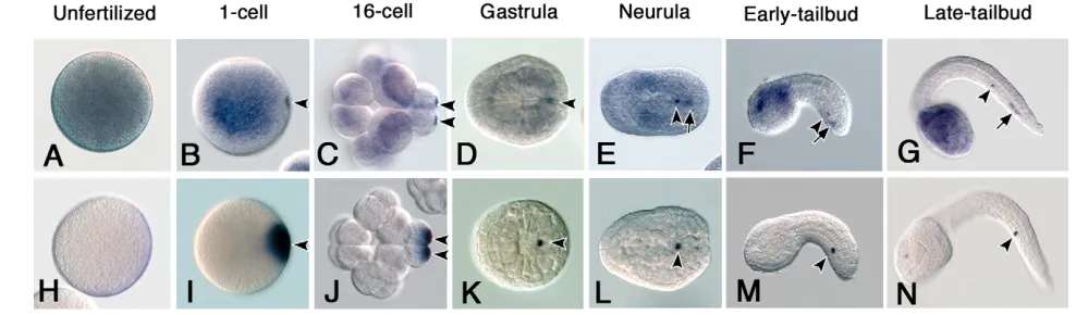
The distribution pattern of CiVHRNA during embryogenesis has been reported (Fujimura and Takamura, 2000). However, we detected a novel CiVH localization pattern through an improved in situ hybridization procedure in the present study. The previous report showed that CiVHRNA is distributed broadly in unfertilized eggs (Fig. 1A) and gradually accumulates at the posterior pole of the embryos during the 4- to 16-cell stage (Fujimura and Takamura, 2000). However, using our improved procedure, we found that a small amount of CiVHRNA had already started to accumulate at the posterior cortex of the fertilized one-cell stage embryo (Fig. 1B). During the cleavage stages, CiVHRNA was incorporated into a pair of blastomeres located at the posterior pole (Fig. 1C). At the gastrula stage, CiVHRNA showed a single dot-like distribution in the embryos (Fig. 1D, arrowheads). However, in the neurula, the dot-like CiVH RNA distribution was found in two points of the posterior part of the embryo (Fig. 1E, arrowhead and arrow). In tailbud embryos, the CiVH RNA signals were clearly segregated into the middle and distal regions of the endodermal strand of the tail (Fig. 1F,G, arrowheads and arrows, respectively). Under our staining conditions, 100% of the tailbud embryos showed this CiVHRNA distribution pattern.
By contrast, Ci-PEMRNA, which accumulated in the postplasm after fertilization and partitioned into the posterior-most blastomeres during the cleavage stage (Fig. 1H-J, arrowheads), was, unlike the
CiVHRNA, detected only in the middle of the tail region in the tailbud embryo (Fig. 1M,N, arrowheads). We found that the maternal Ci-macho-1RNA had the same distribution pattern as the Ci-PEMRNA during embryogenesis (data not shown) (Satou et al., 2002a). Thus, although the distribution patterns of CiVHRNA overlapped with those of the Ci-PEMandCi-macho-1RNAs during the cleavage stages, in subsequent stages, the CiVHRNA, but not the Ci-PEMor Ci-macho-1RNAs, was distributed into two discrete cell masses in the middle and distal regions of the tail.
B7.6 cells undergo an asymmetric cell division during gastrulation
We next examined the spatiotemporal distribution of CiVH protein. For these experiments, we raised an anti-CiVH polyclonal antibody, which recognized a single band in extracts prepared from adult ovaries (data not shown). The size of this band matched the predicted mass for CiVH protein (72 kDa), and the antibody also detected a recombinant CiVH protein expressed inE. coli(data not shown). Thus, the specificity of our new polyclonal antibody was sufficient for analyzing the CiVH protein distribution in embryogenesis.
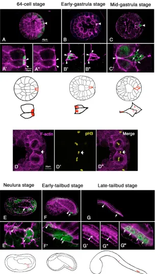
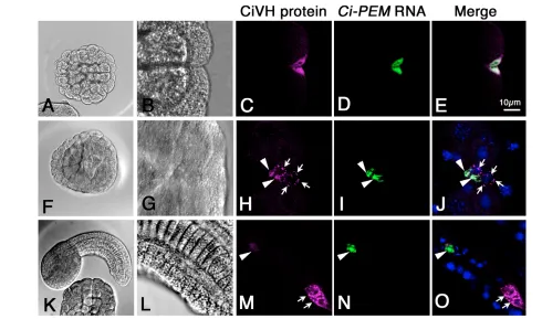
[image:3.612.53.547.555.700.2]In 64-cell stage embryos, the CiVH protein was concentrated at the posterior pole of the posterior-most cells, the B7.6 cells (Fig. 2A,A⬘), where postplasmic/PEM RNAs and the CAB also accumulate (Yoshida et al., 1996; Iseto and Nishida, 1999). Notably, this region was also highly enriched with F-actin, as revealed by phalloidin staining (Fig. 2A⬘,A⬙, arrowheads), suggesting that the CAB associates tightly with an F-actin mass. During the initial stage of gastrulation, the position of the CiVH protein within the B7.6 cells changed from posterior to anterior, owing to the movement of the cells towards the interior of the embryo (Fig. 2B,B⬘). At the beginning of the invagination, the CiVH protein in the B7.6 cells was associated with the F-actin layer at the cortex (Fig. 2B⬘,B⬙ arrowheads), suggesting that CiVH protein remained in the CAB. However, during gastrulation, although some CiVH protein remained anchored to the CAB at the anterior cortex (Fig. 2C⬘, arrowheads), some was released from the anterior cortex and diffused into the bulk cytoplasm to form cytoplasmic granules (Fig. 2C⬘, arrows). At this stage, we found that the chromosomes of the B7.6 cells were positive for phosphohistone H3, a mitosis marker (Fig. 2D⬘,D⬙, arrows), and the B7.6 cells subsequently underwent asymmetric cell division to form small anterior and large posterior daughter cells (Fig. 2E; arrowhead and arrow, respectively). Thus, the B7.6 cells became
Fig. 1. Expression patterns of CiVHand Ci-PEMRNAs during embryogenesis. (A-G) CiVHRNA expression; (H-N) Ci-PEMRNA expression.
D
E
V
E
LO
P
M
E
N
T
mitotically active during early gastrulation to produce two morphologically distinct cells that both contained CiVH protein. Following the previous lineage nomenclature for ascidians (Conklin, 1905), the small anterior daughter cells were named B8.11, and the large posterior daughter cells B8.12.
CiVH protein in the B8.12 cells forms perinuclear granules
After the asymmetric cell division, the level of CiVH staining in the B8.12 cells increased (Fig. 2E⬘,F⬘, arrows), and the CiVH protein formed clear granules around the nucleus at the tailbud stage (Fig.
2F⬘, arrows). Each B8.12 cell then divided once to form four CiVH-positive cells by the late tailbud stage (Fig. 2G, arrows). The CiVH staining in the B8.12 daughter cells remained high during the larval stage (Fig. 3A-D). By contrast, the CiVH staining in the B8.11 cells remained low in the F-actin aggregates (Fig. 2G⬘,G⬙arrowheads), and the anterior CiVH foci in the tail became undetectable by the beginning of metamorphosis (Fig. 3A-D).
[image:4.612.52.362.197.734.2]Although CiVH protein was maintained at a high level in the B8.12 cells and their descendants, a low level of CiVHRNA and protein was also detected throughout the entire egg, and in all cells of cleavage to neurula-stage embryos (Fig. 1A-E, Fig. 2A-E).
Fig. 2. CiVH protein is localized to two discrete
regions through the B7.6 cell division.Embryos
were stained for CiVH protein (green) and for F-actin (magenta) to visualize the cell boundary. A-C, dorsal views; E-G, lateral views. Anterior is towards the left in all panels. (A,B) CiVH protein (green) accumulated at the posterior cortex of the B7.6 cells with an F-actin layer (A⬘,B⬘, arrowheads). (A⬙,B⬙) Phalloidin staining alone highlights a thick layer of cortical F-actin in the CAB region (arrowheads). (C,C⬘) At the mid-gastrula stage, while some CiVH protein remained associated with an F-actin mass (C⬘, arrowhead), some was diffused in the cytoplasm of the B7.6 cells (C⬘, arrows). Diagrams show the position of the B7.6 cells in above embryos (upper row; outlined in red) and distribution of CiVH protein within the B7.6 cells (lower row; red). (D) A mid-gastrula embryo showing the pH3-positive chromosomes in one of the B7.6 cells (D⬘, yellow). pH3-positive chromosomes are aligned, indicating that this pH3-positive B7.6 cell was in metaphase (arrow). (E-G) In the B8.11 cells (E⬘,F⬘,G⬘,
D
E
V
E
LO
P
M
E
N
T
Western blot analysis showed that the total amount of CiVH protein per embryo increased during the cleavage stages and fell after gastrulation (data not shown). During the larval stages, the CiVH signal remained in the trunk region (Fig. 3A-C, black arrows), and the total amount of CiVH protein in the trunk region was much higher than in the tail region (data not shown). However, by the end of metamorphosis, the level of CiVH protein in the trunk region had dropped dramatically and become undetectable (Fig. 3C-F).
B8.12 descendents develop into germ cells
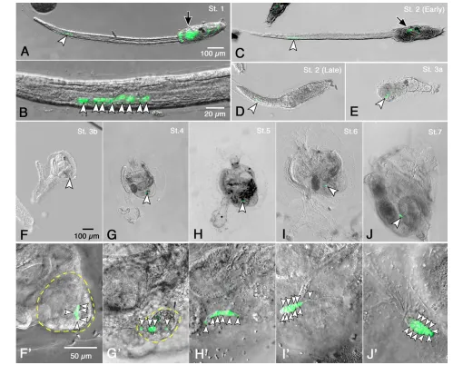
The B8.12 daughter cells divided further in the tail to form 8-16 CiVH-positive cells during the larval stage (Fig. 3A-D, arrowheads). Subsequently, these cells were carried into the trunk region along with other resorbed tail cells, ~24 hours after hatching
[image:5.612.51.550.239.641.2](stage 3a; Fig. 3E, arrowhead). At stage 3b, the CiVH-positive cells (Fig. 3F,F⬘, arrowheads) remained in the tail debris. After the tail debris was mostly absorbed at stage 4 (Fig. 3G,G⬘), the CiVH-positive cells aligned within the sparse remaining tail debris (Fig. 3G⬘, arrowheads). In stage 5 juveniles, the CiVH-positive cells aligned within a tube-like structure that extended from the tail debris toward the stomach (Fig. 3H,H⬘, arrowheads). By stage 6, this tube-like structure had changed into a drop-like shape containing the CiVH-positive cells (Fig. 3I,I⬘). Subsequently, the CiVH-positive cells were surrounded by somatic cells to form the primitive gonad at stage 7 (Fig. 3J,J⬘arrowheads) (Okada and Yamamoto, 1999; Chiba et al., 2004). Within the primitive gonad, the CiVH-positive cells proliferated, and their number increased with subsequent development (Fig. 3J⬘, arrowheads; data not shown).
Fig. 3. CiVH-positive cells in the tail region become germ cells.(A,B) An early tadpole larva. Eight CiVH-positive cells were localized to the
D
E
V
E
LO
P
M
E
N
T
To confirm the origin of the germline cells in the primitive gonads, we traced the lineage of the B7.6 cells with a fluorescent dye, CM-DiI. We labeled the B7.6 cells with DiI immediately after the B6.3 cell division, when the B7.6 cells stuck out from the embryos. After the DiI injection, we confirmed the DiI labeling of the B7.6 cells at the gastrula stage (Fig. 4A), and of the B8.11 (arrowheads) and B8.12 (arrows) cells at the tailbud stage of live specimens (Fig. 4B). Although in some cases the DiI signals were also detected in the sister blastomeres of B7.6 cells, the B7.5 cells, we could nonetheless trace the B7.6 cell fate, as the lineage of the B7.5 cells has been described (Satou et al., 2004).
Even 14 days after the DiI labeling, we successfully detected the DiI signals in the CiVH-positive cells, which assembled in the primitive gonads in stage 6-7 juveniles (Fig. 4C). Notably, juveniles
that showed relatively strong DiI labeling in the B8.11 cells at the tailbud stages (arrowheads in Fig. 4B) had DiI-positive but CiVH-negative cells attached to the hind-gut wall near the anus (data not shown), suggesting that the B8.11 cells never coalesce into the primitive gonad. To confirm the fate of the B8.11 cells in the juveniles, we removed the B8.12 descendants from DiI-labeled larvae by cutting off the distal region of the tail where the B8.12 descendants clustered (Fig. 4D). When such larvae were allowed to develop, the DiI-positive cells were still detected at the gut wall in stage 5 juveniles [Fig. 4E, arrowhead; in the images we obtained, the DiI signal was very faint, and the auto-fluorescence of the tail debris (arrow) was thus relatively greater under microscopic observation]. These results demonstrate that the B8.12 descendants coalesce into the primitive gonads. Although it is still possible that the B8.11 cells migrate to the gonad in later stages, these results strongly suggest that, under normal developmental conditions, the B8.12 descendants, but not the B8.11 cells, develop into germ cells.
CiVHRNA and protein are specifically released
from the CAB
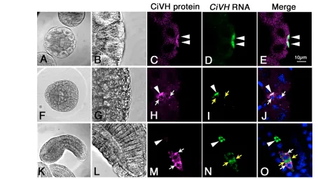
We further examined the CiVHRNA and protein distribution as well as other postplasmic components during the B7.6 cell division. First, we double stained embryos for CiVH protein and CiVHRNA. We found that CiVHRNA was tightly colocalized with CiVH protein in the CAB during the cleavage stages (Fig. 5C-E, arrowheads). However, in gastrula-stage embryos, CiVH RNA and protein diffused together into the cytoplasm just before the B7.6 cell division (Fig. 5H-J, arrows), and were distributed into both the B8.11 and 8.12 cells (Fig. 5M-O, arrowheads and arrows, respectively). In tailbud embryos, the CiVH protein and RNA were separated in the cytoplasm within the B8.12 cells, and the CiVH protein formed peri-nuclear granules (Fig. 5M-O).
We next examined the localization of CiVH protein and Ci-PEM RNA. Although the Ci-PEMRNA was strictly colocalized with the CiVH protein in the CAB during the cleavage stage (Fig. 6C-E), it remained concentrated in the CAB at the gastrula stage (Fig. 6H-J, arrowheads). After the B7.6 cell division, the Ci-PEM RNA segregated only into the B8.11 cells (Fig. 6M-O, arrowheads), while the CiVH protein was distributed to both the B8.11 and 8.12 cells (Fig. 6M, arrows). We also observed that maternal Ci-macho-1RNA never diffused from the CAB and segregated only into the B8.11 cells (data not shown). These observations indicate that CiVHRNA and protein are specifically released from the CAB prior to the B7.6 cell division and that their specific diffusion into the bulk cytoplasm is likely to be crucial for their distribution into the B8.12 cells.
It has remained elusive whether the release of CiVH protein from the CAB is a specific mechanism for certain proteins or a general one for all the protein components in the postplasm. To discriminate between these possibilities, we examined the distribution of another protein component in the postplasm, CiYB1 (Tanaka et al., 2004). We found that CiYB1 protein tightly colocalized with Ci-PEM and Ci-macho-1RNA and was partitioned only into the B8.11 cells (Fig. 7; data not shown). These results indicate that the release of CiVHRNA and protein from the CAB prior to the B7.6 cell division is a specific mechanism that enables their distribution into the B8.12 cells.
CiVH protein is produced from inherited maternal RNA in the B8.12 cells
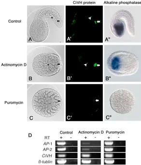
[image:6.612.51.299.236.588.2]We next examined whether CiVH protein production in the B8.12 cell lineage requires zygotic CiVHtranscription, or whether the translation of maternally inherited CiVHRNA is upregulated in the B8.12 cells. For this purpose, we examined the effects of
Fig. 4. B8.12 descendants coalesce into the gonad.(A) A gastrula
D
E
V
E
LO
P
M
E
N
T
transcription and translation inhibitors on CiVH protein production in the B8.12 cells. Embryos at the 110-cell stage were treated with a transcription inhibitor, actinomycin D (AcD), or a translation inhibitor, puromycin, and they were then allowed to develop until the presumptive tailbud stage. Although both AcD and puromycin treatments severely affected embryogenesis, we could still detect the B7.6 cell lineage by observing the CiVH signals in the posterior of the embryos.
We found that, in all AcD-treated embryos, the level of anti-CiVH staining in the presumptive B8.12 cells was as high as in the untreated tailbud embryos (Fig. 8A⬘,B⬘, arrows). Under our experimental conditions, the expression of zygotic alkaline phosphatase (AP) activity in the endoderm was reduced (Fig. 8B⬙), although it was not completely repressed as previously reported (Whittaker, 1977). RT-PCR analysis confirmed that there was a severe reduction in zygotic AP expression in the AcD-treated embryos (Fig. 8D). These data indicate that CiVH protein production in the B8.12 cells was not suppressed by inhibiting the zygotic transcription during embryogenesis.
By contrast, the accumulation of CiVH protein in the presumptive B8.12 cells was severely inhibited by the puromycin treatment (Fig. 8C⬘). When 110-cell stage embryos were treated with 100 g/ml puromycin, only 3% of the embryos had a normal level of CiVH protein in their B8.12 cells at the presumptive tailbud stage, but 43% showed weak production, and in 54% the CiVH granules were undetectable. Furthermore, when embryos at the 110-cell stage were treated with 200 g/ml puromycin, 6% showed a normal level of anti-CiVH staining and 33% showed weak staining (Fig. 8C⬘), and CiVH signals were not detectable in 61% of the embryos. Thus, the reduction of anti-CiVH staining in the B8.12 cells by the puromycin treatment occurred in a
dose-dependent manner. RT-PCR analysis indicated that the level of CiVHRNA was not affected by the inhibitor treatments (Fig. 8D). These results indicated that it is the translation of maternal CiVH RNA, and not zygotic CiVHtranscription, which is responsible for the production of CiVH protein in the B8.12 cells. These results further suggest that the specific release of CiVHRNA and protein from the CAB prior to the B7.6 cell division is crucial for germ cell specification in ascidians.
DISCUSSION
Previous reports of CiVH distribution have shown that the CiVH RNA and protein are highly concentrated in the postplasm, and that the CiVH-positive cells are later incorporated into the primitive gonads of juveniles to form germ cells (Fujimura and Takamura, 2000; Takamura et al., 2002). However, it remained unclear whether B7.6 cells directly coalesce into the gonads to develop into germ cells and whether other postplasmic components with known functions in somatic cell development contribute to germ cell formation. In the present study, by tracing the B7.6 cell fate in detail, we clarify the process by which germ cells in ascidians are specified.
Dynamic redistribution of CiVH protein and germ cell specification in Ciona
We showed here that a dynamic redistribution of CiVH protein occurs following the cleavage stage. First, each of the two posterior-most blastomeres, the B7.6 cells, divided asymmetrically during gastrulation (Fig. 2D⬘,D⬙) to produce two distinct daughter cells, B8.11 and B8.12. Second, although most of the postplasmic components segregated only into the small CAB-inheriting B8.11 cells, the CiVHRNA and protein were inherited by both the
CAB-Fig. 5. CiVHRNA and protein are inherited by the B8.12 cells through their release from the CAB.Embryos were stained for CiVH protein
[image:7.612.55.509.57.313.2]D
E
V
E
LO
P
M
E
N
T
containing B8.11 cells and the CAB-lacking B8.12 cells (Figs 2, 5). Third, the segregation of CiVHRNA and protein into both daughter cells was mediated through their specific diffusion into the cytoplasm prior to the B7.6 cell division (Fig. 2C⬘, Fig. 5H-J). Fourth, after the B7.6 cell division, the CiVH protein in the B8.12 cells formed perinuclear granules (Fig. 2G). As Vasa is an integral component of the germinal granules, or nuage, in a wide range of animals, we propose that the CiVH-containing perinuclear granules are the nuage in C. intestinalis. Finally, by tracing the B8.12 cell fate in detail with DiI labeling and CiVH immunostaining, we found that the B8.12 cells divided further to form 8-16 CiVH-positive cells, which were incorporated into the primitive gonads and developed into germ cells (Figs 3, 4). From these observations, we conclude that B8.12 cells are the PGCs in this organism. Previous electron microscopic observations in C. intestinalis showed nuage-like structures in the putative PGCs located in the tail debris during metamorphosis (Yamamoto and Okada, 1999). Our present observations suggest that the formation of this structure begins in the germline cells at a much earlier stage: immediately after the B7.6 cell division in the gastrula. Electron microscopic analyses of the B7.6 cells and their descendants in the gastrula and tailbud-stage embryos will confirm this issue.
Our immunoblot and immunostaining analyses suggested that CiVHmRNA is translated during cleavage throughout the embryo and in the trunk region of the larva (Figs 2, 3; data not shown). However, CiVH protein in the trunk region was undetectable during early metamorphosis (Fig. 3). Because the level of CiVH protein in the B8.12 descendants was much lower than that in the rest of the embryo and larva, we could not detect a specific increase in the
CiVH protein in B8.12 cells using immunoblotting (data not shown). However, immunostaining and the lineage-tracing study demonstrated that only B8.12 and its descendants maintained the CiVH protein during development and that they were incorporated into the primitive gonad. Thus, even though CiVH is not a germline-specific protein in embryos and early larvae, it is still a suitable marker for tracing the germline cells throughout development.
Most postplasmic components are excluded from the germline lineage
In contrast to the B8.12 cells, we found that the B8.11 cells never coalesced into the primitive gonads, but rather associated with the gut wall in juveniles, where they were without CiVH expression (Fig. 4E; data not shown). The B8.11 cells contained a mass of F-actin, which was probably the CAB remnant. Furthermore, maternal Ci-PEM and Ci-macho-1 RNAs, which are involved in unequal cleavage and somatic cell differentiation, and a putative translational repressor, CiYB1 protein, were never detected in the B8.12 cells, but remained in the B8.11 cells with the putative CAB remnant (Fig. 6; data not shown). Although it is formally possible that protein products of these postplasmic/PEM RNAs were partitioned into the B8.12 cells, we favor the idea that these postplasmic components of the B8.11 cells have no role in germ cell specification. Rather, it may be that these postplasmic components and/or the CAB structure itself could interfere with proper germ cell specification, and are therefore excluded from the B8.12 cells.
[image:8.612.53.543.57.346.2]The mechanism by which the CiVH RNA and protein are specifically released from the CAB prior to the B7.6 cell division remains uncertain. It is reported that the postplasmic/PEM RNAs
Fig. 6. Ci-PEMRNA and CiVH protein show different distributions after the gastrula stage.Embryos were stained for CiVH protein
D
E
V
E
LO
P
M
E
N
T
contain cis-acting signals in their 3⬘untranslated region (UTR) that direct their localization to the CAB (Sasakura and Makabe, 2002). Similarly, it is likely that the CiVHRNA contains a specialized cis-acting sequence for its specific release from the CAB prior to the B7.6 cell division.
[image:9.612.51.461.61.312.2]The CAB is composed of two distinct domains, the EDM and cER (Hibino et al., 1998; Iseto and Nishida, 1999). It is reported that in both Cionaand Halocynthia, macho1, PEMand several other postplasmic/PEM mRNAs associate with the cER in the CAB to form the cER/mRNA domain (Sardet et al., 2005; Prodon et al.,
Fig. 7. CiYB1 protein is inherited only by the
B8.11 cells, along with
Ci-PEMRNA.Embryos were stained for CiYB1 protein (magenta) and Ci-REMRNA (green). (A-E) A 32-cell embryo; (F-J) a gastrula embryo; (K-O) a mid-tailbud embryo. (B-E,G-J,
[image:9.612.50.342.396.737.2]L-O) Enlarged views of A,F,K. (A,B,F,G,K,L) Nomarski optics. The CiYB1 protein tightly colocalized with Ci-PEMRNA and segregated only into the B8.11 cells (E,J,O).
D
E
V
E
LO
P
M
E
N
T
2005; Nakamura et al., 2005). Although the precise distribution of CiVHRNA within the CAB remains unknown, the specific release of CiVHRNA and protein from the CAB may be due to a difference in the strength of their association with the CAB; e.g. they might be associated with the EDM rather than the cER.
Recently, the aPKC-PAR6-PAR3 complex (PAR complex) has been identified as a component of the CAB (Patalano et al., 2006). The PAR complex associates with the cortical F-actin under the plasma membrane and is thought to regulate unequal cleavages in the ascidian embryo. As the PAR complex is crucial for polarized RNA and protein distribution within a cell and for asymmetric cell divisions in a wide range of cell types (Macara, 2004), the PAR proteins may be involved in the CiVH partitioning into the B8.12 cells.
Translational upregulation of maternal RNA in the B8.12 cells may promote germ cell formation After the B7.6 cell division, CiVH protein production was dramatically increased in the B8.12 cells, resulting in the formation of perinuclear granules (Fig. 2G). We found that the maternal CiVH RNA, released from the CAB prior to the B7.6 cell division, was responsible for the CiVH protein production in the B8.12 cells (Figs 5, 8). These data suggest that the translation of CiVHRNA is partially repressed in the B7.6 cells but is upregulated in the B8.12 cells, where the RNA is no longer associated with the CAB. CiYB1 protein, which binds to and represses the translation of the Ci-PEM and Ci-macho-1 RNAs in vitro (Tanaka et al., 2004), may be involved in the storage and/or translational control of CiVH RNA in the postplasm. After the cleavage stage, when somatic cell-fate determination is completed, the CiVH RNA was specifically released from the CAB, distributed to the B8.12 cells, and translated to form perinuclear CiVH granules. Thus, the CAB appears to be required for the storage of CiVHRNA as well as for the repression of its translation during the cleavage stage, when somatic cell-fate determination proceeds.
We have found that several other postplasmic/PEM RNAs are partitioned into the B8.12 cells in a manner similar to that of CiVH RNA during Ciona embryogenesis (M.S.-K. and A.N., unpublished). Furthermore, in Halocynthia, it has also been observed that some postplasmic/PEM RNAs are segregated into two regions within the embryos after gastrulation (Nakamura et al., 2003) (H. Nishida, personal communication). Thus, we propose that the dynamic redistribution of specific postplasm components through the B7.6 cell division is conserved mechanisms underlying germ cell specification in ascidians.
We thank K. Hirayama, S. Yamada, T. Kusakabe and S. Fujiwara for supplying live materials, members of the Onagawa Marine Biological Station for their hospitality during part of this work, and the Suma Aqualife Park for the maintenance of live C. intestinalis. We also thank N. Satoh, Y. Satou and Y. Kohara for cDNA clones, and S. Chiba for staging the juvenile animals. We are also grateful to H. Nishida, M. Royle and K. Hanyu-Nakamura for comments on the manuscript. This work was supported in part by a Grant-in-Aid from the Ministry of Education, Culture, Sports, Science and Technology, Japan.
References
Chiba, S., Sasaki, A., Nakayama, A., Takamura, K. and Satoh, N.(2004). Development of Ciona intestinalisjuveniles (through 2nd ascidian stage). Zool. Sci. 21, 285-298.
Conklin, E. G.(1905). The organization and cell lineage of the ascidian egg. J. Acad. Nat. Sci. Phila.13, 1-119.
Etani, K. and Nishikata, T.(2002). Novel G-protein-coupled receptor gene expressed specifically in the entire neural tube of the ascidian Ciona intestinalis.
Dev. Genes Evol. 212, 447-451.
Fujimura, M. and Takamura, K.(2000). Characterization of an ascidian DEAD-box gene, Ci-DEAD1: specific expression in the germ cells and its mRNA
localization in the posterior-most blastomeres in early embryos. Dev. Genes Evol.
210, 64-72.
Hibino, T., Nishikata, T. and Nishida, H.(1998). Centrosome-attracting body: a novel structure closely related to unequal cleavages in the ascidian embryo. Dev. Growth Differ. 40, 85-95.
Houston, D. W. and King, M. L.(2000). Germ plasm and molecular determinants of germ cell fate. Curr. Top. Dev. Biol. 50, 155-181.
Imai, K., Takada, N., Satoh, N. and Satou, Y.(2000). -catenin mediates the specification of endoderm cells in ascidian embryos. Development127, 3009-3020.
Imai, K. S., Hino, K., Yagi, K., Satoh, N. and Satou, Y.(2004). Gene expression profiles of transcription factors and signaling molecules in the ascidian embryo: towards a comprehensive understanding of gene networks. Development131, 4047-4058.
Iseto, T. and Nishida, H.(1999). Ultrastructural studies on the centrosome-attracting body: electron-dense matrix and its role in unequal cleavages in ascidian embryos. Dev. Growth Differ. 41, 601-609.
Kobayashi, K., Sawada, K., Yamamoto, H., Wada, S., Saiga, H. and Nishida, H.(2003). Maternal macho-1is an intrinsic factor that makes cell response to the same FGF signal differ between mesenchyme and notochord induction in ascidian embryos. Development130, 5179-5190.
Kondoh, K., Kobayashi, K. and Nishida, H.(2003). Suppression of macho-1 -directed muscle fate by FGF and BMP is required for formation of posterior endoderm in ascidian embryos. Development130, 3205-3216.
Leatherman, J. L. and Jongens, T. A.(2003). Transcriptional silencing and translational control: key features of early germline development. BioEssays 25, 326-335.
Macara, I. G.(2004). Parsing the polarity code. Nat. Rev. Mol. Cell Biol. 5, 220-231.
Matsumoto, K. and Wolffe, A. P.(1998). Gene regulation by Y-box proteins: coupling control of transcription and translation. Trends Cell Biol. 8, 318-323.
Nakamura, Y., Makabe, K. W. and Nishida, H.(2003). Localization and expression pattern of type I postplasmic mRNAs in embryos of the ascidian
Halocynthia roretzi. Gene Expr. Patterns3, 71-75.
Nakamura, Y., Makabe, K. W. and Nishida, H.(2005). POPK-1/Sad-1 kinase is required for the proper translocation of maternal mRNAs and putative germ plasm at the posterior pole of the ascidian embryo. Development132, 4731-4742.
Nishida, H.(1987). Cell lineage analysis in ascidian embryos by intracellular injection of a tracer enzyme. III. Up to the tissue restricted stage. Dev. Biol. 121, 526-541.
Nishida, H.(2005). Specification of embryonic axis and mosaic development in ascidians. Dev. Dyn. 233, 1177-1193.
Nishida, H. and Kumano, G.(1997). Analysis of the temporal expression of endoderm-specific alkaline phosphatase during development of the ascidian Halocynthia roretzi. Dev. Growth Differ. 39, 199-205.
Nishida, H. and Sawada, K.(2001). macho-1encodes a localized mRNA in ascidian eggs that specifies muscle fate during embryogenesis. Nature409, 724-729.
Nishikata, T., Hibino, T. and Nishida, H.(1999). The centrosome-attracting body, microtubule system, and posterior egg cytoplasm are involved in positioning of cleavage planes in the ascidian embryo. Dev. Biol. 209, 72-85.
Okada, T. and Yamamoto, M.(1999). Differentiation of the gonad rudiment into ovary and testis in the solitary ascidian, Ciona intestinalis. Dev. Growth Differ. 41, 759-768.
Patalano, S., Pruliere, G., Prodon, F., Paix, A., Dru, P., Sardet, C. and Chenevert, J.(2006). The aPKC-PAR-6-PAR-3 cell polarity complex localizes to the centrosome attracting body, a macroscopic cortical structure responsible for asymmetric divisions in the early ascidian embryo. J. Cell Sci. 15, 1592-1603.
Prodon, F., Dru, P., Roegiers, F. and Sardet, C.(2005). Polarity of the ascidian egg cortex and relocalization of cER and mRNAs in the early embryo.J. Cell Sci.
118, 2393-2404.
Raz, E.(2000). The function and regulation of vasa-like genes in germ-cell development. Genome Biol. 1, Reviews1017.1-Reviews1017.6.
Sardet, C., Dru, P. and Prodon, F.(2005). Maternal determinants and mRNAs in the cortex of ascidian oocytes, zygotes and embryos. Biol. Cell. 97, 35-49. Sasakura, Y. and Makabe, K. W.(2002). Identification of cis elements which
direct the localization of maternal mRNAs to the posterior pole of ascidian embryos. Dev. Biol. 250, 128-144.
Sasakura, Y., Ogasawara, M. and Makabe, K. W.(1998). HrWnt-5: a maternally expressed ascidian Wnt gene with posterior localization in early embryos. Int. J. Dev. Biol. 42, 573-579.
Sasakura, Y., Ogasawara, M. and Makabe, K. W.(2000). Two pathways of maternal RNA localization at the posterior-vegetal cytoplasm in early ascidian embryos. Dev. Biol. 220, 365-378.
D
E
V
E
LO
P
M
E
N
T
Satou, Y., Yagi, K., Imai, K. S., Yamada, L., Nishida, H. and Satoh, N.(2002a).
macho-1-Related genes in Ciona embryos. Dev. Genes Evol. 212, 87-92. Satou, Y., Yamada, L., Mochizuki, Y., Takatori, N., Kawashima, T., Sasaki, A.,
Hamaguchi, M., Awazu, S., Yagi, K., Sasakura, Y. et al.(2002b). A cDNA resource from the basal chordate Ciona intestinalis. Genesis33, 153-154. Satou. Y., Imai, K. S. and Satoh, N.(2004). The ascidian Mesp gene specifies
heart precursor cells. Development. 131, 2533-2541.
Sommerville, J.(1999). Activities of cold-shock domain proteins in translation control. BioEssays 21, 319-325.
Takamura, K., Fujimura, M. and Yamaguchi, Y.(2002). Primordial germ cells originate from the endodermal strand cells in the ascidian Ciona intestinalis. Dev. Genes Evol. 212, 11-18.
Tanaka, K. J., Matsumoto, K., Tsujimoto, M. and Nishikata, T.(2004). CiYB1 is a major component of storage mRNPs in ascidian oocytes: implications in translational regulation of localized mRNAs. Dev. Biol. 272, 217-230.
Tokuoka, M., Imai, K. S., Satou, Y. and Satoh, N.(2004). Three distinct lineages
of mesenchymal cells in Ciona intestinalisembryos demonstrated by specific gene expression Dev. Biol. 274, 211-224.
Tomioka, M., Miya, T. and Nishida, H.(2002). Repression of zygotic gene expression in the putative germline cells in ascidian embryos. Zool. Sci. 19, 49-55.
Whittaker, J. R.(1977). Segregation during cleavage of a factor determining endodermal alkaline phosphatase development in ascidian embryos. Exp. Zool.
202, 139-154.
Yamada, L., Kobayashi, K., Satou, Y. and Satoh, N.(2005). Microarray analysis of localization of maternal transcripts in eggs and early embryos of the ascidian,
Ciona intestinalis. Dev. Biol. 284, 536-550.
Yamamoto, M. and Okada, T.(1999). Origin of the gonad in the juvenile of a solitary ascidian, Ciona intestinalis. Dev. Growth Differ. 41, 73-79. Yoshida, S., Marikawa, Y. and Satoh, N.(1996). Posterior end mark, a novel
maternal gene encoding a localized factor in the ascidian embryo. Development