4921
Introduction
Different species of animals show large variations in size, even within the same systematic group. For example, the size of Dipteran flies may range between a 1.5 mm long Drosophila and a 25 mm long Volucella. Given the general conservation of major biological processes, the overall mechanism controlling size is likely to be conserved, at least for closely related species, e.g. Drosophila and Volucella. Yet the final size may be very different, indicating that the same mechanism may produce very different outcomes.
In normal circumstances, the different parts of an organism grow in a coherent manner: each organ reaches a size related to the overall size. When, after experimental (e.g. malnutrition) or genetic [mutations defective in the Insulin pathway; reviewed by Stocker and Hafen (Stocker and Hafen, 2000)] manipulations, the overall body size of Drosophila is altered, all organs are correspondingly modified, indicating the existence of a general mechanism that controls growth.
Superimposed with this overall mechanism there have to be other local processes controlling growth in individual organs and tissues. For example, the imaginal discs of Drosophila grow by active cell division during most of the larval period and stop growing at the beginning of pupation (García-Bellido and Merriam, 1971a). By contrast, the abdominal histoblasts do not divide during the larval period and start rapid proliferation at the beginning of pupation (García-Bellido and Merriam, 1971b; Madhavan and Madhavan, 1984). These two organs use different modes of growth.
The imaginal discs of Drosophila provide a convenient model with which to study growth and size control. The wing disc begins cell proliferation at the first larval instar when it contains ~30-50 cells (Lawrence and Morata, 1977; Morata and Garcia-Bellido, 1976) and reaches the final size at the onset
of pupation, with about 50,000 cells. The proliferation rate appears to be uniform in the different regions of the disc, and is about 9 hours per division cycle (Garcia-Bellido and Merriam, 1971a; Johnston and Sanders, 2003).
The wing disc contains endogenous factors that promote, as well as others arrest, growth (Bryant and Simpson, 1984). For example, a young disc will continue growing when cultured in vivo but will not grow beyond the size corresponding to the mature disc, even if it is maintained in in vivo culture for several additional days (Bryant, 1975; Kirby et al., 1982). This is in contrast to the behaviour of dissociated disc cells or disc fragments under similar culture conditions, which can grow indefinitely and often transdetermine (Gehring, 1976). This indicates the existence of some internal mechanism, presumably related with the dimensions and the physical integrity of the disc, that stops growth at the appropriate developmental stage.
The Dpp signalling pathway is a key factor involved in establishing pattern and growth in the wing disc (Podos and Ferguson, 1999; Strigini and Cohen, 1999). The dpp gene is expressed in a narrow stripe close to the AP compartment boundary, but the Dpp protein diffuses in anterior and posterior directions forming a concentration gradient (Entchev et al., 2000; Lecuit et al., 1996; Nellen et al., 1996; Teleman and Cohen, 2000; Zecca et al., 1995). Through a well-characterised transduction pathway (reviewed by Raftery and Sutherland, 1999; Tabata, 2001), the Dpp signal activates different target genes according to its local concentration. The local values of Dpp therefore reflect a measure of the distance relative to the AP border, thus providing a positional cue. Various Dpp targets already identified, such as spalt (sal), optomotor blind (omb), vestigial (vg), are positively regulated by Dpp and appear to be involved in the patterning of specific regions of the wing (de Celis et al., 1996; Grimm and
The Decapentaplegic (Dpp) morphogen gradient controls growth and patterning in the Drosophila appendages. There is recent evidence indicating that the Dpp gradient is converted into an inverse gradient of activity of the gene brinker (brk), which encodes a transcriptional repressor and is negatively regulated by the Dpp pathway. We have studied how alterations in the Brk gradient affect the growth of the wing disc. We find that there is a negative correlation between brk activity and growth of the disc: high levels of brk prevent or reduce growth, whereas loss of brk activity results in excessive growth. This effect is
concentration dependent: different amounts of Brk produce distinct rates of growth. Furthermore, our results demonstrate that although brk is able to induce apoptosis where there is a sharp difference in Brk levels, its role as a growth repressor is not achieved by inducing apoptosis but by reducing cell proliferation. Brk appears to downregulate the activity of genes that control cell proliferation, such as bantam.
Key words: Dpp pathway, Brinker, Growth control, Wing disc, Drosophila
Summary
The brinker gradient controls wing growth in Drosophila
Francisco A. Martín, Ainhoa Pérez-Garijo, Eduardo Moreno* and Ginés Morata†Centro de Biología Molecular CSIC-UAM, Universidad Autónoma de Madrid, Madrid 28049, Spain
*Present address: Institut für Molecularbiologie, Universitat Zurich, Winterthurerstrasse 190, 8057 Zürich, Switzerland †Author for correspondence (e-mail: [email protected])
Accepted 29 July 2004
Development 131, 4921-4930
Published by The Company of Biologists 2004 doi:10.1242/dev.01385
Pflugfelder, 1996; Kim et al., 1996; Lecuit et al., 1996; Podos and Ferguson, 1999; Sturtevant et al., 1997). One particular target is the transcriptional repressor brinker (brk), which is negatively regulated by Dpp, but, where active, is able to block the expression of Dpp target genes (Campbell and Tomlinson, 1999; Jazwinska et al., 1999; Minami et al., 1999); brk behaves as a general antagonist of the Dpp pathway. Recent evidence indicates that the Dpp gradient is converted into an inverse gradient of brk (Muller et al., 2003). As the Dpp targets can be activated in absence of Dpp activity (Marty et al., 2000), it can be argued that it is the local levels of brk that determine the pattern and growth of the disc. In this report, we refer to the Dpp/Brk gradient as a single biological function, assuming that the intracellular concentrations of Dpp are converted in the nuclei of the cells into the corresponding levels of the transcriptional repressor Brk.
One of the functions of Dpp is to stimulate growth: cells deficient for the activity of the Dpp receptor thick veins (tkv) do not proliferate, even when they are located away from the Dpp source (Burke and Basler, 1996), indicating that it stimulates growth at a distance. Conversely, cells with unrestricted activity of the Tkv receptor proliferate in excess (Martín-Castellanos and Edgar, 2002). Other additional evidence for the growth-promoting role of Dpp comes from experiments in which Dpp activity is forced outside its normal domain (Burke and Basler, 1996; Capdevila and Guerrero, 1994; Haerry et al., 1998; Zecca et al., 1995). The usual outcome is the appearance of outgrowths associated with local duplications.
As Dpp functions may be mediated by brk, it follows that the latter has a role in growth control. Indeed, there is evidence that alterations in brk activity affect growth: brk mutant discs are bigger than wild type (Campbell and Tomlinson, 1999), and clones of brk–cells produce local outgrowths (Campbell and
Tomlinson, 1999; Jazwinska et al., 1999; Minami et al., 1999). In addition, recent work (Moreno et al., 2002) has shown that in certain circumstances brk is able to trigger programmed cell death (apoptosis) to eliminate slow dividing cells, a property that may play a role in regulating growth.
Recent reports (Brennecke et al., 2003; Harvey et al., 2003; Hipfner et al., 2002; Jia et al., 2003; Kango-Singh et al., 2002; Pantalacci et al., 2003; Tapon et al., 2002; Udan et al., 2003; Wu et al., 2003) have identified several genes involved in the control of cell proliferation, notably bantam, hippo (hpo), salvador (sav) and warts (wts). bantam encodes a 21 nucleotide microRNA that promotes cell division and prevents apoptosis (Hipfner et al., 2002; Brennecke et al., 2003). Genes encoding miRNAs are supposed to be post-transcriptional regulators, interfering with the function of their target genes by a mechanism similar to RNA-mediated interference (Ruvkun, 2001). Thus, bantam would be expected to suppress target genes that repress cell proliferation and promote apoptosis. Indeed, Brennecke et al. (Brennecke et al., 2003) have shown that bantam suppresses the pro-apoptotic gene hid.
In this report, we study the role of the Dpp pathway and brk in the growth of the wing disc. We show that the growth-promoting activity of the Dpp pathway is achieved by repression of brk, which functions as a growth repressor in a concentration-dependent manner. We also show that although brk is able to induce apoptosis, its role in preventing growth in the wing disc is not mediated by massive apoptosis, but by arresting cell proliferation. We present evidence that brk downregulates bantam
Materials and methods
Fly strainsThe following Drosophila lines were used to generate loss-of-function clones: y w brkM68f36FRT18A/FM6 (Jazwinska et al., 1999), w
arm-LacZ FRT18A/FM6; hsFlp/CyO, y w ubi-GFP FRT18A; hsFlp/TM6B (Bloomington Stock center), y w hsGFP hsFlp FRT18 (Moreno et al., 2002). Clones were induced by larval heat shock carried out at 37°C for 30 minutes at 48-72 hours after egg laying.
For gain-of-function experiments, the GAL4 lines used were: nub-Gal4, C765-nub-Gal4, en-nub-Gal4, ap-nub-Gal4, omb-Gal4 (Calleja et al., 1996) (M. Calleja and G.M., unpublished) and hh-Gal4 (a gift from T. Tabata). The UAS lines were: UAS-GFP (Bloomington Stock Center), UAS-dppD, UAS-dppG; UAS-dppD (Capdevila and Guerrero, 1994),
UAS-tkvQD(Nellen et al., 1996), UAS-tkvDN (Haerry et al., 1998),
UAS-dad (Tsuneizumi et al., 1997), UAS-brk (Jazwinska et al., 1999), UAS-p35 (Bloomington Stock center) and UAS-puc2A (Martín-Blanco et al., 1998). Other strains were pucE69(Martín-Blanco et al.,
1998), bantam sensor (Brennecke et al., 2003) and the B40 transgene (Muller et al., 2003) that reproduced brk expression faithfully. To induce brk–clones in territories where Dpp pathway is inactivated,
larvae of y w brkM68f36FRT18A/y w hs-GFP hsFlp FRT18A;
nub-Gal4/UAS-dad were heat shocked at 37°C for 15 minutes at 48-72 hours after egg-laying.
Histochemistry
Fixation and inmunohistochemistry of imaginal discs were carried out as described (Aldaz et al., 2003). The following antibodies and dilutions were used: rabbit anti-cleaved caspase 3, 1:50 (Cell Signalling Technology); mouse anti-wg, 1:50 (Hybridoma Center); rabbit anti-β-Gal, 1:2000 (Cappel); and rabbit anti-Phospho-Histone H3, 1:400 (Cell Signalling Technology). Secondary Antibodies used were purchased from Jackson Inmunoresearch.
The TdT-mediated dUTP nick end-labelling (TUNEL) assay was performed following the in situ cell death detection kit as described (Wang et al., 1999). BrdU staining was carried out as described (Udan et al., 2003).
Images were taken in confocal microscopes MicroRadiance (BioRad) or LSM510 META (Zeiss), and subsequently processed using Zeiss LSM Image Browser or MetaMorph and Adobe Photoshop.
Preparation of adult cuticles
The adult flies were dissected in water and cut into pieces. They were then treated with 10% KOH at 95°C for 3-5 minutes to digest internal tissues, washed with water, rinsed in ethanol and mounted in Euparal. The preparations were studied and photographed using a Zeiss photomicroscope.
Bantam sequence analysis
By using the Target Explorer tool (Sosinsky et al., 2003), we generated a weight matrix with a set of sequences that have been shown to interact physically and functionally with Brk protein (Barrio and de Celis, 2004; Rushlow et al., 2001; Saller et al., 2002; Sivasankaran et al., 2000). We searched for these binding sites in a 20 kb fragment of DNA containing the bantam sequence and found two possible sites (GCAGCGCCAC and TCAGCGCCAC), 700 bp and 500 bp upstream bantam.
Results
Wing size correlates with the activity of the Dpp gradient
transduce the Dpp signal fail to proliferate (Burke and Basler, 1996). Moreover, overexpression of the Dpp pathway produces excessive growth in some wing regions (Capdevila and Guerrero, 1994; Martín-Castellanos and Edgar, 2002).
We have tested in detail how the growth of the wing disc is affected by modifications of the Dpp pathway. We have used the Gal4/UAS method (Brand and Perrimon, 1993) to alter the active levels of the pathway and have examined their effects on the size of the disc or of the adult wing. Some constructs allow modification of the amount or the distribution of the Dpp signal (UAS-dpp), whereas others permit the interference with Dpp transduction: UAS-tkvQD, UAS-tkvDNand UAS-dad. TkvQDis a modified form of the Tkv receptor that causes a constitutive activity of the pathway (Nellen et al., 1996), whereas TkvDNis a dominant-negative form that causes a reduction of activity (Haerry et al., 1998). daughters against dpp (dad) is a negative modulator of the pathway; it encodes a Smad protein that interferes with the phosphorylation of the Mad protein, a Dpp transducer, and with its interaction with the co-factor Medea (Inoue et al., 1998; Tsuneizumi et al., 1997). Raising dad levels produces a debilitation or inactivation of the Dpp pathway (Inoue et al., 1998; Muller et al., 2003; Tsuneizumi et al., 1997). We have used Gal4 lines that permit the discrimination of the major regions of the wing. The nub-Gal4 and C765 lines drive expression uniformly in the wing region, so we can examine the response of all wing cells to alterations of ligand concentration or of other components of the pathway. One advantage of the use of these lines is that, as alterations are mostly restricted to the wing blade, nearly all the combinations are viable or produce pharate adults, so that the effects can be examined in differentiated wings and in imaginal discs
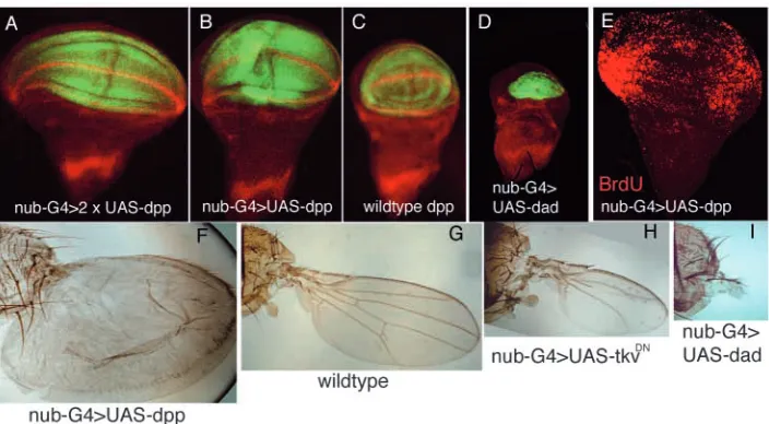
The general result is that the size of the wing correlates with the activity of the Dpp pathway. Some of the results are shown in Fig. 1. The increase of Dpp signal in nub-Gal4>UAS-dpp
(Fig. 1A,B) results in discs in which the wing pouch is bigger than the wild type (Fig. 1C), whereas the inhibition of Dpp activity in nub-Gal4>UAS-dad produces a very small wing pouch (Fig. 1D). The comparison of Fig. 1A,B is of interest because the only difference between the two discs is the amount of Dpp signal; their difference in size illustrates clearly the dependence of growth on the levels of Dpp activity. The effect observed in the discs can also be visualised in adult wings. In the series of genotypes shown in Fig. 1F-I, the gradual decrease in the size of the adult wing correlates with the levels of activity of the Dpp pathway.
A significant finding is that in the cases in which the wing pouch becomes bigger than the wild type (Fig. 1A,B), the additional growth appears to be due to excessive cell proliferation in the lateral region of the disc: while the incorporation of BrdU in the wild-type disc is homogenous (not shown), in all the discs examined (n=11) of genotype nub-Gal4>UAS-dpp BrdU incorporation is much more intense in the lateral region, although we still observe incorporation in the central region (Fig. 1E). The zone of increased proliferation coincides approximately with the brk domain and suggests that the size increase corresponds mostly or entirely to expansion of the brk domain.
[image:3.612.133.485.467.661.2]The previous result suggests that the Dpp pathway affects wing size by regulating brk activity, and is coherent with the fact that it represses brk expression (Campbell and Tomlinson, 1999; Jazwinska et al., 1999; Minami et al., 1999). Then it would be expected that there should be a negative correlation between wing size and brk levels. This is indeed the case, as illustrated in Fig. 2. This observation suggests that brk functions as a growth repressor and that the excessive growth observed in genotypes with high levels of Dpp activity (Fig. 1A,B,F; Fig. 2A,B) is due to suppression of brk in the wing pouch.
Fig. 1. Wing size correlates with Dpp activity. (A-D) Wing discs containing different levels of Dpp activity in the wing pouch. All the discs are
Brk as a growth repressor
We have examined directly the role of brk on growth by altering its normal function, either eliminating or inducing high levels of brk activity. There is evidence that mutant brk discs grow in excess (Campbell and Tomlinson, 1999) and that clones of brk mutant cells produce outgrowths (Campbell and Tomlinson, 1999; Jazwinska et al., 1999; Minami et al., 1999). We have generated a large number of brk–clones and compared
their size with control clones. A typical feature of these clones is that they produce outgrowths (Fig. 3A,B), which can be observed both in discs and in differentiated wings. They develop independently from surrounding wild-type cells; many form vesicles that sort out from the rest of the disc, while others develop outgrowths that recreate the wing pattern. Unlike the clones of cells expressing dpp ectopically (Zecca et al., 1995), brk– clones do not produce mirror-image duplications, as
caused by the formation of ectopic sources of the Dpp signal (Zecca et al., 1995). The best description of brk mutant clones is simply that they grow more than surrounding brk+cells and
therefore tend to make more pattern elements. The clones producing outgrowths are restricted to the lateral region of the disc, the normal brk expression domain, whereas those in the central region are of normal size. In accordance with this, brk–clones in the lateral region incorporate more BrdU
than does the surrounding zone: in a sample of nine discs containing 19 brk–clones, 15 are more densely labelled than
surrounding cells (Fig. 3C). These observations indicate that the repressor role of brk is restricted to the lateral region of the disc; the central region is regulated independently (see Discussion).
We have also examined whether the loss of brk activity can induce additional growth in the absence (or low levels) of Dpp pathway activity. Thus, we induced brk– clones in discs of
genotype nub-Gal4>UAS-dad in which the high levels of dad impede normal transduction of the Dpp signal (Tsuneizumi et al., 1997). As shown in Fig. 1D,I there is very little growth in the wing pouch of such discs (Fig. 2H). The significant result (illustrated in Fig. 3D) is that brk–clones are able to overgrow
in the wing pouch. We examined 13 discs of this genotype, that contained 26 brk–clones, all of which showed overgrowth in
comparison with control clones. In seven cases in which we could unambiguously identify a brk– clone and its twin, the
average size of the former was 5.36 times bigger. This result emphasises the role of brk as a growth repressor and also indicates that Dpp activity per se does not promote growth (see Discussion).
[image:4.612.82.259.73.403.2]The role of brk on growth can also be demonstrated in misexpression experiments. We have forced brk activity in Fig. 2. Wing size correlates negatively with brk expression. All the
discs are doubly stained for GFP and brk (anti-βgal). The right panels show brk expression in red. For simplification, the labels present only a partial notation of the genotype (see Materials and methods). (A,B) A nub-Gal4>UAS-tkvQDUAS-GFP disc with a large
wing pouch that has little or no brk activity. (C,D) nub-Gal4>UAS-GFP of normal size and normal brk activity. (E,F) The reduction of Dpp pathway activity in nub-Gal4>UAS-tkvDNUAS-GFP results in a
small wing pouch associated with high brk levels. (G,H) Further diminution of Dpp pathway activity in nub-Gal4>dad UAS-GFP produces a smaller wing pouch associated with an expansion of brk expression that covers the whole of the wing pouch.
Fig. 3. Extra growth produced by brk mutant clones. (A,B) brk–
clones (A, yellow; B, forked36) in the anterior (A) and posterior (B)
compartments. The clones do not produce pattern duplications but do produce additional tissue. (C) A disc doubly labelled arm-lacZ (green) and BrdU (red) with a brk–clone (arrow) showing greater
BrdU incorporation than surrounding cells. (D,E) Disc of genotype nub-Gal4>UAS-dad stained for wg (red) containing two brk–clones
[image:4.612.313.561.75.244.2]various regions of the disc using the Gal4 lines described above. In the combinations C765-Gal4>UAS-brk or nub-Gal4>UAS-brk, there is brk expression in the whole of the wing blade. In all these combinations, it can be observed that the size of the wing is greatly reduced (Fig. 4). The degree of the diminution correlates with the amount of Brk, as illustrated in Fig. 4 for the combination C765-Gal4>UAS-brk. At 17°C, the activity of the Gal4 protein is lower than at 25°C or 29°C (Brand and Perrimon, 1993), and this is reflected in the amount of Brk protein synthesised. We observe a clear difference of size both in differentiated wings (Fig. 4A,B) and in discs (Fig. 4C-E) grown at different temperatures. This result is significant, for it indicates that the Brk protein represses growth in a concentration-dependent manner.
The repressing role of brk is also demonstrated by the fact that it can suppress the growth of nub-Gal4>UAS-tkvQD(Fig.
5) wings; flies of genotype nub-Gal4>UAS-tkvQD UAS-brk
develop vestigial wings that are indistinguishable from those of nub-Gal4>UAS-brk. Again, the implication is that the excessive growth induced by the constitutive function of Dpp is mediated by inactivation of brk. In fact, no brk activity is detected in the wing pouch of nub-Gal4>UAS-tkvQDdiscs (Fig.
2A).
Mode of action of brk: apoptosis or growth retardation?
The preceding results demonstrate that Brk protein can block growth, but are not informative about its mode of action. Brk may act through two different mechanisms. The first is that it triggers apoptosis, which may result in reduced growth. In principle, this possibility does not appear likely because there is little apoptosis in normal development of wing discs, even in lateral regions where brk is active (Milan et al., 1997; Wolff
and Ready, 1991). However, a certain amount of apoptosis may have passed unnoticed, especially because it has only been looked at in mature discs. Moreover, recent experiments (Adachi-Yamada et al., 1999; Adachi-Yamada and O’Connor, 2002; Moreno et al., 2002) have shown that upregulation of brk or disruptions in Dpp signalling induce JNK-mediated apoptosis. The other possible mechanism is that Brk represses growth by reducing cell proliferation. We have tested these two possibilities.
We first checked the occurrence of apoptosis in cases in which elevated levels of brk cause a large reduction in wing size. In normal wing discs, the levels of apoptosis markers such as TUNEL and the cleaved (active) form of caspase 3 is variable, but low and scattered. In the wing pouch of mature nub-Gal4>UAS-brk wing discs, we find a slight increase of caspase 3 (Fig. 6A,B) and TUNEL (Fig. 6C,D), but most of brk-expressing cells fail to show these markers.
The previous experiments suggested that apoptosis is not a major factor in the growth repression caused by Brk. However, as these experiments were carried out in mature discs, there was the possibility that Brk may have induced apoptosis in earlier phases of development. If this were the case, it would be expected that apoptosis inhibitors should rescue partially or totally the effect of Brk. We used the baculovirus protein P35 (Hay et al., 1994) to prevent the death of cells that contain high levels of Brk. As shown in Fig. 6E,F the presence of the P35 protein in nub-Gal4>UAS-brk UAS-p35 discs suppresses the basal apoptosis in the wing pouch. The comparison of nub-Gal4>UAS-brk and omb-nub-Gal4>UAS-brk flies with their sibs nub-Gal4>brk p35 and omb-Gal4>brk UAS-p35, respectively, reveals that P35 does not rescue the effect of Brk (not shown). To strengthen this observation, we performed additional experiments generating flies of similar genotypes but containing two doses of UAS-p35. The extra dose of P35 did not modify the phenotype.
[image:5.612.337.548.73.239.2]The results of the previous experiments were intriguing, because there is evidence that alterations in brk levels cause JNK-mediated apoptosis (Adachi-Yamada and O’Connor, Fig. 4. Brk represses growth in a concentration-dependent manner.
[image:5.612.52.298.453.653.2](A,B) Adult wings and thoraces of the same genotype C765-Gal4>UAS-brk grown at 17°C (A) and 25°C (B). The greater activity of Gal4 at 25°C (B) produces more Brk protein, which results in less growth than at 17°C (A). (C-E) Sets of mature C765-Gal4>UAS-brk UAS-GFP wing, haltere and leg discs dissected from larvae grown at 17°C (C), 25°C (D) and 29°C (E). The size of the discs inversely correlates with temperature.
Fig. 5. Brk suppresses the excessive growth caused by the
2002; Moreno et al., 2002). The former authors have proposed that this form of apoptosis occurs when there is a disruption in the normal Dpp signalling gradient (and hence of the Brk gradient). This apoptosis aims to eliminate cells with disparate Dpp activity levels in order to restore the normal smooth gradient. The implication is that brk induces JNK activity only where there is a discontinuity of expression. We tested this by inducing brk activity with the hh-Gal4, en-Gal4 and ap-Gal4 drivers, and have monitored JNK activity with the puc-lacZ insertion (Martín-Blanco et al., 1998). These experiments generate a sharp discontinuity of brk at the AP (hh-Gal4>UAS-brk, en-Gal4>UAS-brk) or the DV (ap-Gal4>UAS-brk) borders. Some of the results are illustrated in Fig. 7: in hh-Gal4>UAS-brk puc-lacZ there is a line of puc activity close to the AP border. Similarly, in ap-Gal4>UAS-brk puc-lacZ there is puc activity close to the DV boundary, as previously described by Adachi-Yamada and O’Connor (Adachi-Yamada and O’Connor, 2002). The activation of puc in these cases appears to be non-autonomous, as it affects cells that do not possess brk activity (Fig. 7C,D).
In another set of experiments, we examined puc expression in discs containing brk– clones. These clones generate a
discontinuity in their borders as they confront cells containing high and null levels of brk activity. The clones in the brk domain (n=27) are associated with a complete (20 cases) or
incomplete ring of JNK activation in the border, which affects cells outside the clones as well as inside (Fig. 7E,F). In some cases (16 out of a sample of 40) there is also caspase 3 activity in cells in the borders (Fig. 7G,H). The variations of brk levels in this experiment are, unlike the high levels often obtained using the Gal4/UAS method, within the physiological range of brk activity. Thus, the induction of JNK-mediated apoptosis does not depend on absolute brk levels, but on the formation of a sharp interface.
We tested the possibility that the JNK-mediated apoptosis described above may contribute to the reduction in wing size. Overexpression of puc has been shown to downregulate the activity of the JNK pathway (Martín-Blanco et al., 1998), and also to reduce apoptosis of cells containing high brk levels (Moreno et al., 2002). We therefore constructed flies of genotypes nub-Gal4>UAS-brk UAS-puc and omb-Gal4>UAS-brk UAS-puc, and compared them with nub-Gal4>UAS-omb-Gal4>UAS-brk and omb-Gal4>UAS-brk. We failed to observe any difference in wing size.
Brk inhibits cell division and downregulates bantam
[image:6.612.54.279.71.315.2]All the preceding results suggest that the growth inhibition induced by brk is not mediated by massive apoptosis, but more likely by reducing the rate of cell proliferation. We have checked the division rate of cells containing high levels of brk Fig. 6. Brk induces low levels of apoptosis in the wing disc. (A) A
wing disc of genotype nub-Gal4>UAS-brk UAS-GFP doubly stained for GFP (green) and caspase 3 activity (red). The green fluorescence marks where brk is expressed. (B) Low level of caspase 3 activity (red) in brk-expressing cells. (C,D) A disc of the same genotype but stained for GFP and TUNEL. Only a minority of brk-expressing cells undergo apoptosis (red in D). (E,F) Wing disc of genotype nub-Gal4>UAS-brk UAS-p35 UAS-GFP stained for GFP and TUNEL (red). The green fluorescence marks the cell containing brk and p35 activity. This disc (F) shows unusually high apoptotic levels, but was chosen to illustrate the effectiveness of p35 in suppressing cell death (absence of red staining in area corresponding to green staining in E).
Fig. 7. Discontinuous levels of Brk give rise to JNK-mediated
apoptosis. The activity of the JNK pathway is monitored by the expression of the puc-lacZ insert. (A,B) Control hh-Gal4>UAS-GFP disc showing normal JNK activity, which is restricted to the proximal thoracic region. pucZ expression is shown in red. (C,D) Disc of hh-Gal4>UAS-brk UAS-GFP. There is puc expression (red) at the border of the brk-expressing cells (green), along the AP compartment boundary (arrows). There appears to be puc expression at both sides of the border, suggesting a non-autonomous effect. (E,F) Clone of brk–cells labelled by the loss of GFP (green) showing puc activity at
the border (arrows). (G,H) Caspase 3 activity (red, arrows) in the border of a brk–clone (shown by loss of green staining). There are
also some scattered caspase-positive cells in the vicinity of other brk–
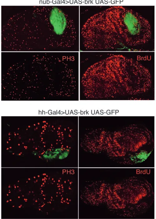
[image:6.612.313.561.75.294.2]using two different markers of cell division: the incorporation of BrdU and the staining with an antibody that recognises the phosphorylated form of Histone 3 (Su et al., 1998). In wild-type discs, the levels of BrdU and PH3 staining are uniform over the disc. In nub-Gal4>UAS-brk discs (n=10), both proliferation markers are less expressed in the wing pouch in comparison with other regions of the disc (Fig. 8). Similar results are obtained with hh-Gal>UAS-brk discs (n=27) in which brk is expressed at high levels in the posterior compartment (Fig. 8). These results strongly suggest that the principal function of brk is to reduce the rate of cell proliferation.
We tried to identify genes involved in cell proliferation as possible targets of Brk. A candidate is the gene bantam, which encodes a small RNA and has been shown to promote proliferation and to prevent apoptosis (Brennecke et al., 2003). To monitor bantam expression, we have used the bantam sensor developed by Brennecke et al. (Brennecke et al., 2003). The expression of the sensor can be taken as the negative of the levels of proliferation in the wing disc. We have examined bantam expression in clones of brk mutant cells, as well as in
genetic combinations with altered brk activity. In nub-Gal4>UAS-tkvQDdiscs, the levels of the sensor are reduced in
the lateral region of the disc (Fig. 9A-D), indicating a raise of bantam expression, in correspondence with the increased proliferation levels observed in this zone. Similarly, brk–clones
show a diminution of sensor level (Fig. 9E,F), also indicating a raise in bantam expression. These experiments clearly indicate that bantam is downregulated by brk, although the regulation may not be direct. However, using a collection of published DNA sequences containing Brk-binding sites we constructed a matrix (see Materials and methods) to identify potential sites in the vicinity of the bantam gene. We have found two sites in a 20 kb fragment that includes bantam, suggesting the possibility of direct regulation by Brk.
Discussion
[image:7.612.319.564.306.632.2]Our experiments deal with the roles of the Dpp signalling pathway and brk in the control of growth of the Drosophila wing disc. As the Dpp gradient is transformed into a
Fig. 8. Brk reduces the rates of cell proliferation. The upper panels
[image:7.612.50.299.321.672.2]show two discs of genotype nub-Gal4>UAS-brk UAS-GFP doubly stained for GFP and PH3 (left) and GFP and BrdU (right). In both cases, there is a marked reduction of both PH3 or BrdU (red) in the cells expressing brk. In the lower panels, two discs of genotype hh-Gal4>UAS-brk UAS-GFP show a similar result.
Fig. 9. Brk downregulates bantam. (A,B) Wing disc with normal brk
activity stained for PH3 and the bantam sensor: the brighter green colour corresponds to low bantam expression (see main text). The arrow indicates a characteristic zone of low bantam levels located in the brk domain. The distribution of the PH3 dots in the disc is uniform, indicating the uniform cell proliferation levels in the disc. (C,D) Disc of nub-Gal4>UAS-tkvQDgenotype showing more PH3
staining in the lateral region, which is associated with partial loss of bantam. (E,F) A brk–clone showing greater bantam activity
complementary Brk gradient (Muller et al., 2003), the issue of how the Brk gradient regulates wing growth can be addressed. We will first discuss some aspects of its mode of action and then we will deal with its overall function in growth control.
Two different functions of brk
We find that alterations of brk expression may have two different consequences.
Activation of the JNK pathway
This occurs when an alteration of brk expression generates a sharp border of brk activity. We have observed this phenomenon both in experiments inducing ectopic brk activity and in others in which brk function is eliminated in clones of cells (see Fig. 7). The local induction of JNK results in apoptosis that can be visualised by the activation of caspase 3 (Fig. 7G,H).
This local apoptosis induced by Brk is probably the mechanism of cell elimination during cell competition (Morata and Ripoll, 1975; Moreno et al., 2002) and suggests that brk is involved in the elimination of slow dividing cells or of cells that are not able to read or interpret efficiently the Dpp pathway. This function may be aimed to keep the general fitness of the cell population (Moreno et al., 2002). However, it does not appear to be involved in growth control, because apoptosis inhibition (by means of puc or p35 overexpression) does not eliminate the effect on size caused by Brk.
Alterations of cell proliferation rate
Previous work has already shown that loss of brk activity results in increased growth: in mutant brk discs there is an enlargement of the lateral region (Campbell and Tomlinson, 1999), and cells mutant for brk produce outgrowths (Campbell and Tomlinson, 1999; Jazwinska et al., 1999; Minami et al., 1999) (this work). We show that the cause for the additional growth associated with the loss or reduction of brk activity is due to an increase in the cell proliferation rate: brk– clones
incorporate BrdU more actively than surrounding cells (Fig. 3C). Conversely, the repression of growth caused by elevated levels of Brk is associated with reduced mitotic activity and BrdU incorporation (Fig. 8).
Given the nature of the Brk protein, it would be expected that its role in growth be mediated by transcriptional repression of genes involved in cell division and proliferation. Our results indicate that it acts as a repressor of bantam (Fig. 9), although this control may not be direct. Given that Bantam protein is itself a post-transcriptional regulator of cell division genes (Brennecke et al., 2003), this observation suggests that Brk occupies a high position in the genetic hierarchy controlling cell proliferation. Its activity links Dpp signalling and cell proliferation.
Control of growth by the Brk gradient
Our results, and those of others (Burke and Basler, 1996; Martín-Castellanos and Edgar, 2002), have established that the Dpp pathway is involved in the control of growth of the wing (and of other appendages; data not shown). The activity of the Dpp pathway has a positive effect on growth, and, furthermore, we find that the growth response of the disc correlates with its levels of activity. This graded response is of interest, as it suggests that growth control mechanisms recognise different
concentrations of inducing or repressing factors. This result has implications in the understanding of these mechanisms; classically, it has been argued that proliferation in the imaginal discs is a response to confrontation of cells with different positional values (French et al., 1976; Haynie and Bryant, 1976). Our results in the wing disc do not support this view, as they suggest that growth is a lineal response to Dpp/Brk activity.
Our results also indicate that the role of Dpp on growth is mediated by brk. The simplest view is that as the Dpp gradient is converted into an inverse Brk gradient, the concentration-dependent stimulus of Dpp on growth should be converted into a concentration-dependent repression by Brk. Our demonstration (Fig. 4) that the effect of Brk on wing size depends on the amount of protein supports this view.
There are several arguments that implicate brk as a principal factor controlling growth. First, loss of brk activity leads to increased proliferation (Fig. 3A-C). This is consistent with previous observations (Campbell and Tomlinson, 1999) showing that brk wing discs are bigger than wild-type discs. Furthermore, this excessive proliferation can occur in absence of Dpp activity. Fig. 3D shows two overgrowing brk mutant clones originated from the wing pouch of nub-Gal4>UAS-dad discs in which Dpp function is obliterated or much reduced. Second, increased or ectopic brk levels block or reduce growth, even though brk does not alter dpp expression (Fig. 4A-E). And, third, the stimulation caused by the Dpp pathway on growth requires repression of brk. This is demonstrated by our finding that the presence of Brk protein suppresses the excessive growth caused by Dpp hyperactivity (Fig. 5).
Together, these observations indicate that growth does not require direct input from Dpp, but simply its repression of brk. However, the repression of brk by Dpp is an important developmental phenomenon because in the absence of such control brk would become constitutively active, thus repressing all or the majority of Dpp targets. Recent work (Muller et al., 2003) has identified two control elements in the brk regulatory region: a Dpp-regulated silencer that contains binding sites for the Mad/Medea complex; and a constitutive enhancer. This enhancer is probably responsible of the generalised brk expression in the absence of Dpp activity.
What is the role of brk in normal development? Our results demonstrate that Brk has the properties of a growth repressor and can perform this function all over the wing. However, in wild-type wing discs, brk is expressed only in the lateral region and therefore its repressing role is limited to this region. This is agreement with the observation that brk–clones overgrow
only on the sides of the disc (Campbell and Tomlinson, 1999; Jazwinska et al., 1999; Minami et al., 1999) (this work).
In principle, a candidate could be daughters against dpp (dad), a Dpp target that is expressed at high levels in the centre of the disc. We have observed that dad overexpression reduces growth. However, this appears to be achieved by allowing high brk levels (Fig. 2G,H) subsequent to slackening of Dpp activity (Tsuneizumi et al., 1997), indicating that the effect of dad is mediated by brk. Thus, dad appears to be a Dpp modulator with no direct role in growth. Our finding that brk– clones
containing high levels of dad activity can overgrow (Fig. 3D) also supports this view.
We thank Ernesto Sanchez-Herrero and José Felix de Celis for comments on the manuscript, Luis F. de Navas for help with the crosses, Rosa Gonzalez and Angélica Cantarero for technical help, and T. Tabata and S. Cohen for sending fly stocks. F.A.M. is supported by a fellowship of the Comunidad Autónoma de Madrid. This work was supported by grants from the Ministry of Science and Technology.
References
Adachi-Yamada, T. and O’Connor, M. B. (2002). Morphogenetic apoptosis:
a mechanism for correcting discontinuities in morphogen gradients. Dev.
Biol. 251, 74-90.
Adachi-Yamada, T., Fujimura-Kamada, K., Nishida, Y. and Matsumoto, K. (1999). Distorsion of proximodistal information causes JNK-dependent
apoptosis in Drosophila wing. Nature 400, 166-169.
Aldaz, S., Morata, G. and Azpiazu, N. (2003). The Pax-homeobox gene
eyegone is involved in the subdivision of the thorax of Drosophila.
Development 130, 4473-4482.
Barrio, R. and de Celis, J. F. (2004). Regulation of spalt expression in the
Drosophila wing blade in response to the Decapentaplegic signaling pathway. Proc. Natl. Acad. Sci. USA 101, 6021-6026.
Brand, A. H. and Perrimon, N. (1993). Targeted gene expression as a means
of altering cell fates and generating dominant phenotypes. Development 118, 401-415.
Brennecke, J., Hipfner, D. R., Stark, A., Russell, R. B. and Cohen, S. M.
(2003). bantam encodes a developmentally regulated microRNA that controls cell proliferation and regulates the proapoptotic gene hid in Drosophila. Cell 113, 25-36.
Bryant, P. (1975). Pattern formation in the imaginal wing disc of Drosophila
melanogaster: fate map, regeneration and duplication. J. Exp. Zool. 193, 49-78.
Bryant, P. J. and Simpson, P. (1984). Intrinsic and extrinsic control of growth
in developing organs. Q. Rev. Biol. 59, 387-415.
Burke, R. and Basler, K. (1996). Dpp receptors are autonomously required
for cell proliferation in the entire developing Drosophila wing. Development
122, 2261-2269.
Calleja, M., Moreno, E., Pelaz, S. and Morata, G. (1996). Visualization of
gene expression in living adult Drosophila. Science 274, 252-255.
Campbell, G. and Tomlinson, A. (1999). Transducing the Dpp morphogen
gradient in the wing of Drosophila: regulation of Dpp targets by brinker.
Cell 96, 553-562.
Capdevila, J. and Guerrero, I. (1994). Targeted expression of the signaling
molecule decapentaplegic induces pattern duplications and growth alterations in Drosophila wings. EMBO J. 13, 4459-4468.
de Celis, J. F., Barrio, R. and Kafatos, F. C. (1996). A gene complex acting
downstream of dpp in Drosophila wing morphogenesis. Nature 381, 421-424.
Entchev, E. V., Schwabedissen, A. and Gonzalez-Gaitan, M. (2000).
Gradient formation of the TGF-beta homolog Dpp. Cell 103, 981-991.
French, V., Bryant, P. J. and Bryant, S. V. (1976). Pattern regulation in
epimorphic fields. Science 193, 969-981.
García-Bellido, A. and Merriam, J. (1971a). Parameters of the wing
imaginal disc development of Drosophila melanogaster. Dev. Biol. 24, 61-87.
García-Bellido, A. and Merriam, J. R. (1971b). Clonal parameters of tergite
development in Drosophila. Dev. Biol. 26, 264-276.
Gehring, W. (1976). Developmental genetics of Drosophila. Ann. Rev. Genet. 10, 209-252.
Grimm, S. and Pflugfelder, G. O. (1996). Control of the gene
optomotor-blind in Drosophila wing development by dacapentaplegic and wingless.
Science 271, 1601-1604.
Haerry, T. E., Khalsa, O., O’Connor, M. B. and Wharton, K. A. (1998).
Synergistic signaling by two BMP ligands through the SAX and TKV receptors controls wing growth and patterning in Drosophila. Development
125, 3977-3987.
Harvey, K. F., Pfleger, C. M. and Hariharan, I. K. (2003). The Drosophila
Mst ortholog, hippo, restricts growth and cell proliferation and promotes apoptosis. Cell 114, 457-467.
Hay, B. A., Wolff, T. and Rubin, G. M. (1994). Expression of baculovirus
P35 prevents cell death in Drosophila. Development 120, 2121-2129.
Haynie, J. L. and Bryant, P. J. (1976). Intercalary regeneration in imaginal
wing disk of Drosophila melanogaster. Nature 259, 659-662.
Hipfner, D. R., Weigmann, K. and Cohen, S. M. (2002). The bantam gene
regulates Drosophila growth. Genetics 161, 1527-1537.
Inoue, H., Imamura, T., Ishidou, Y., Takase, M., Udagawa, Y., Oka, Y., Tsuneizumi, K., Tabata, T., Miyazono, K. and Kawabata, M. (1998).
Interplay of signal mediators of decapentaplegic (Dpp): molecular characterization of mothers against dpp, Medea, and daughters against dpp.
Mol. Biol. Cell 9, 2145-2156.
Jazwinska, A., Kirov, N., Wieschaus, E., Roth, S. and Rushlow, C. (1999).
The Drosophila gene brinker reveals a novel mechanism of Dpp target gene regulation. Cell 96, 563-573.
Jia, J., Zhang, W., Wang, B., Trinko, R. and Jiang, J. (2003). The
Drosophila Ste20 family kinase dMST functions as a tumor suppressor by restricting cell proliferation and promoting apoptosis. Genes Dev. 17, 2514-2519.
Johnston, L. A. and Sanders, A. L. (2003). Wingless promotes cell survival
but constrains growth during Drosophila wing development. Nat. Cell Biol.
5, 827-833.
Kango-Singh, M., Nolo, R., Tao, C., Verstreken, P., Hiesinger, P. R., Bellen, H. J. and Halder, G. (2002). Shar-pei mediates cell proliferation
arrest during imaginal disc growth in Drosophila. Development 129, 5719-5730.
Kim, J., Sebring, A., Esch, J. J., Kraus, M. E., Vorwerk, K., Magee, J. and Carroll, S. B. (1996). Integration of positional signals and regulation of
wing formation and identity by Drosophila vestigial gene. Nature 382, 133-138.
Kirby, B., Bryant, P. and Schneiderman, H. (1982). Regeneration following
duplication in imaginal wing disc fragments of Drosophila melanogaster.
Dev. Biol. 90, 259-271.
Lawrence, P. and Morata, G. (1977). The early development of mesothoracic
compartments in Drosophila. An analysis of cell lineage and fate mapping and an assesment of methods. Dev. Biol. 56 40-51.
Lecuit, T., Brook, W. J., Ng, M., Calleja, M., Sun, H. and Cohen, S. M.
(1996). Two distinct mechanisms for long-range patterning by Decapentaplegic in the Drosophila wing. Nature 381, 387-393.
Madhavan, M. M. and Madhavan, K. (1984). Do larval epidermal cells
possess the blueprint for adult pattern in Drosophila? J. Embryol. Exp.
Morph. 82, 1-8.
Martín-Blanco, E., Gampel, A., Ring, J., Virdee, K., Kirov, N., Tolkovsky, A. M. and Martinez, A. A. (1998). puckered encodes a phosphatase that
mediates a feedback loop regulating JNK activity during dorsal closure in Drosophila. Genes Dev. 12, 557-570.
Martín-Castellanos, C. and Edgar, B. A. (2002). A characterization of the
effects of Dpp signaling on cell growth and proliferation in the Drosophila wing. Development 129, 1003-1013.
Marty, T., Muller, B., Basler, K. and Affolter, M. (2000). Schnurri mediates
Dpp-dependent repression of brinker transcription. Nat. Cell Biol. 2, 745-749.
Milan, M., Campuzano, S. and García-Bellido, A. (1997). Developmental
parameters of cell death in the wing disc of Drosophila. Proc. Natl. Acad.
Sci. USA 94, 5691-5696.
Minami, M., Kinoshita, N., Kamoshida, Y., Tanimoto, H. and Tabata, T.
(1999). brinker is a target of Dpp in Drosophila that negatively regulates Dpp- dependent genes. Nature 398, 242-246.
Morata, G. and García-Bellido, A. (1976). Developmental analysis of some
mutants of the bithorax sistem of the Drosophila. Rouxs Arch. Dev. Biol.
179, 125-143.
Morata, G. and Ripoll, P. (1975). Minutes: mutants of drosophila
autonomously affecting cell division rate. Dev. Biol. 42, 211-221.
Moreno, E., Basler, K. and Morata, G. (2002). Cells compete for
Muller, B., Hartmann, B., Pyrowolakis, G., Affolter, M. and Basler, K.
(2003). Conversion of an extracellular Dpp/BMP morphogen gradient into an inverse transcriptional gradient. Cell 113, 221-233.
Nellen, D., Burke, R., Struhl, G. and Basler, K. (1996). Direct and
long-range action of a DPP morphogen gradient. Cell 85, 357-368.
Pantalacci, S., Tapon, N. and Leopold, P. (2003). The Salvador partner Hippo
promotes apoptosis and cell-cycle exit in Drosophila. Nat. Cell Biol. 5, 921-927.
Podos, S. D. and Ferguson, E. L. (1999). Morphogen gradients: new insights
from DPP. Trends Genet. 15, 396-402.
Raftery, L. A. and Sutherland, D. J. (1999). TGF-beta family signal
transduction in Drosophila development: from Mad to Smads. Dev. Biol.
210, 251-268.
Rushlow, C., Colosimo, P. F., Lin, M. C., Xu, M. and Kirov, N. (2001).
Transcriptional regulation of the Drosophila gene zen by competing Smad and Brinker inputs. Genes Dev. 15, 340-351.
Ruvkun, G. (2001). Molecular biology. Glimpses of a tiny RNA world. Science 294, 797-799.
Saller, E., Kelley, A. and Bienz, M. (2002). The transcriptional repressor
Brinker antagonizes Wingless signaling. Genes Dev. 16, 1828-1838.
Sivasankaran, R., Vigano, M. A., Muller, B., Affolter, M. and Basler, K.
(2000). Direct transcriptional control of the Dpp target omb by the DNA binding protein Brinker. EMBO J. 19, 6162-6172.
Sosinsky, A., Bonin, C. P., Mann, R. S. and Honig, B. (2003). Target
Explorer: an automated tool for the identification of new target genes for a specified set of transcription factors. Nucleic Acids Res. 31, 3589-3592.
Stocker, H. and Hafen, E. (2000). Genetic control of cell size. Curr. Opin. Genet. Dev. 10, 529-535.
Strigini, M. and Cohen, S. M. (1999). Formation of morphogen gradients in
the Drosophila wing. Semin. Cell Dev. Biol. 10, 335-344.
Sturtevant, M. A., Biehs, B., Marin, E. and Bier, E. (1997). The spalt gene
links the A/P compartment boundary to a linear structure in the Drosophila wing. Development 124, 21-32.
Su, T. T., Sprenger, F., DiGregorio, P. J., Campbell, S. D. and O’Farrell, P. H. (1998). Exit from mitosis in Drosophila syncytial embryos requires
proteolysis and cyclin degradation, and is associated with localized dephosphorylation. Genes Dev. 12, 1495-1503.
Tabata, T. (2001). Genetics of morphogen gradients. Nat. Rev. Genet. 2,
620-630.
Tapon, N., Harvey, K. F., Bell, D. W., Wahrer, D. C., Schiripo, T. A., Haber, D. A. and Hariharan, I. K. (2002). salvador Promotes both cell cycle exit
and apoptosis in Drosophila and is mutated in human cancer cell lines. Cell
110, 467-478.
Teleman, A. and Cohen, S. (2000). Dpp gradient formation in the Drosophila
wing imaginal disc. Cell 103, 971-980.
Tsuneizumi, K., Nakayama, T., Kamoshida, Y., Kornberg, T., Christian, J. and Tabata, T. (1997). Daughters against dpp modulates dpp organizing
activity in Drosophila wing development. Nature 389, 627-631.
Udan, R. S., Kango-Singh, M., Nolo, R., Tao, C. and Halder, G. (2003).
Hippo promotes proliferation arrest and apoptosis in the Salvador/Warts pathway. Nat. Cell Biol. 5, 914-920.
Wang, S. L., Hawkins, C. J., Yoo, S. J., Muller, H. A. and Hay, B. A. (1999).
The Drosophila caspase inhibitor DIAP1 is essential for cell survival and is negatively regulated by HID. Cell 98, 453-463.
Wolff, T. and Ready, D. F. (1991). Cell death in normal and rough eye mutants
of Drosophila. Development 113, 825-839.
Wu, S., Huang, J., Dong, J. and Pan, D. (2003). hippo encodes a Ste-20
family protein kinase that restricts cell proliferation and promotes apoptosis in conjunction with salvador and warts. Cell 114, 445-456.
Zecca, M., Basler, K. and Struhl, G. (1995). Sequential organizing activities
of engrailed Hedhehog and decapentaplegic in the Drosophila wing.