CLINICAL REPORT
ADULT BRAIN
Diffusion-Weighted Zonal Oblique Multislice–EPI Enhances the
Detection of Small Lesions with Diffusion Restriction in the
Brain Stem and Hippocampus: A Clinical Report of
Selected Cases
XT. Sartoretti,XE. Sartoretti,XC. Binkert,XA. Gutzeit,XC. Reischauer,X D. Czell,XM. Wyss,X E. Bru¨llmann, and XS. Sartoretti-Schefer
ABSTRACT
SUMMARY: Diffusion restriction is the morphologic hallmark of acute ischemic infarcts and excitotoxic brain injury in various cerebral pathologies. Diffusion restriction is visible as hyperintensity on DWI and as hypointensity on ADC maps. Due to the vicinity of multiple anatomic structures in the brain stem and hippocampus, very small lesions with diffusion restriction may result in severe clinical symp-tomatology, but these small lesions easily go undetected on standard cerebral DWI due to insufficient spatial resolution, T2* blurring, and image artifacts caused by susceptibility-related image distortions. Diffusion-weighted zonal oblique multislice–EPI with reduced FOV acquisition permits a considerable increase in spatial resolution and enhances the visualization of very small pathologic lesions in the brain stem and hippocampus. Improved performance in the depiction of different pathologic lesions with diffusion restriction in the brain stem and hippocampus using this sequence compared with standard DWI in selected cases is presented.
ABBREVIATIONS:CN III⫽oculomotor nerve nucleus area; CN VI⫽abducens nerve nucleus area; DW⫽diffusion-weighted; sshot SE⫽single-shot spin-echo; TGA⫽transient global amnesia; ZOOM⫽zonal oblique multislice
D
iffusion restriction is caused by different cerebral pathologies leading to cytotoxic edema by energy failure, altered intra- and extracellular ion and water concentrations, and excitotoxic brain in-jury due to extracellular increase of glutamate.1,2These mechanismsmay overlap in certain pathologies. Diffusion restriction is the main characteristic feature of an acute ischemic event2; therefore, DWI is
the only reliable sequence for detecting an acute ischemic event. DWI is routinely used in brain protocols for the evaluation of various cerebral pathologies characterized by diffusion restriction. The limited spatial resolution of a standard single-shot spin-echo (sshot SE) EPI DWI, called “standard DWI” in our work, is an enormous disadvantage if the clinical symptomatology indicates a lesion in the brain stem and hippocampus.
This clinical report shows how diffusion-weighted (DW) zonal oblique multislice (ZOOM)-EPI can improve the detection of DWI abnormalities in pathologies in the brain stem and hippocampus by increasing spatial resolution, allowing the visualization of small lesions with diffusion restriction not visible on standard DWI.
MATERIALS AND METHODS
In this clinical report, the MR images of 7 patients of a group with 44 patients (23 women and 21 men; 40 – 89 years of age; mean age, 76.4 years) are depicted. Informed and written consent for publi-cation of patient data and images was obtained by all patients described in this study. All patients with various acute clinical symptoms all indicating a brain stem pathology (mainly charac-terized by nausea, vertigo, ataxia, nystagmus, gaze disorders) or hippocampal pathology with amnestic syndrome were examined by both standard transverse sshot SE EPI DWI (called “standard DWI”) and transverse DW ZOOM-EPI during the same exami-nation. The acquisition of DW ZOOM-EPI followed immediately after the acquisition of the standard DWI on the same MR imag-ing scanner with an identical field strength. Identical image plane angulation was used in both transverse diffusion-weighted sequences.
Patient examinations were performed from October 2016 to August 2017 on a 1.5T Ingenia (Philips Healthcare, Best, the Netherlands) or a 3T Achieva (Philips Healthcare) scanner. Im-aging parameters of DW ZOOM-EPI and of standard DWI are shown inTables 1and2. In all patients, 3D FLAIR imaging and transverse T2 TSE sequences were added for possible confirma-tion of the suspected pathology.
In each patient, the presence or absence of acute ischemic lesions in the brain stem on standard DWI and on DW ZOOM-EPI was determined retrospectively. In 27 patients, no patho-logic lesion was visible on both standard DWI and DW
Received October 19, 2017; accepted after revision March 5, 2018.
From the Institut fu¨r Radiologie (T.S., E.S., C.B., M.W., S.S.-S.), Kantonsspital Winter-thur, WinterWinter-thur, Switzerland; Institut fu¨r Radiologie und Nuklearmedizin (A.G., C.R.), Hirslanden Klinik St. Anna, Luzern, Switzerland; Medizinische Klinik (D.C.), Abteilung fu¨r Neurologie, Uznach, Switzerland; and Philips HealthSystems (M.W., E.B.), Zu¨rich, Switzerland.
Please address correspondence to Sabine Sartoretti-Schefer, MD, Institut fu¨r Radi-ologie, Kantonsspital Winterthur, Brauerstr 15, 8401 Winterthur, Switzerland; e-mail: [email protected]
ZOOM-EPI. In 9 patients both standard DWI and DW ZOOM-EPI showed an acute ischemic infarct with diffusion restriction, involving both the brain stem and cerebellum in 6 pa-tients, only the cerebellum in 2 papa-tients, and only the brain stem in 1 patient. All infarcts visible on both standard DWI and DW ZOOM-EPI had a minimum size of 1 cm. In 8 patients, mostly punctate acute ischemic foci, either in the brain stem, cerebellum, or hippocampi, were only visible on DW ZOOM-EPI.
Despite the acquisition time of the DW ZOOM-EPI (3 min-utes to 5 minmin-utes and 30 seconds), no visually evident movement artifacts in the patients examined were observed, but a quantifi-cation of movement artifacts was not performed.
Case Series
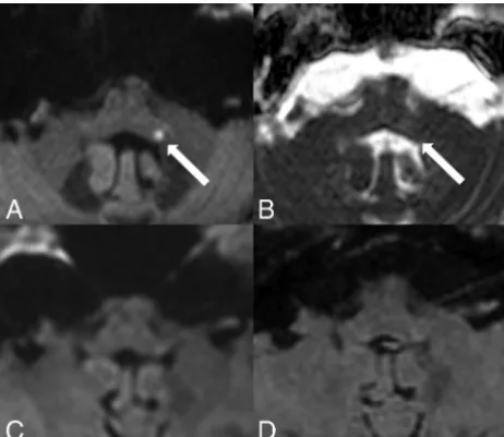
Transient Global Amnesia. A 65-year-old female patient pre-sented with antegrade and retrograde amnesia due to clinically suspected transient global amnesia (TGA) lasting for 18 hours (Fig 1). Punctate diffusion restriction was demonstrated on DW ZOOM-EPI with ADC maps in the lateral hippocampal body as a specific TGA-associated lesion. No abnormality was visible on standard DWI or on FLAIR.
Hippocampal Ischemia. An 83-year-old woman with atrial fibrillation presented with acute hemiparesis on the left side and antegrade amnesia (Fig 2). Multiple punctate acute embolic ischemic in-farcts with diffusion restriction were visible on DW ZOOM-EPI with ADC maps, namely in the mesencephalon along the pyramidal tract, in the left me-dial occipitotemporal gyrus, in the left hippocampus, and bilaterally in the pre-and postcentral area on the right. The punctate hippocampal infarct was not visible on standard DWI or FLAIR.
Acute Internuclear Ophthalmoplegia and Partial Oculomotor Nerve Palsy. A 44-year-old woman presented with acute internuclear ophthalmoplegia and a partial oculomotor nerve palsy on the left. The acute ischemic infarct in the oc-ulomotor nerve nuclear area (CN III) and the medial longitudinal fascicle was visible on DW ZOOM-EPI and ADC maps but not on standard DW and was only faintly apparent on FLAIR (Fig 3). The ischemic infarct was caused by an acute embolus from a pulmonary arte-riovenous malformation.
Acute Internuclear Ophthalmoplegia and Acute Abducens Palsy. A 71-year-old man presented with acute internuclear ophthalmoplegia/abducens nerve palsy of the left side, skew deviation, and nystag-mus to the right side. An acute ischemic infarct with diffusion restriction was visi-ble on DW ZOOM-EPI and ADC maps involving the left medial longitudinal fascicle and the abducens nerve nuclear area (CN VI). Standard DWI and FLAIR images did not show any ab-normality (Fig 4).
Acute Ischemic Infarct in the Left Inferior Cerebellar Peduncle. A 57-year-old woman presented with severe intractable vertigo, nystagmus, and vomiting due to isolated vestibular syndrome lasting 3 days. A punctate diffusion abnormality on DW ZOOM-EPI and ADC maps (Fig 5) in the left inferior cerebellar peduncle was present, not visible on standard DWI or FLAIR.
Acute Ischemic Infarct in the Right Nucleus Prepositus Hypo-glossi. A 40-year-old female patient presented with acute forward and backward vertigo and horizontal nystagmus. A punctate dif-fusion abnormality was visible on DW ZOOM-EPI and ADC maps (Fig 6) in the right nucleus prepositus hypoglossi; the lesion was not depicted on standard DWI and on FLAIR.
[image:2.594.54.374.55.231.2]Acute Ischemic Infarct in the Right Flocculus. A 51-year-old woman presented with vertigo and saccadic eye movements during the last 5 days. A punctate diffusion abnormality representing her infarction
Table 1: Imaging parameters of DW ZOOM-EPI
DW ZOOM-EPI on 1.5T MRI
DW ZOOM-EPI on 3T MRI
Sequence type sshot SE-EPI sshot SE-EPI
Slice orientation Transversal Transversal
FOV (RL⫻AP) 97⫻92 mm2 180⫻64 mm2
Acquisition voxel size (RL⫻AP⫻FH) 1.2⫻1.2⫻3.0 mm3 1.0⫻1.15⫻3.0 mm3
No. of slices 19 25
TR 3545 ms 2899 ms
TE 85 ms 64 ms
Flip angle 90° 90°
Half scan factor 0.6 0.6
Bandwidth 11.5 Hz/pixel 12.9 Hz/pixel
EPI factor 75 81
No. of signal averages 5 2
Acquisition duration (minutes:seconds) 05:39 03:04
Fat suppression SPAIR SPIR
b-values 0, 1000 s/mm2 0, 1000 s/mm2
Note:—SPIR indicates spectral presaturation with inversion recovery; SPAIR, spectral attenuated inversion recovery; RL, right left; AP, anterior posterior; FH, feet head.
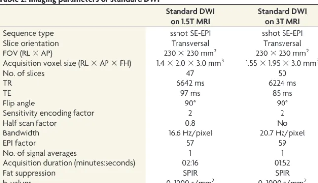
Table 2: Imaging parameters of standard DWI
Standard DWI on 1.5T MRI
Standard DWI on 3T MRI
Sequence type sshot SE-EPI sshot SE-EPI
Slice orientation Transversal Transversal
FOV (RL⫻AP) 230⫻230 mm2 230⫻230 mm2
Acquisition voxel size (RL⫻AP⫻FH) 1.4⫻2.0⫻3.0 mm3 1.55⫻1.95⫻3.0 mm3
No. of slices 47 50
TR 6642 ms 6224 ms
TE 97 ms 85 ms
Flip angle 90° 90°
Sensitivity encoding factor 2 2
Half scan factor 0.8 No
Bandwidth 16.6 Hz/pixel 20.7 Hz/pixel
EPI factor 57 59
No. of signal averages 1 1
Acquisition duration (minutes:seconds) 02:16 01:52
Fat suppression SPIR SPIR
b-values 0, 1000 s/mm2 0, 1000 s/mm2
[image:2.594.55.378.269.455.2]was present in the right flocculus on DW ZOOM-EPI and ADC maps (Fig 7). Standard DWI and FLAIR findings were normal.
DISCUSSION
Standard sshot SE EPI DWI is prone to geometric distortions, mainly due to the long readout time and low bandwidth in the
phase-encoding direction. Reduced-FOV imaging allows acquisi-tion of a small FOV with either reduced geometric distoracquisi-tion (shorter readout time) or higher spatial resolution (same readout time) compared with standard EPI DWI. An FOV smaller than the object typically induces foldover artifacts. Numerous different techniques have been introduced in the past to prevent these, especially selective excitation methods that excite only the ROI such as “inner volume imaging” and suppression-based methods that apply bands to saturate signal external to the target FOV such as “outer volume suppression” or a combination of both.3-7
Reduced-FOV imaging or ZOOM imaging or small-FOV im-aging is a commonly used technique in the context of data
acqui-FIG 1. TGA. MR imaging was performed on a 1.5T scanner 24 hours after the beginning of the clinical symptomatology. On DWI ZOOM-EPI (white arrowinA). a punctate hyperintensity and, on DWI ZOOM-EPI ADC map (white arrowinB), a punctate hypointensity with an ADC of 0.915⫻10⫺3mm2/second were demonstrated in the left lateral hippocampal body as a specific TGA-associated lesion. ADC in the normal right hippocampus was 1.093⫻10⫺3mm2/second. Stan-dard DWI (C) did not show the abnormality nor was any signal inten-sity change visible on transverse FLAIR (D).
FIG 2. Hippocampal embolic ischemia. MR imaging was performed on a 1.5T scanner. On DW ZOOM-EPI, 3 acute ischemic infarcts in the right mesencephalon along the pyramidal tract (white arrow), in the left me-dial occipitotemporal gyrus (white arrow), and in the left hippocampus (white arrowhead) were visible as hyperintense foci inA. On DW ZOOM-EPI ADC, the same infarcts were hypointense inBmarked withblack arrowsand ablack arrowhead(with an ADC of 0.817⫻10⫺3mm2/ second compared with the ADC of 1.03⫻10⫺3mm2/second in the nor-mal right hippocampus). The punctate snor-mall hippocampal infarct on the left side was not visible on standard DWI (C) or FLAIR (D).
FIG 3. Acute internuclear ophthalmoplegia with partial oculomotor nerve palsy. MR imaging was performed on a 3T scanner. The acute ischemic infarct in left CN III and the medial longitudinal fascicle was visible as a hyperintensity (white arrow) on DW ZOOM-EPI (A), as a hypointensity (white arrow) on DW ZOOM-EPI ADC (B) with an ADC of 0.6⫻10⫺3mm2/second (compared with an ADC of 0.985⫻10⫺3 mm2/second in the normal right CN III), and as very faintly hyperin-tense on transverse FLAIR (D,white arrowhead). No pathologic find-ings were seen on standard DWI (C).
[image:3.594.302.531.46.216.2] [image:3.594.54.286.47.259.2] [image:3.594.300.534.318.482.2] [image:3.594.54.284.359.547.2]sition of small FOVs within larger objects. In this work, a non-coplanar excitation combined with outer volume suppression, originally presented by Wilm et al,8was used. This technique is
now referred to as DW ZOOM-EPI, and the main applications using small-FOV imaging are imaging of the prostate, spinal cord, pancreas, breast, and heart,5-11where a relatively small
area of interest surrounded by tissue of less interest is depicted with high resolution. However, to date, the use of reduced-FOV imaging in the depiction of very small pathologic lesions with diffusion restriction in the brain stem and hippocampus has not yet been systematically evaluated.12-18The imaging
examples presented in this clinical report stress but do not prove the importance of reduced-FOV imaging for the visual-ization of small lesions in the brain stem and hippocampus.
The increased detectability of small lesions with diffusion re-striction in the brain stem and hippocampus on DW ZOOM-EPI in comparison with standard DWI might have been influenced by several technical factors:
1) The registration effect: The standard DWI and the DW ZOOM-EPI slice selections may be slightly different. As a result, the comparison of visual lesion detectability may not entirely rep-resent an effect of the sequence in these patients.
2) The difference in scan time: The total scan time and thus the signal-to-noise ratio differed between standard DWI and DW ZOOM-EPI. The increased scan time in DW ZOOM-EPI auto-matically increased the signal-to-noise ratio and could also affect the visibility of lesions.
3) The difference in the display window/level settings: Due to the differences in the imaging parameters between standard DWI and DW ZOOM-EPI, it was not possible to keep an identical display window/level. Window and level selection was at the dis-cretion of the reviewer to optimally depict pathology.
Therefore, a next step might be a prospective study of standard DWI and DW ZOOM-EPI with constant imaging parameters to make a precise comparison of the 2 sequences. This would help clarify whether the improved visibility of small lesions originates from a higher SNR, higher resolution, reduced image distortion, or a combination of these factors.
FIG 5. Acute ischemic infarct in the left inferior cerebellar peduncle. MR imaging was performed on a 3T scanner. Note a punctate area with hyperintensity in the inferior cerebellar peduncle on the left side on DW ZOOM-EPI (white arrowinA) and as a slight hypointensity on DW ZOOM-EPI ADC with an ADC of 0.562⫻10⫺3 mm2/second (white arrowinB) compared with the ADC of 0.841⫻10⫺3mm2/ second in the normal right inferior cerebellar peduncle. This diffusion abnormality was not visible on transverse standard DWI (C) or the transverse FLAIR image (D). Severe chronic microangiopathy was pres-ent in the supra- and infratpres-entorial areas.
FIG 6. Acute ischemic infarct in the nucleus prepositus hypoglossi on the right side. MR imaging was performed on a 1.5T scanner. A punc-tate area with diffusion restriction was visible in the right nucleus prepositus hypoglossi. This lesion was hyperintense on DW ZOOM-EPI (white arrowinA) and slightly hypointense on DW ZOOM-EPI ADC with an ADC of 0.701⫻10⫺3mm2/second (white arrowinB) compared with an ADC of 0.822⫻10⫺3mm2/second in the normal left nucleus prepositus hypoglossi. The lesion was not visible on trans-verse standard DWI (C) and FLAIR (D) and was thought to be of local microangiopathic origin.
[image:4.594.54.285.47.247.2] [image:4.594.302.532.47.229.2] [image:4.594.54.285.357.546.2]CONCLUSIONS
Pathologies with diffusion restriction are routinely depicted by standard DWI. However, the visualization of very small lesions with diffusion restriction in the brain stem and hippocampus may be difficult on standard DWI. Tailored DW ZOOM-EPI enhances the visualization of very small lesions in the brain stem and hip-pocampus and thus can be the only MR image depicting the pa-thology. The acquisition of DW ZOOM-EPI may be recom-mended in selected patients presenting with specific brain stem syndromes or with clinical suspicion of hippocampal pathologies such as TGA or hippocampal ischemia and with negative standard DWI findings. The correct diagnosis of an ischemic-pathologic lesion can expedite correct subsequent therapy.
ACKNOWLEDGMENTS
The authors thank Dr Simon Rauch, Institute of Radiology, Kan-tonsspital Winterthur, Winterthur, Switzerland, for his support in the preparation of the manuscript.
Disclosures: Christoph Binkert—UNRELATED:Consultancy: Merit Medical Systems, Biotronik;Grants/Grants Pending: Cook, Merit Medical Systems. David Czell— UNRELATED:Payment for Development of Educational Presentations: University of Zürich. Michael Wyss—UNRELATED:Employment: My position in the hospital is paid by Philips within the framework of a collaboration agreement,Comments:€4000 per year.* Erika Bru¨llmann—OTHER RELATIONSHIPS: Philips HealthSystems employee. *Money paid to the institution.
REFERENCES
1. Moritani T, Smoker WRK, Sato Y, et al.Diffusion-weighted imaging of acute excitotoxic brain injury.AJNR Am J Neuroradiol2005;26: 216 –28Medline
2. Schaefer PW, Grant PE, Gonzalez RG.Diffusion-weighted MRI im-aging of the brain.Radiology2000;217:331– 45CrossRef Medline
3. Wargo CJ, Moore J, Gore JC.A comparison and evaluation of re-duced-FOV methods for multi-slice 7T human imaging.Magn Reson Imaging2013;31:1349 –59CrossRef Medline
4. Wheeler-Kingshott CA, Parker GJ, Symms MR, et al.ADC mapping of the human optic nerve: increased resolution, coverage, and reli-ability with CSF-suppressed ZOOM-EPI.Magn Reson Med2002;47: 24 –31CrossRef Medline
5. Andre JB, Bammer R.Advanced diffusion-weighted magnetic reso-nance imaging techniques of the human spinal cord.Top Magn Reson Imaging2010;21:367–78CrossRef Medline
6. Dowell NG, Jenkins TM, Ciccarelli O, et al.Contiguous-slice zonally oblique multislice (COZOOM) diffusion tensor imaging: examples of in vivo spinal cord and optic nerve applications.J Magn Reson Imaging2009;29:454 – 60CrossRef Medline
7. Samson RS, Le´vy S, Schneider T, et al.ZOOM or non-ZOOM? As-sessing spinal cord diffusion tensor imaging protocols for multi-centre studies.PLoS One2016;11:e0155557CrossRef Medline
8. Wilm BJ, Svensson J, Henning A, et al.Reduced field-of-view MRI using outer volume suppression for spinal cord diffusion imaging. Magn Reson Med2007;57:625–30CrossRef Medline
9. Dong H, Li Y, Li H, et al.Study of the reduced field-of-view diffu-sion-weighted imaging of the breast.Clin Breast Cancer2014;14: 265–71CrossRef Medline
10. Kim H, Lee JM, Yoon JH, et al.Reduced field-of-view diffusion-weighted magnetic resonance imaging of the pancreas: comparison with conventional single-shot echo-planar imaging.Korean J Radiol
2015;16:1216 –25CrossRef Medline
11. Reischauer C, Wilm BJ, Froehlich JM, et al.High-resolution diffu-sion tensor imaging of prostate cancer using a reduced FOV tech-nique.Eur J Radiol2011;80:34 – 41CrossRef Medline
12. Fo¨rster A, Griebe M, Gass A, et al.Diffusion-weighted imaging for the differential diagnosis of disorders affecting the hippocampus. Cerebrovasc Dis2012;33:104 –15CrossRef Medline
13. Bartsch T, Deuschl G.Transient global amnesia: functional anat-omy and clinical implications.Lancet Neurol2010;9:205–14CrossRef Medline
14. Weon YC, Kim JH, Lee JS, et al.Optimal diffusion-weighted imag-ing protocol for lesion detection in transient global amnesia.AJNR Am J Neuroradiol2008;29:1324 –28CrossRef Medline
15. Dekezyer S, De Kock S, Nikoubashman O, et al. “Unforgettable”: a pictorial essay on anatomy and pathology of the hippocampus. In-sights Imaging2017;8:199 –212CrossRef Medline
16. Szabo K, Fo¨rster A, Ja¨ger Th, et al.Hippocampal lesion patterns in acute posterior cerebral artery stroke: clinical and MRI findings. Stroke2009;40:2042– 45CrossRef Medline
17. Kim HJ, Lee SH, Park JH, et al. Isolated vestibular nuclear infarction: report of two cases and review of the literature.J Neurol
2014;261:121–29CrossRef Medline