ORIGINAL RESEARCH
PEDIATRICS
Choice of Diffusion Tensor Estimation Approach Affects Fiber
Tractography of the Fornix in Preterm Brain
A. Plaisier, K. Pieterman, M.H. Lequin, P. Govaert, A.M. Heemskerk, I.K.M. Reiss, G.P. Krestin, A. Leemans, and J. Dudink
ABSTRACT
BACKGROUND AND PURPOSE: Neonatal DTI enables quantitative assessment of microstructural brain properties. Although its use is increasing, it is not widely known that vast differences in tractography results can occur, depending on the diffusion tensor estimation methodology used. Current clinical work appears to be insufficiently focused on data quality and processing of neonatal DTI. To raise awareness about this important processing step, we investigated tractography reconstructions of the fornix with the use of several estimation techniques. We hypothesized that the method of tensor estimation significantly affects DTI tractography results.
MATERIALS AND METHODS: Twenty-eight DTI scans of infants born⬍29 weeks of gestation, acquired at 30-week postmenstrual age and without intracranial injury observed, were prospectively collected. Four diffusion tensor estimation methods were applied: 1) linear least squares; 2) weighted linear least squares; 3) nonlinear least squares, and 4) robust estimation of tensors by outlier rejection. Quality of DTI data and tractography results were evaluated for each method.
RESULTS: With nonlinear least squares and robust estimation of tensors by outlier rejection, significantly lower mean fractional anisot-ropy values were obtained than with linear least squares and weighted linear least squares. Visualized quality of tract reconstruction was significantly higher by use of robust estimation of tensors by outlier rejection and correlated with quality of DTI data.
CONCLUSIONS: Quality assessment and choice of processing methodology have considerable impact on neonatal DTI analysis. Dedi-cated acquisition, quality assessment, and advanced processing of neonatal DTI data must be ensured before performing clinical analyses, such as associating microstructural brain properties with patient outcome.
ABBREVIATIONS:LLS⫽linear least squares; WLLS⫽weighted linear least squares; NLLS⫽nonlinear least squares; RESTORE⫽robust estimation of tensors by outlier rejection; FA⫽fractional anisotropy
D
TI enables in vivo assessment of WM microstructure and has become essential for quantification of brain abnormalities because it has been suggested to provide early biomarkers of neu-rodevelopment.1Fiber tractography has the unique property todelineate specific WM pathways and is rapidly gaining in popu-larity because it may reveal substantial insights into disturbed brain connectivity and functionality of infants born preterm.2-4
There are many technical issues that may complicate the anal-ysis of DTI data, including scanner type, hardware setup,
acqui-sition parameters, and processing methodology.5,6In addition,
DTI applied in preterm infants is especially challenging because of specific clinical factors, such as the increased risk of subject mo-tion, hemodynamic vulnerability, smaller head sizes, and higher heart and breathing rates compared with healthy adults. There-fore, before associations between tractography results and neuro-developmental outcome can be established, it is of paramount importance that acquisition and processing of DTI data are per-formed with the highest standards possible.7,8For example,
dif-ferent algorithms to estimate the diffusion tensor have been de-veloped. These methods differ considerably in processing speed and dealing with data outliers. For instance, the linear least squares (LLS) method is widely used to estimate diffusion param-eters but may lead to inaccuracy as it incorrectly assumes that data outliers are homogeneously distributed. Furthermore, there seems to be no consensus on how to practically define and handle data outliers. Awareness of these matters is essential because im-proper use may lead to inaccuracy; especially if data are compared when different estimation methods have been used.
Unfortu-Received September 13, 2013; accepted after revision October 24.
From the Division of Neonatology, Department of Pediatrics (A.P., K.P., P.G., A.M.H., J.D.), Erasmus Medical Center–Sophia, Rotterdam, The Netherlands; De-partments of Radiology (A.P., M.H.L., A.M.H., G.P.K., J.D.) and Neonatology (I.K.M.R.), Erasmus Medical Center, Rotterdam, The Netherlands; Department of Pediatrics (P.G.), Koningin Paola Children’s Hospital, Antwerp, Belgium; and Image Sciences Institute (A.L.), University Medical Center Utrecht, Utrecht, The Netherlands. Please address correspondence to J. Dudink, MD, PhD, Erasmus Medical Center– Sophia, dr. Molewaterplein 60, 3015 GJ Rotterdam, The Netherlands; e-mail: [email protected]
nately, however, most studies using preterm brain DTI data have hardly focused on these important aspects, calling for a thorough investigation.
In the present study, trajectories of the fornix were recon-structed with fiber tractography for 28 preterm infants and com-pared when different diffusion tensor estimation approaches were used. Our hypothesis was that the chosen tensor estimation meth-odology significantly affects results of fiber tractography. This would demonstrate that an informed choice of diffusion tensor estimation is crucial for a reliable tractography analysis, which is especially relevant when artifact-sensitive DTI data of the preterm brain are involved.
MATERIALS AND METHODS
This study was approved by the institutional review board. Writ-ten informed parental consent was obtained for all subjects.
Patients
Between February 2011 and December 2012, preterm infants born before a gestational age of 29 weeks were recruited prospec-tively. MR imaging data were acquired at a postmenstrual age of 30 weeks (29 4/7 to 30 4/7 weeks). To avoid unnecessary data heterogeneity, infants with evidence of intracranial injury (intra-ventricular or cerebellar hemorrhage, WM abnormalities) ob-served with conventional MR imaging (see T1WI and T2WI pro-tocols below) were excluded. Of the 217 eligible infants, 36 died before 30-week postmenstrual age; in 82 infants, the MR imaging scan could not be performed at 30-week postmenstrual age be-cause of hemodynamic instability or logistic circumstances; and informed parental consent was not obtained for 20 infants. Of the remaining 79 infants, 36 had intracranial abnormalities, and 15 others were excluded from further analysis because different DTI acquisition settings were applied. This eventually resulted in 28 usable datasets.
MR Imaging
MR imaging procedures were carried out according to protocol9:
all infants were accompanied by trained staff only and were posi-tioned in an MR imaging– compatible incubator (Lammers Medical Technology, Luebeck, Germany) that provided con-trolled temperature and humidity, MR-compatible pulse oxime-try, and MR-compatible ventilation. Moldable earplugs and neo-natal earmuffs protected the infants from auditory noise; no sedation was given.
Imaging data were acquired with a 1.5T EchoSpeed scanner (GE Healthcare, Milwaukee, Wisconsin). Axial T2WI FSE was obtained with the following parameter settings: TR: 13,100 ms; TE: 139 ms; flip angle: 90°; section thickness: 1.2 mm; FOV: 190⫻ 190 mm2; acquisition matrix: 256⫻224; reconstruction matrix:
256⫻256 (voxel size: 0.74⫻0.74⫻1.23 mm3); acquisition time
was 2:58 minutes. Axial 3D T1-spoiled gradient-echo MR imag-ing data were acquired by use of: TR: 9 ms; TE: 3 ms; flip angle: 15°; section thickness: 1.6 mm; FOV: 150⫻150 mm2; acquisition
matrix: 224⫻224; reconstruction matrix 256⫻256 (voxel size: 0.59⫻0.59⫻1.6 mm3); acquisition time was 3:10 minutes. DTI
was performed with the use of a single-shot EPI sequence with diffusion gradients in 25 noncollinear directions, TR: 11,725 ms;
TE: 85.6 ms; section thickness: 3 mm; FOV: 220⫻220 mm2;
acquisition matrix: 128⫻64; reconstruction matrix 256⫻256 (voxel size: 0.86⫻0.86⫻3 mm3);bvalue: 750 seconds/mm2;
number of non– diffusion-weighted images: 3; acquisition time was 5:17 minutes.
DTI Data Processing
DTI data were analyzed with the use of ExploreDTI (http://www. exploredti.com)10version 4.8.3. The diffusion-weighted images
were first corrected for eddy currents, EPI distortion, and patient movement.11,12The diffusion tensor was then estimated
accord-ing to 4 different methods: 1) LLS; 2) weighted linear least squares (WLLS); 3) nonlinear least squares (NLLS), and 4) robust estima-tion of tensors by outlier rejecestima-tion (RESTORE).13-15Next,
whole-brain tractography was performed for all datasets with the follow-ing parameters: fractional anisotropy (FA) threshold: 0.08; fiber length range: 15–500 mm; angle threshold 30°; and step size: 1 mm.16Without loss of generality of this work, a single WM
struc-ture was investigated. Because of its important relation to cogni-tion17,18 and high tracking reproducibility as the result of its
unique shape, we performed tractography of the fornix. ROI placement was performed on the color-coded FA maps19: 1) 1
“OR” ROI was placed in the axial plane at the level of the bilateral columns of the fornix, above the mammillary bodies; 2) 2 “AND” ROIs were placed: in the coronal plane to encompass the corpus of the fornix and in the axial plane to include both crura of the fornix in the same section where the “OR” ROI was placed, and 3) 2 “NOT” ROIs were placed in the sagittal plane laterally to the seed region to exclude fibers from the anterior commissure20(Fig
1). For each subject, tractography was repeated for each method of tensor estimation while the same subject-specific ROIs were used.
Data Analysis
Quality of the diffusion-weighted images was assessed with the outlier profiles of each dataset, after diffusion tensor estimation by use of RESTORE (Fig 2). The mean percentage of outliers per dataset was calculated by averaging the percentage artifacted vox-els across the diffusion gradient orientations.6In addition, tract
parameters, including mean FA, mean diffusivity, mean fiber tra-jectory length (in mm), and number of fiber trajectories were computed for each dataset.
The quality of tractography was visually and systematically evaluated (Table) by 2 authors independently. Both reviewers were blinded to the method of tensor estimation. The final score was the average of both total scores and ranged from 0 –10 (Ta-ble). Statistical analysis was performed by use of SPSS version 20.0.0.1 (IBM, Armonk, New York). Intraclass correlations be-tween both observers were calculated by use of a 2-way mixed model. Coefficients⬍0 were considered as no agreement; 0 – 0.20 as slight; 0.21– 0.40 as fair; 0.41– 0.60 as moderate; 0.61– 0.80 as substantial; and 0.81–1 as almost perfect agreement.21The
Repeated-measures ANOVA served to test differences in tract parameters between the diffusion tensor estimation methods, respectively. Difference in variability of tract parameters between the estima-tion techniques was tested with the Levene test for equality of variances. A value ofP⬍.05 (2-sided) was considered statistically significant.
RESULTS
Descriptive Statistics
Twenty-eight infants (15 boys) were included in this study. Mean gestational age and birth weight were 27.7 weeks (SD: 1.1 weeks) and 1053 g (SD: 256 g), respectively. Mean postmenstrual age at image acquisition was 30.0 weeks (SD: 0.3 weeks). The mean per-centage of outliers per dataset was 10.1% (SD: 1.3%).
Outlier Evaluation
Figures 3 and 4 are characteristic repre-sentations of data with poor and good quality of data, respectively. Interclass correlation between observers showed ex-cellent agreement with high significance (intraclass correlation coefficient: 0.87; 95% confidence interval: 0.82– 0.91;P⬍
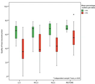
.01). Although there was some overlap among the methods of tensor estimation, visualized quality of reconstruction of the fornix was significantly higher with the use of the RESTORE algorithm, particu-larly in datasets with a high percentage (⬎10%,n⫽13) of data outliers (Fig 5). Furthermore, visualized quality of tract reconstruction across all tensor estima-tion methods depended also on the pres-ence of data artifacts because tract quality
FIG 1. Placement of ROIs. Tractography of the fornix was performed by placing 1 “OR” ROI (in blue), 2 “AND” ROIs (in green), and 2 “NOT” ROIs (in red) on color-coded fractional anisotropy maps.
[image:3.594.55.534.401.713.2]was significantly correlated to the mean outlier percentage per dataset (Spearman correlation coefficient:⫺0.46;P⬍.01). This correlation was also tested for each tensor estimation method separately. The following Spearman coefficients were found: LLS:
⫺0.48 (P⫽.01); WLLS:⫺0.47 (P⫽.01); NLLS:⫺0.57 (P⬍.01), and RESTORE:⫺0.36 (P⫽.06).
Tract Parameters
The impact of the diffusion tensor estimation method used on tract parameters is shown in Fig 6. There was a significant differ-ence in mean FA value through the use of different diffusion esti-mation algorithms. Significantly lower mean FA values were ob-tained with NLLS and RESTORE than with LLS and WLLS. Furthermore, application of the RESTORE approach resulted in the lowest standard deviation of the mean FA value; LLS: 0.059; WLLS: 0.054; NLLS: 0.052; RESTORE: 0.051 (repeated-measures ANOVA,
P⬍.05). Although not statistically significant, there was a trend to-ward an increased number of fiber trajectories in the following order of tensor estimation approaches: LLS, WLLS, NLLS, and RESTORE (Spearman correlation coefficient: 0.10;P⫽.32).
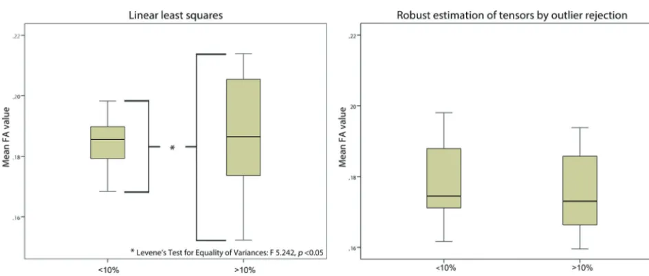
With the use of LLS, variability of mean FA values was signif-icantly higher in datasets with more than 10% outliers compared with datasets with less than 10% outliers. With WLLS, NLLS, or
RESTORE, there was no difference in variability of mean FA values with regard to the quality of the diffusion-weighted images (Fig 7).
DISCUSSION
This study emphasizes the paramount importance of quality assessment and dedicated use of processing methodology of neonatal DTI data before performing analysis. With our work, we demon-strated that 1) tract parameters are signif-icantly affected by the chosen tensor esti-mation method and are estimated more reliably if data outliers are handled care-fully; 2) robust estimation of the diffusion tensor results in significantly improved visualized quality of fiber reconstruction; 3) the mean percentage of data outliers of the diffusion-weighted images correlates significantly to visualized quality of tract reconstruction; and 4) data outliers are common and significantly affect subse-quent DTI analysis if they are not taken into account.
Although the incidence of destructive types of brain injury with subsequent seri-ous deficits is decreasing, preterm infants remain at considerable risk to develop cognitive and socio-emotional disabilities that persist into adolescence.22,23Advanced MR imaging techniques, such as DTI,
have already provided important valuable insights into WM micro-structural properties of these “subtle” types of brain injury.1Still, the
neuropathologic correlates show high variation and inconsistency,23
and their workings are not completely understood.24Moreover,
pre-term infants are vulnerable to respiratory and hemodynamic insta-bility and movement artifacts.7,9Therefore, acquisition and
process-ing of DTI data must be handled with dedicated care, which is essential to avoid misinterpretation. This is appropriately described in technical DTI reports,5-7,25,26but, paradoxically, such work
re-ceives little attention in clinical research. This could be because of their emphasis on clinical results, or, more importantly, because of the lack of awareness that the choice of diffusion tensor estimation approach may affect the subsequent reconstruction of fiber pathways.
As shown in the present study, the choice of tensor estimation algorithms can significantly affect DTI tractography results, which may complicate the interpretation of specific findings. In this context, study populations can only be compared reliably when identical processing pipelines have been applied, necessitating
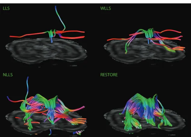
FIG 3. Impact of diffusion tensor estimation method on tract reconstruction of poor-quality DTI data. Characteristic representations illustrate the effect of tensor estimation methodology on reconstruction of the fornix with high percentage of data outliers (⬎10%). Note that recon-struction is not possible with the use of the linear least squares (LLS) and weighted linear least-squares (WLLS) methods and appears to be slightly possible with nonlinear least squares (NLLS) but is very well performed if the robust estimation of tensors by outlier rejection (RESTORE) approach is used.
Scoring system for visual evaluation of tract reconstruction of the fornix
0 Points 1 Point 2 Points
Shape of the fornixa No recognition of shape Partially abnormal shape Normal shape
Orientation of fibersa Complete disorientation Partially abnormal orientation Normal orientation
Symmetry of crura One missing crus Partially asymmetric Normal symmetry
Presence of non-realistic fibersa Outweighing the total number of fibers Less than the number of realistic fibers None
No. of fiber trajectories ⬍10 10–100 ⬎100
a
[image:4.594.54.372.139.367.2]the use of standardized guidelines before drawing conclusions with regard to outcome. Thus, strategies to limit image corruption should be incorporated into setups to acquire neonatal DTI data27; this
in-cludes 1) prevention of motion by comforting the infant and pro-moting natural sleep28; 2) adjustment of parameter settings, by
shortening diffusion time, applying stron-ger gradients, or by use of lower b-values7;
3) oversampling gradient-sensitizing direc-tions and removing corrupted diffusion-weighted images6; and 4) applying more
advanced tensor estimation methods.5
Be-cause diffusion tensor estimation tech-niques differ considerably in principle, speed, and accuracy,29 awareness of the
benefits and pitfalls is essential: the LLS method is fast and mostly used but assumes that errors are identically distributed, which can result in inaccurate estimation of the tensor.30 The WLLS method is slightly
slower but provides more accurate results because it considers errors to be hetero-geneously distributed.31 NLLS iteratively
minimizes errors and results in more reli-able estimation but needs considerably longer processing time and may get stuck in local optima during optimiza-tion.5,14The RESTORE approach
automat-ically detects and removes outliers before tensor estimation. This avoids manual and subjective identification of cor-rupted diffusion-weighted images and appears to be particularly valuable for data with frequent motion corrup-tion.13,26In summary, the reliability of
DTI analyses is drastically improved when handling data outliers in an ap-propriate way. However, additional re-search is needed to determine what types of data processing can reliably be performed without affecting data qual-ity. This report presses the need for careful data handling because corrupted data can significantly affect the final re-sult. Although this will require longer processing time and perhaps the need to remove datasets completely, it will probably decrease the spread in the final analysis and therefore improve statisti-cal significance and reduce sample size. Limitations of this study are important and must be addressed. First, only datasets without evidence of injury were used, and this may have resulted in a selection bias. Although this policy provided a homoge-neous study population, we did not investi-gate the impact of processing datasets with brain injury. Second, we applied an arbi-trary boundary to define “good-quality” and “poor-quality” data: 10% data outliers. We used this threshold solely to illustrate the im-pact of poor data quality on DTI analysis; hence, we do not suggest that this 10% level should be used as a threshold for future studies to define poor data quality.
FIG 4. Impact of diffusion tensor estimation method on tract reconstruction of good-quality DTI data. Characteristic representations illustrate the effect of the tensor estimation on fiber tracking of the fornix with low percentage of data outliers (⬍10%). Note the more accurate tract reconstruction with the use of the robust estimation of tensors by outlier rejection approach.
[image:5.594.54.371.47.273.2] [image:5.594.52.369.329.610.2]FIG 6. Impact of diffusion tensor estimation method on tract parameters. Tract parameters, such as fractional anisotropy (FA)(A), mean diffusivity(B), mean fiber trajectory length(C),and number of fiber trajectories(D)were affected by the tensor estimation method; mean FA value was significantly lower with use of the nonlinear least squares and robust estimation of tensors by outlier rejection techniques (paired samplettest,P⬍.05).
[image:6.594.56.532.49.433.2] [image:6.594.55.527.487.688.2]CONCLUSIONS
As demonstrated with our tractography analysis of the fornix in the preterm brain, it is clear that the choice of diffusion tensor estimation methodology is crucial and that it has a considerable impact on subsequent analyses for studying microstructural brain properties. Given the insufficient attention in most clinical stud-ies to date, this work raises the urgency to comply with the re-quirements to include state-of-the-art standardized research methodology wherever and whenever possible. Future studies should apply dedicated acquisition setups, standardized evalua-tion of data quality, and reliable processing of neonatal DTI data before performing analyses, such as associating microstructural brain properties with outcome.
Disclosures: Gabriel P. Krestin—UNRELATED: Consultancy:GEHC,* Comments: Stra-tegic consultancy to GEHC Europe;Grants/Grants Pending:GEHC,* Siemens,* Bayer,* Bracco,* Philips* (*money paid to institution).
REFERENCES
1. Ment LR, Hirtz D, Huppi PS.Imaging biomarkers of outcome in the developing preterm brain.Lancet Neurol2009;8:1042–55
2. Bassi L, Ricci D, Volzone A, et al.Probabilistic diffusion tractogra-phy of the optic radiations and visual function in preterm infants at term equivalent age.Brain2008;131:573– 82
3. Ball G, Boardman JP, Aljabar P, et al.The influence of preterm birth on the developing thalamocortical connectome.Cortex2013;49: 1711–21
4. van der Aa NE, Leemans A, Northington FJ, et al.Does diffusion tensor imaging-based tractography at 3 months of age contribute to the prediction of motor outcome after perinatal arterial ischemic stroke?Stroke2011;42:3410 –14
5. Jones DK, Cercignani M.Twenty-five pitfalls in the analysis of dif-fusion MRI data.NMR Biomed2010;23:803–20
6. Tournier JD, Mori S, Leemans A.Diffusion tensor imaging and be-yond.Magn Reson Med2011;65:1532–56
7. Heemskerk AM, Leemans A, Plaisier A, et al.Acquisition guidelines and quality assessment tools for analyzing neonatal diffusion ten-sor MRI data.AJNR Am J Neuroradiol2013;34:1496 –1505 8. Kozak LR, David S, Rudas G, et al.Investigating the need of
trigger-ing the acquisition for infant diffusion MRI: a quantitative study including bootstrap statistics.Neuroimage2013;69:198 –205 9. Plaisier A, Raets MM, van der Starre C, et al.Safety of routine early
MRI in preterm infants.Pediatr Radiol2012;42:1205–11
10. Leemans A, Jeurissen B, Sijbers J, et al.ExploreDTI: a graphical tool-box for processing, analyzing, and visualizing diffusion MR data. 17th Annual Meeting of the International Society of Magnetic Reso-nance Medicine, Honolulu, Hawaii. April 18 –24, 2009:3537 11. Leemans A, Jones DK.The B-matrix must be rotated when
correct-ing for subject motion in DTI data.Magn Reson Med2009;61: 1336 – 49
12. Irfanoglu MO, Walker L, Sarlls J, et al.Effects of image distortions
originating from susceptibility variations and concomitant fields on diffusion MRI tractography results.Neuroimage2012;61:275– 88 13. Chang LC, Jones DK, Pierpaoli C.RESTORE: robust estimation of
tensors by outlier rejection.Magn Reson Med2005;53:1088 –95 14. Jones DK, Basser PJ.“Squashing peanuts and smashing pumpkins”:
how noise distorts diffusion-weighted MR data.Magn Reson Med
2004;52:979 –93
15. Veraart J, Sijbers J, Sunaert S, et al.Weighted linear least squares estimation of diffusion MRI parameters: strengths, limitations, and pitfalls.Neuroimage2013;81C:335– 46
16. Basser PJ, Pajevic S, Pierpaoli C, et al.In vivo fiber tractography using DT-MRI data.Magn Reson Med2000;44:625–32
17. Nagy Z, Ashburner J, Andersson J, et al.Structural correlates of pre-term birth in the adolescent brain.Pediatrics2009;124:e964 –72 18. Zhuang L, Sachdev PS, Trollor JN, et al.Microstructural white
mat-ter changes, not hippocampal atrophy, detect early amnestic mild cognitive impairment.PLoS One2013;8:e58887
19. Catani M, Thiebaut de Schotten M.A diffusion tensor imaging tractography atlas for virtual in vivo dissections.Cortex2008;44: 1105–32
20. Nieuwenhuys R, Voogd J, van Huijzen C.The Human Central Ner-vous System.Berlin: Springer-Verlag; 2008
21. Landis JR, Koch GG.The measurement of observer agreement for categorical data.Biometrics1977;33:159 –74
22. Volpe JJ.Brain injury in premature infants: a complex amalgam of destructive and developmental disturbances.Lancet Neurol2009; 8:110 –24
23. Plaisier A, Govaert P, Lequin MH, et al.Optimal timing of cerebral MRI in preterm infants to predict long-term neurodevelopmental outcome: a systematic review.AJNR Am J Neuroradiol 2014;35: 841– 47
24. Dyet LE, Kennea N, Counsell SJ, et al.Natural history of brain le-sions in extremely preterm infants studied with serial magnetic res-onance imaging from birth and neurodevelopmental assessment.
Pediatrics2006;118:536 – 48
25. Mukherjee P, Chung SW, Berman JI, et al.Diffusion tensor MR im-aging and fiber tractography: technical considerations.AJNR Am J Neuroradiol2008;29:843–52
26. Morris D, Nossin-Manor R, Taylor MJ, et al.Preterm neonatal dif-fusion processing using detection and replacement of outliers prior to resampling.Magn Reson Med2011;66:92–101
27. Malamateniou C, Malik SJ, Counsell SJ, et al.Motion-compensation techniques in neonatal and fetal MR imaging.AJNR Am J Neurora-diol2013;34:1124 –36
28. Mathur AM, Neil JJ, McKinstry RC, et al.Transport, monitoring, and successful brain MR imaging in unsedated neonates.Pediatr Radiol2008;38:260 – 64
29. Veraart J, Rajan J, Peeters RR, et al.Comprehensive framework for accurate diffusion MRI parameter estimation.Magn Reson Med
2013;70:972– 84
30. Koay CG, Chang LC, Carew JD, et al.A unifying theoretical and algorithmic framework for least squares methods of estimation in diffusion tensor imaging.J Magn Reson2006;182:115–25