With remarkable foresight, Davson and Danielli (1952) clearly articulated the importance of cell membranes: ‘It can truely be said of living cells, that by their membranes ye shall know them’. Cell membranes were known then to be composed of lipids and proteins: lipids making membranes nearly impermeable to most water-soluble solutes; proteins serving as transporters and signaling devices. The classic deductions of Gorter and Grendel (1925) became the dogma that membrane lipids are arranged in a bilayer configuration: parallel sheets of phospholipids with polar or charged head groups oriented towards the aqueous environment and acyl chains interacting within the hydrophobic membrane core. The acceptance of the idea that cell membranes were not merely static barriers containing immobile proteins but dynamic lipid–protein matrices contributing to the regulation of cell function can be attributed to publication of the fluid mosaic model of cell membranes (Singer and Nicolson, 1972). This model viewed membrane lipids as a viscous matrix in which
transmembrane proteins have a degree of motion which, in turn, can have a dramatic impact upon protein activity. The characteristics of the lipid matrix depend upon the physical properties of the individual lipid components in the membrane. Membrane protein activity is influenced by the surrounding lipid matrix and specifically by the association between lipids and proteins at the lipid–protein interface. At any instant, a population of lipids is always associated with a membrane protein. Depending on the time these lipids spend associated with the protein, they are classified as either restricted or interfacial lipids. Lipids that have a long residence time on the protein and exchange with lipids in the surrounding membrane at a slow rate are termed motionally restricted. The nature of the association between this group of lipids and proteins and the type of lipid that has access to the protein in this restricted domain modulates the activity of the protein. Several examples of membrane proteins that are influenced by specific restricted lipids are presented in Table 1. In some of these cases, protein JEB1204
Photoreceptor rod cells and blood platelets are remarkably different, yet both illustrate a similar phenomenon. Both are strongly affected by membrane cholesterol, and the distribution of cholesterol in the membranes of both cell types is determined by the lipid composition within the membranes. In rod cells, cholesterol strongly inhibits rhodopsin activity. The relatively higher level of cholesterol in the plasma membrane serves to inhibit, and thereby conserve, the activity of rhodopsin, which becomes fully active in the low-cholesterol environment of the disk membranes of these same cells. This physiologically important partitioning of cholesterol between disk membranes and plasma membranes occurs because the disk membranes are enriched with phosphatidylethanolamine, thus
providing a thermodynamically unfavorable environment for the sterol. Cholesterol enrichment of platelets renders these cells more responsive to stimuli of aggregation. Stimuli for platelet aggregation cause a rapid transbilayer movement of cholesterol from the outer monolayer. This stimulus-dependent redistribution of cholesterol appears to result from the concomitant movement of phosphatidylethanolamine into the outer monolayer. The attractive, yet still unproven, hypothesis is that cholesterol translocation plays an important role in the overall platelet response and is intimately related to the sensitizing actions of cholesterol on these cells.
Key words: lipid, cell membrane, photoreceptor, rod cell, phosphatidylethanolamine, platelet, cholesterol.
Summary
REVIEW
CELL MEMBRANE LIPID COMPOSITION AND DISTRIBUTION: IMPLICATIONS
FOR CELL FUNCTION AND LESSONS LEARNED FROM PHOTORECEPTORS
AND PLATELETS
KATHLEEN BOESZE-BATTAGLIA1,* ANDRICHARD J. SCHIMMEL2
1Department of Molecular Biology and 2Department of Cell Biology, University of Medicine and Dentistry of New
Jersey, School of Osteopathic Medicine, Stratford, NJ 08084, USA
Accepted 17 September 1997
*Author for correspondence and present address: Department of Molecular Biology, 2 Medical Center Drive, UMDNJ School of Osteopathic Medicine, Stratford, NJ 08084, USA (e-mail: [email protected]).
activity is subject to additional modulation by a second class of lipids, the interfacial lipids.
Membrane proteins may also have associated with them a sufficient number of specific or non-specific lipids which form a coat or annulus around the circumference of the protein. Such interfacial lipids exchange rapidly with the surrounding lipids and determine the bulk properties of the lipid matrix. Phosphatidylserine and stearic acid show a high affinity for contact with the transmembrane portion of the Na+/K+
-ATPase, while rhodopsin requires an unsaturated fatty-acid-rich phospholipid annulus for function (Watts et al. 1979). It is important to point out that, in other regions of the membrane, lipid–lipid or protein–protein interactions predominate and modulate protein function by changing the properties of the lipid–protein interface.
Phosphatidylcholine, phosphatidylethanolamine, phosphatidylserine and phosphatidylinositol are the constitutive lipids that provide a structural framework and the environment that defines the functional parameters associated with individual cell types. Phosphatidylcholine, phosphatidylserine and phosphatidylinositol provide for hydrated or charged membrane surfaces, allowing water and/or ions to bind to their polar headgroups. In contrast, surfaces rich in phosphatidylethanolamine are hydrophobic, poorly hydrated and promote surface-to-surface interactions without direct protein binding. Because phosphatidylethanolamine is not readily hydrated, it promotes the formation of non-bilayer structures (i.e. hexagonal II phase) to compensate for the hydrophobic effect. The inherent instability of this phospholipid is necessary in such cellular functions as membrane fusion.
Cell membranes also contain trace amounts of transient lipids, which exist in the membrane for only brief periods, serving predominantly as second messengers (for a review, see Ghosh et al. 1997). Phospholipids are asymmetrically distributed between the inner and outer monolayer of cell membranes (for a review, see Op den Kamp, 1979);
choline-containing lipids, phosphatidylcholine and sphingomyelin, are found preferentially in the outer monolayer and amine-containing lipids, phosphatidylethanolamine and phosphatidylserine, are localized to the inner monolayer. The establishment and maintenance of phospholipid asymmetry occurs through a specific ATP-dependent transport protein, the aminophospholipid translocase (for a review, see Devaux, 1991), which translocates phosphatidylethanolamine and phosphatidylserine between the monolayers.
After phospholipids, cholesterol is the next most abundant constitutive lipid. The planar sterol ring and the hydrophobic tail orient cholesterol within the membrane core, while the
3-β-hydroxyl group allows cholesterol to contribute to the surface properties of the bilayer. The structure of cholesterol allows it to reduce the freedom of movement of phospholipid acyl chains, thus rigidifying the membrane, which can have a dramatic impact upon membrane function. This phenomenon is referred to as a ‘condensing effect’ (Demel et al. 1972). The importance of the contribution of cholesterol to membrane properties is reflected in its ubiquitous distribution and its necessity for normal cell growth and function. However, high levels of membrane cholesterol, often secondary to high levels of serum cholesterol, exert deleterious effects on cells.
Like phospholipids, cholesterol is also distributed non-uniformly in cell membranes (Dawidowicz, 1987; Boesze-Battaglia et al. 1996; Lange, 1992). An extreme example of non-uniform cholesterol distribution is in platelet membranes which, it is suggested, contain regions devoid of cholesterol (i.e. ‘cholesterol-free patches’) (Gordon et al. 1983). Sterol carrier proteins have been identified that deliver sterol to the plasma membrane from the endoplasmic reticulum (for a review, see Schroeder et al. 1996), but there is no compelling evidence for a role of these proteins in transmembrane cholesterol distribution; similarly, an intramembrane cholesterol-specific translocase has not been identified.
[image:2.609.45.560.84.280.2]With the acceptance of the fluid mosaic model, the concept of ‘membrane dynamics’ added a new dimension to the
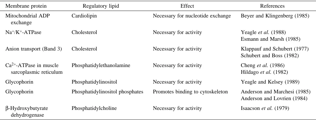
Table 1. Examples of membrane proteins that are influenced by specific restricted lipids
Membrane protein Regulatory lipid Effect References
Mitochondrial ADP Cardiolipin Necessary for nucleotide exchange Beyer and Klingenberg (1985) exchange
Na+/K+-ATPase Cholesterol Necessary for activity Yeagle et al. (1988) Esmann and Marsh (1985)
Anion transport (Band 3) Cholesterol Necessary for activity Klappauf and Schubert (1977) Schubert and Boss (1982)
Ca2+-ATPase in muscle Phosphatidylethanolamine Necessary for activity Cheng et al. (1986)
sarcoplasmic reticulum Hildago et al. (1982)
Glycophorin Phosphatidylinositol Necessary for activity Yeagle and Kelsey (1989)
Glycophorin Phosphatidylinositol phosphates Promotes binding to cytoskeleton Anderson and Marchesi (1985) Anderson and Lovrien (1984) β-Hydroxybutyrate Phosphatidylcholine Necessary for activity Isaacson et al. (1979)
consideration of the functional regulation of proteins. For the purposes of this review, membrane dynamics is defined as the culmination of the motions of individual membrane components and their interrelationships in non-repeating units of the membrane. The distribution of lipids within the lateral and transverse planes of the bilayer adds another dimension to the dynamic environment of the membrane. The asymmetric bilateral organization of lipids differing in the polarity, size, charge and reactivity of their polar headgroups suggests different chemical and different dynamic environments within the two monolayers. Such differences between the two monolayers open the possibility of differential regulation of transmembrane protein activity on either side of the cell membrane. This review selects two cell types, platelets and photoreceptor cells, which provide unique model systems illustrating how changes in membrane lipid composition and membrane dynamics alter cell function. The lipid composition and distribution within the membrane of these cells can be altered, in the case of platelets quite rapidly following cell stimulation, and these lipid alterations exert control over cell function.
Platelets
The plasma membrane of platelets has a lipid composition similar to that of other cells (Lagarde et al. 1982; Fauvel et al. 1986). Phosphatidylcholine and phosphatidylethanolamine are the most abundant glycerophospholipids, each accounting for nearly 35 % of the phospholipid mass. Phosphatidylserine is less abundant, accounting for approximately 13 %, and phosphatidylinositol accounts for less than 5 %. The sphingolipid sphingomyelin contributes approximately 20 % of the lipid mass. Phosphatidylcholine and sphingomyelin are found preferentially in the outer monolayer, while phosphatidylserine and phosphatidylethanolamine are found in the inner monolayer. Platelet membranes also contain cholesterol, and the molar ratio of cholesterol to phospholipid varies, with a mean value of approximately 0.50 (Marcus et al. 1969; Shattil et al. 1975, 1977). The platelet membrane cholesterol concentration usually reflects the plasma cholesterol concentration and is substantially higher in hypercholesterolemic individuals (Carvalho et al. 1974; Shattil et al. 1977). Platelets are incapable of synthesizing cholesterol, and the content and localization of the sterol in platelet membranes is probably established during their formation from megakaryocytes (Schick and Schick, 1985). Platelets may also acquire cholesterol through exchange with plasma lipoproteins (Schick and Schick, 1985; Aviram and Brook, 1980).
Platelet membranes demonstrate a unique phenomenon: upon exposure to stimuli of aggregation, membrane phospholipids move rapidly between the two monolayers and the asymmetric distribution of phospholipids in the platelet plasma membrane is disrupted (for reviews, see Schroit and Zwaal, 1991; Zwaal and Schroit, 1997). The seminal observation supporting this proposal is that phosphatidylethanolamine and phosphatidylserine are poorly
hydrolyzed by exogenous phospholipase A2 except when
platelets are exposed to collagen, thrombin or ionomycin, which greatly increase the rate of hydrolysis of these lipids by phospholipase A2 (Bevers et al. 1982, 1983). Schick et al.
(1976) showed that 2,4,6-trinitrobenzenesulfonic acid reacted with a much greater percentage of the phosphatidylethanolamine following incubation with thrombin. They proposed that phosphatidylethanolamine is rapidly translocated from the inner to the outer monolayer upon stimulation. More contemporary studies incorporated either fluorescent (Gaffet et al. 1995; Smeets et al. 1994; Tilly et al. 1990; Williamson et al. 1995) or spin-labeled (Basse et al. 1993; Sune et al. 1987) phospholipid probes into platelet membranes. Stimulation of platelets caused these probes to translocate from one monolayer to the other and, on this basis, investigators inferred that the distribution of endogenous membrane lipids changed in a qualitatively similar manner. The disruption of phospholipid asymmetry in the platelet plasma membrane may involve a bidirectional movement of phospholipids between the membrane monolayers, a so-called scrambling of lipids (Smeets et al. 1994; Williamson et al. 1995). Alternatively, Gaffet et al. (1995) proposed a vectorial efflux of phosphatidylserine and phosphatidylethanolamine from the inner to the outer monolayer without an accompanying reciprocal influx of choline phospholipids. The loss of phospholipid asymmetry is not the result of inhibition of the aminophospholipid translocase activity (Basse et al. 1993; Comfurius et al. 1990), and investigators have proposed that a separate enzyme activity, termed a scramblase (Zwaal and Schroit, 1997), catalyzes the transmembrane redistribution of phospholipids in platelets. Similar evidence has been presented for erythrocytes (Smeets et al. 1994). This putative activity appears to be activated in response to the elevation of intracellular [Ca2+] (Williamson et al. 1992; Dachary-Prigent et al. 1995; Smeets et al. 1994; Zwaal and Schroit, 1997), which simultaneously inhibits the aminophospholipid translocase (Comfurius et al. 1990). Persuasive evidence supporting the presence of a scramblase in platelet membranes was recently provided by Comfurius et al. (1996). These investigators reconstituted a platelet membrane protein preparation into lipid vesicles containing 7-nitrobenz-2-oxa-1.3-diazol-4-yl (NBD)-labeled phospholipids. The addition of Ca2+ and a Ca2+ ionophore to the vesicles induced the
process (i.e. a thermodynamic process). Model membrane (Yeagle and Young, 1986; Backer and Dawidowicz, 1981) and biological membrane (House et al. 1989) studies have found that the introduction of phosphatidylethanolamine into membranes creates a thermodynamically unfavorable environment for cholesterol. The thermodynamic constraints when cholesterol and phosphatidylethanolamine coexist predict an unfavorable entropy, causing the translocation of cholesterol from the phosphatidylethanolamine-rich lipid environment as a compensatory mechanism. It is proposed that such partitioning of cholesterol is due to the increased ordering of water molecules on the membrane surface. Chaotropic agents relieve the constraint on the membrane by disrupting the interstitial hydrogen-bonded water and, thereby, allow both lipids to coexist within the same bilayer. In fact, cholesterol translocation is blocked by exposure of platelets to the chaotropic agents urea and guanidine–HCl (Boesze-Battaglia et al. 1996). However, the participation of an enzymatic activity in cholesterol translocation has not been investigated. Platelet responses culminating in aggregation are strongly influenced by the cholesterol content of the platelet membrane and are positively correlated with the membrane cholesterol content. Carvalho et al. (1974) reported increased sensitivity to epinephrine, ADP- and collagen-initiated aggregation and increased nucleotide release in platelets from patients with hypercholesterolemia. Supporting the view that the increased sensitivity of platelets removed from hypercholesterolemic individuals is a consequence of their elevated cholesterol levels are data from studies by Shattil et al. (1975) and Tomizuka et al. (1990) reporting increased sensitivity of platelets to epinephrine, ADP and thromboxane A2when the platelets had
been enriched with cholesterol in vitro. Shattil et al. (1975) also reported a reduction of platelet sensitivity to epinephrine following the loss of platelet cholesterol. A related study noted that hypercholesterolemia was associated with an increased number of lower-density platelets, which were more responsive to thrombin stimulation (Opper et al. 1995).
The mechanisms underlying cholesterol modulation of platelet function are not well established. A number of signal transduction events have been shown to be increased following cholesterol enrichment of platelets. These events include increased release of arachidonic acid, indicative of phospholipase A2 activity (Sorisky et al. 1990; Stuart et al.
1980), increased adrenergic and thrombin receptor numbers (Insel et al. 1978; Tandon et al. 1983), and higher receptor-stimulated Ca2+ and inositol phosphate levels (Sorisky et al.
1990). While cholesterol may be acting as a restricted lipid directly modifying the function of integral membrane proteins mediating signal transduction events, anisotropy and polarization data suggest that cholesterol behaves like an interfacial lipid modulating the bulk properties of the bilayer. In this regard, Shattil and Cooper (1976) reported an increased viscosity of platelet membranes upon enrichment with cholesterol, and this fundamental relationship has been confirmed by others in platelets (Hochgraf et al. 1994). Alternatively, the mechanism of cholesterol action may be
related to cholesterol-induced changes in membrane thickness (Chen et al. 1995).
The rapid reorganization of membrane lipids, including cholesterol, in platelets makes it likely that the dynamic properties of platelet membranes change as a result of stimulation. The bulk properties of membranes can be assessed using fluorescence polarization and anisotropy measurements, which reveal the mean angular displacement of a fluorophore that occurs between the absorption and subsequent emission of a photon. This angular displacement is dependent on the rate and extent of rotational diffusion during the lifetime of the excited state. These diffusive motions in turn depend on the viscosity of the solvent, in this case the lipid bilayer. The often-used term for this property of the lipid bilayer is ‘fluidity’. The higher the polarization or anisotropy value, the lower the membrane fluidity and the more ordered the membrane.
A number of studies employing fluorescent membrane probes have reported decreases in platelet membrane fluidity following platelet stimulation. Nathan et al. (1979) reported an increase in 1,6-diphenyl-1,3,5-hexatriene (DPH) fluorescence polarization coincident with shape change and aggregation in thrombin- or ADP-stimulated platelets and concluded that platelet activation is accompanied by an increased rigidity of the membrane lipids. Steiner and Luscher (1984) and Kowalska and Cierniewski (1983) reported similar findings using 4,4′ -diisothiocyano-2,2′-stilbenedisulfonic acid (DIDS) or DPH, respectively, in platelets stimulated with thrombin, ADP or fibrinogen. Steiner and Luscher (1984) detected a decrease in anisotropy which preceded the rise in anisotropy of thrombin-stimulated but not ADP-thrombin-stimulated platelets. The increase in anisotropy in response to thrombin could be prevented by pretreatment with cytochalasin B, suggesting a role of the extensive platelet cytoskeleton in thrombin-induced changes in anisotropy. Nathan et al. (1980) subsequently reported similar findings using N-carboxymethylisatoic anhydride, a fluorescent probe which binds covalently to membrane proteins. The similar changes in polarization using the two probes suggested that the changes in the microenvironment surrounding the membrane proteins are analogous to the changes in the overall dynamic properties of the bulk membrane lipids. Taken together, these findings suggest an increased ordering of platelet membrane lipids following stimulation.
efforts to resolve the relative changes in fluidity of each monolayer of the membrane.
Although the unique rearrangement of membrane lipids in stimulated platelets has been appreciated for two decades, its significance in terms of platelet responses leading to aggregation has eluded investigators. However, another role has been ascribed to stimulus-dependent phospholipid reorganization: in addition to promoting aggregation, platelet activation also alters the platelet membrane to provide a catalytic surface for the conversion of factor X to factor Xa and of prothrombin to thrombin, which then catalyzes fibrinogen formation. The formation of this catalytic surface requires exposure of the anionic phospholipid phosphatidylserine (Bevers et al. 1982). Patients with Scott syndrome, a rare bleeding disorder (Weiss et al. 1979), have an isolated deficiency in the formation of a platelet procoagulant activity due to a failure to expose phosphatidylserine on the outer membrane monolayer (Bevers et al. 1992; Sims et al. 1989). Platelets from patients with Scott syndrome exhibit apparently normal aggregation in response to stimuli and have a lipid composition indistinguishable from that of normal platelets. Erythrocytes from patients with Scott syndrome have a similar defect (Bevers et al. 1992). The molecular basis for Scott syndrome is not known, but the possibility that platelets and erythrocytes from these patients are deficient in the putative ‘scrambalase’ enzyme has been excluded (Stout et al. 1997).
Photoreceptors
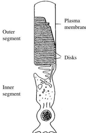
Photoreceptor rod cells provide a unique window in which the relationship between physiological function and membrane lipid composition may be visualized. These cells convert light energy into a change in electrical potential, triggering a nerve impulse that is ultimately delivered to the visual cortex. The biochemical events initiating this process occur within the disk membranes located in the interior of the rod outer segment (ROS, Fig. 1). The disks are closed, flattened membranous sacs organized in a discontinuous stacked array along the length of the outer segment. Nascent disks are formed from evaginations of the surrounding ROS plasma membrane at the base of the cell and are then progressively displaced towards the apical tip of the outer segment as additional new disks are formed. Old
disks are shed at the apical tip and phagocytosed by the overlying retinal pigment epithelium. The transition of disks from the base to the tip of the outer segment requires approximately 10 days in mammals and maintains the ROS at a constant length.
Disks and plasma membranes differ in their lipid composition (Table 2). The plasma membrane is enriched in cholesterol (Boesze-Battaglia and Albert, 1989) and the sterol precursor squalene (Fliesler et al. 1997) relative to the disk membrane. In plasma membrane, the ratio of phosphatidylethanolamine to phosphatidylcholine is 0.16, while in disks this ratio is 0.92 (Boesze-Battaglia and Albert, 1992) (Table 2). The fatty acid composition (Boesze-Battaglia et al. 1989) and the ratio of saturated to unsaturated fatty acids
Plasma membrane
Disks Outer
segment
[image:5.609.366.510.72.295.2]Inner segment
Fig. 1. Schematic representation of a retinal rod cell.
Table 2. Cholesterol and phospholipid headgroup composition of rod outer segment membranes
C/P* PE PS PI PC
Disk membrane 0.11 41.6±2.6 13.7±2.1 2.5±0.8 45.3±3.2 Plasma membrane 0.38 10.6±2.8 24.1±2.8 <1.0 65.1±3.8
*C/P refers to the cholesterol to phospholipid ratio (mole:mole). Data are shown as mean mol % of total phosphate ± the standard deviation for four determinations on two separate preparations.
PE, phosphatidylethanolamine; PS, phosphatidylserine; PI, phosphatidylinositol; PC, phosphatidylcholine.
Table 3. Fatty acid composition of rod outer segment membranes
Fatty acid species Disk membrane Plasma membrane
14:0 1.4 19.9
16:0 13.2 17.9
18:0 18.8 8.6
18:1 5.2 8.2
18:2 1.1 7.6
18:3 1.1 10.2
18:4 ND 1.3
20:4 7.8 7.0
22:n* 8.0 7.1
22:6 35.4 4.9
Data are represented as percentage of total mass; in the case of the plasma membrane, the values shown are the mean of two preparations (Boesze-Battaglia and Albert, 1989).
[image:5.609.315.567.543.677.2]is also markedly different (Lamba et al. 1994) (Table 3). The most prominent difference is in docosahexaenoic acid (DHA, 22:6), which accounts for 5 % of the total in plasma membrane but 35 % in disk membrane. Collectively, these findings suggest a tremendous sorting of lipid constituents at the base of the ROS upon disk biogenesis.
As the disk membranes are apically displaced, their cholesterol content decreases from 30 mol % at the base of the ROS to 5 mol % at the apical tip (Boesze-Battaglia et al. 1989, 1990). There is no corresponding change in fatty acid or phospholipid composition among disks at different locations in the ROS. The loss of cholesterol as the disk membranes age can be explained by a cholesterol-partitioning model similar to that invoked to account for translocation of cholesterol in collagen-stimulated platelets. In disks, it is proposed that cholesterol is exchanged out of the phosphatidylethanolamine-rich disk membrane into the phosphatidylcholine-phosphatidylethanolamine-rich plasma membrane. Thus, on the basis of the relative differences in the phosphatidylethanolamine/phosphatidylcholine ratio between the disk and the plasma membranes, a gradient is formed which favors the movement of cholesterol from the disk membrane to the plasma membrane during the lifetime of a disk (Yeagle and Young, 1986; House et al. 1989).
Aberrant lipid sorting between disk and plasma membranes is associated with disease states, as illustrated by the Royal College of Surgeons (RCS) strain of rats. These rats carry a recessive mutation which results in a degeneration of the retinal photoreceptor cells (Dowling and Sidman, 1962; Noell, 1965). The rhodopsin content (Organisciak et al. 1982) and the glycosylation pattern (Endo et al. 1996) of this protein are similar in both the normal and diseased animals. One characteristic of this disease is an apparent absence of lipid sorting during disk biogenesis (Boesze-Battaglia et al. 1994). As a result, the plasma membrane phospholipid composition is virtually identical to that of the disk membranes. It is hypothesized that, in the absence of a phosphatidylethanolamine/phosphatidylcholine gradient between the disk and plasma membranes of the RCS rat, there is no partitioning of the cholesterol and, in fact, disk membrane cholesterol level in the RCS rat remains constant at 15 mol % as a function of disk age. The photoreceptors of these animals exhibit abnormal growth, leading to the accumulation of membrane debris and ultimately to blindness, thus providing compelling evidence of the need for the photoreceptor membranes to maintain their distinct lipid composition for normal cell function.
The light sensor, rhodopsin, initiates the visual response as a transmembrane protein in a disk lipid matrix. The sequence of intracellular events triggered by rhodopsin activation has been extensively characterized (for a review, see Stryer, 1986) and is analogous to the β-adrenergic receptor-dependent activation of adenylate cyclase (Oprian, 1992). The absorption of a photon of light results in the photoisomerization of 11-cis retinal, the retinal chromophore of rhodopsin, to all-trans retinal, leading to a series of conformational changes necessary for the formation of Metarhodopsin II, the activated form of
rhodopsin (Bennet et al. 1982). The coupling of the photoreceptor G-protein transducin with activated rhodopsin (Metarhodopsin II) facilitates the activation of the cyclic-GMP-dependent phosphodiesterase. Because rhodopsin is the predominant protein in the disk membrane (95 % of total protein; Papermaster and Dreyer, 1972) and the surrounding plasma membrane (80–85 % of total protein; Molday and Molday, 1987), studies directed towards investigating rhodopsin function in two compositionally distinct membranes from the same cell are possible.
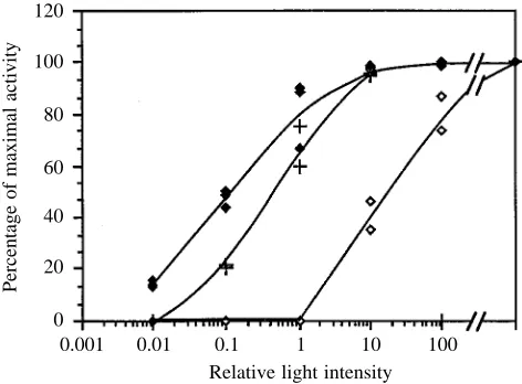
Rhodopsin activation of the cyclic-GMP-dependent phosphodiesterase (PDEase) differs in disk and plasma membrane preparations (Boesze-Battaglia and Albert, 1990). As shown in Fig. 2, plasma membrane rhodopsin requires a light intensity at least two orders of magnitude greater than disk membrane rhodopsin to achieve the same maximal PDEase activity. Because of the profound influence of cholesterol upon the acetycholine receptor (Criado et al. 1982; Dreger et al. 1997) and the Na+/K+-ATPase (Yeagle et al.
1988), and since the plasma membrane contains three times more cholesterol than the disks, we investigated the effect of cholesterol on rhodopsin activation of PDEase. When the cholesterol content of ROS plasma membranes preparations was reduced by incubation with cholesterol oxidase, the PDEase activity was restored, suggesting that the high cholesterol content of these membranes inhibits the activation of phosphodiesterase by rhodopsin.
The molecular mechanism by which cholesterol modulates PDEase activity through the formation of Metarhodopsin II has been extensively characterized. In a series of elegant
120
100
80
60
40
20
0
Percentage of maximal activity
[image:6.609.319.555.72.246.2]0.001 0.01 0.1 1 10 100 Relative light intensity
reconstitution experiments, Litman and his colleagues demonstrated that increasing membrane cholesterol level shifts the equilibrium between Metarhodopsin I and Metarhodopsin II (Straume and Litman, 1987, 1988; Mitchell et al. 1990) towards Metarhodopsin I. Thus, Metarhodopsin II formation is inhibited in the presence of high levels of membrane cholesterol. Cholesterol exerts this effect by modulating the ‘free volume’ of the bilayer. The unsaturated acyl chains of the phospholipids assume a cis/trans conformation, which produces ‘kinks’ in the membrane bilayer. Such kinks take up space or, in three dimensions, ‘volume’, allowing for transient packing defects within the bilayer. These small volume defects are decreased in the presence of cholesterol, thereby inhibiting Metarhodopsin II formation since Metarhodopsin II requires volume expansion by the protein. This series of studies provides compelling support for the modulation of rhodopsin function through changes in the bulk dynamic properties of the ROS membrane bilayer.
In a recent series of experiments, Albert et al. (1996a,b) suggested that cholesterol stabilizes and interacts directly with rhodopsin. Using the fluorescent sterol probe cholestatrienol and fluorescence energy transfer techniques, they proposed a direct interaction between cholesterol and rhodopsin (Albert et al. 1996b). This conclusion is supported by an analysis of spin-labeling experiments, which suggest that a non-phospholipid moiety, probably cholesterol, is found at the lipid–protein interface (Watts et al. 1979). Rhodopsin is thus modulated not only by the bulk membrane properties of the disks (i.e. through changes in free volume) but also potentially through a direct interaction with cholesterol. Additional experiments are needed to determine the mechanism by which cholesterol acting as a restricted lipid modulates rhodopsin function.
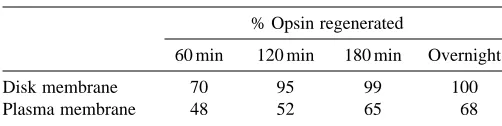
After bleaching, the apo-protein opsin recombines with 11-cis retinal to form newly activatable rhodopsin. Evidence has accumulated over the last decade implicating DHA in rhodopsin regeneration. DHA or its precursor (18:3n-3) must be furnished in the diet (Tinoco, 1982; Scott and Bazan, 1989). In animal models, deprivation of these fatty acids for two generations results in a loss of visual function (Benolken et al. 1973; Wheeler et al. 1975; Dudley et al. 1975) and ROS disk disruptions (Futterman et al. 1971; Bush et al. 1991). Bush et al. (1994) have shown that rats with decreased levels of DHA
showed a slower rate of rhodopsin regeneration compared with controls. Interestingly, plasma membrane rhodopsin does not regenerate to the same extent as disk membrane rhodopsin (Table 4). In fact, the plasma membrane contains only 5 % DHA while the disk membrane contains 35 %. Given that DHA is the predominant fatty acid on phosphatidylethanolamine and that rhodopsin maintains a phospholipid annulus whose composition has not been determined, it is tempting to speculate that the differences in rhodopsin regeneration are due to differences in the phosphatidylethanolamine composition of the lipid annulus of rhodopsin.
By utilizing different lipid components to regulate various aspects of rhodopsin function in conjunction with cytoplasmic components such as kinases, photoreceptors provide for various levels of functional control. The need for such complexity in this regulation can be appreciated if one considers the range of light intensities to which rhodopsin must respond both continuously and intermittently. To conserve rhodopsin function, and hence photoreceptor function, rhodopsin is maintained in a relatively inactive state in the plasma membrane. In this membrane, there is a high level of cholesterol (thus rhodopsin activation is inhibited) and low levels of DHA (thus rhodopsin regeneration occurs slowly). Upon entering the lower-cholesterol, higher-DHA environment of the disk through disk biogenesis, inhibition is removed. Thus, it can be said that the plasma membrane lipid environment may ‘protect’ rhodopsin from bleaching by light, thereby allowing for the maximal amount of activatable rhodopsin to be incorporated into newly formed disks.
The molecular mechanism by which DHA maintains visual function can be explained if one considers how changes in this lipid affect free volume. The presence of cis double bonds in unsaturated fatty acids will increase free volume by introducing ‘kinks’ within the membrane bilayer. DHA contributes as many as six cis bonds. It can be hypothesized that, as the cholesterol/DHA ratio in the disk membranes decreases as the disk ages, a lipid environment is established that favors Metarhodopsin II formation (and therefore favors rhodopsin activation of PDEase) and the subsequent regeneration of rhodopsin (high DHA levels) for continual activation. In fact, Metarhodopsin II formation is favored in the presence of unsaturated fatty acids (Mitchell et al. 1992) such as DHA. The delicate balance between a molecule that enhances free volume (DHA) and one that restricts free volume (cholesterol) allows the photoreceptor to function under what may seem to be adverse light conditions. In general, the larger free volume component associated with the disks as they age contributes to the effective overall functioning of the ROS as a transducer of light.
References
[image:7.609.47.298.613.676.2]ALBERT, A. D., BOESZE-BATTAGLIA, K., PAW, Z., WATTS, A. AND EPAND, R. M. (1996a). Effect of cholesterol on rhodopsin stability in disk membranes. Biochim. biophys. Acta 1297, 77–82. ALBERT, A. D., YOUNG, J. E. AND YEAGLE, P. L. (1996b).
Table 4. Opsin regeneration in rod outer segment membranes
% Opsin regenerated
60 min 120 min 180 min Overnight
Disk membrane 70 95 99 100
Plasma membrane 48 52 65 68
Rhodopsin–cholesterol interactions in bovine rod outer segment disk membranes. Biochim. biophys. Acta 1285, 47–55.
ANDERSON, R. A. ANDLOVRIEN, R. E. (1984). Glycophorin is linked by band 4.1 protein to the human erythrocyte membrane cytoskeleton. Nature 307, 655–658.
ANDERSON, R. A. ANDMARCHESI, V. T. (1985). Association between glycophorin and protein 4.1 are modulated by polyphosphoinositides, a mechanism for membrane skeleton regulation. Nature 318, 295–298.
AVIRAM, M. ANDBROOK, J. G. (1980). Platelet interaction with high and low density lipoproteins. Atherosclerosis 46, 259–268. BACKER, J. M. AND DAWIDOWICZ, E. A. (1981). Mechanism of
cholesterol exchange between phospholipid vesicles. Biochemistry
20, 3805–3810.
BASSE, F., GAFFET, P., RENDU, F. AND BIENVENUE, A. (1993). Translocation of spin labeled phospholipids through plasma membrane during thrombin and ionophore A23187 induced platelet activation. Biochemistry 32, 2337–2344.
BENNET, N., MICHEL-VILLAY, M. ANDKUHN, H. (1982). Light induced interaction between rhodopsin and the GTP binding protein. Metarhodopsin II is the major photoproduct involved Eur. J. Biochem. 127, 97–103.
BENOLKEN, R. M., ANDERSON, R. E. AND WHEELER, T. G. (1973). Membrane fatty acids associated with the electrical response in visual excitation. Science 182, 1253–1254.
BEVERS, E. M., COMFURIUS, P., VANRIJN, J. L. M. L., HENKER, H. C. AND ZWAAL, R. F. A. (1982). Generation of prothrombin-converting activity of PS at the outer surface of the platelets. Eur. J. Biochem. 122, 429–436.
BEVERS, E., COMFURIUS, P. ANDZWAAL, R. F. A. (1983). Changes in membrane phospholipid distribution during platelet activation. Biochim. biophys. Acta 736, 57–66.
BEVERS, E. M., WIEDMER, T., COMFURIUS, P., SHATTIL, S. J., WEISS, H. J., ZWAAL, R. F. A. AND SIMS, P. J. (1992). Defective Ca2+ induced microvesiculation and deficient expression of procoagulant activity in erythrocytes from a patient with a bleeding disorder: A study of the red blood cells of Scott Syndrome. Blood 79, 380–388. BEYER, K. ANDKLINGENBERG, M. (1985). ADP/ATP carrier protein from beef heart mitochondria has high amounts of tightly bound cardiolipin, as revealed by 31P NMR. Biochemistry 24, 3821–3826. BOESZE-BATTAGLIA, K. AND ALBERT, A. D. (1989). Fatty acid composition of bovine rod outer segment plasma membrane. Exp. Eye Res. 49, 699–701.
BOESZE-BATTAGLIA, K. AND ALBERT, A. D. (1990). Cholesterol modulation of photoreceptor function in bovine rod outer segments. J. biol. Chem. 265, 20727–20730.
BOESZE-BATTAGLIA, K. AND ALBERT, A. D. (1992). Phospholipid distribution among bovine rod outer segment plasma membrane and disk membranes. Exp. Eye Res. 54, 821–823.
BOESZE-BATTAGLIA, K., ALBERT, A. D. AND ORGANISCIAK, D. T. (1994). Cholesterol and phospholipid distribution in ROS of normal and RCS rats. Exp. Eye Res. 58, 293–300.
BOESZE-BATTAGLIA, K., CLAYTON, S. T. ANDSCHIMMEL, R. J. (1996). Cholesterol redistribution within human platelet plasma membrane: evidence for a stimulus dependent event. Biochemistry 35, 6664–6673.
BOESZE-BATTAGLIA, K., FLIESLER, S. J. ANDALBERT, A. D. (1990). Relationship of cholesterol content to spatial distribution and age of disk membranes in retinal rod outer segments. J. biol. Chem.
265, 18867–18870.
BOESZE-BATTAGLIA, K., HENNESEY, T. AND ALBERT, A. D. (1989).
Cholesterol heterogeneity in bovine rod outer segment disk membranes. J. biol. Chem. 264, 8151–8155.
BUSH, R. A., MALNOE, A., REME, C. ANDWILLIAMS, T. P. (1994). Dietary deficiency of N-3 fatty acids alters rhodopsin content and function in the rat retina. Invest. Ophthalmol. vis. Sci. 35, 91–100. BUSH, R. A., REME, C. E. ANDMALNOE, A. (1991). Light damage in the rat retina: the effect of dietary deprivation of n-3 fatty acids on acute structural alterations. Exp. Eye Res. 53, 741–752.
CARVALHO, A. C. A., COLMAN, R. W. ANDLEES, R. S. (1974). Platelet function in hyperlipoproteinemia. New Engl. J. Med. 290, 434–438. CHEN, M., MASON, R. P. ANDTULENKO, T. N. (1995). Atheroclerosis alters the composition, structure and function of arterial smooth muscle cell plasma membranes. Biochim. biophys. Acta 1272, 101–112.
CHENG, K., LEPOCK, J. R., HUI, S. W. ANDYEAGLE, P. L. (1986). The role of cholesterol in the activity of the reconstituted Ca-ATPase vesicles containing unsaturated phosphatidylethanolamine. J. biol. Chem. 261, 5081–5087.
COMFURIUS, P., SENDEN, J. M. G., TILLY, R. H. J., SCHROIT, A. J., BEVERS, E. M. ANDZWAAL, R. F. A. (1990). Loss of membrane phosholipid asymmetry in platelets and red cells may be associated with calcium induced shedding of plasma membrane and inhibition of aminophospholipid translocase. Biochim. biophys. Acta 1026, 153–160.
COMFURIUS, P., WILLIAMSON, P., SMEETS, E. F., SCHLEGEL, R. A., BEVERS, E. M. AND ZWAAL, R. F. A. (1996). Reconstitution of phospholipid scramblase activity from human blood platelets. Biochemistry 35, 7631–7636.
CRIADO, M., EIBL, H. ANDBARRANTES, F. J. (1982). Effects of lipids on acetylcholine receptor. Essential need for cholesterol for maintance of agonist induced transitions in lipid vesicles. Biochemistry 21, 3622–3629.
DACHARY-PRIGENT, J., PASQUET, J. M., FREYSSINET, J. M. ANDNURDEN, A. T. (1995). Calcium involvement in aminophospholipid exposure and microparticle formation during platelet activation: A study using Ca2+-ATPase inhibitors. Biochemistry 34, 11625–11634.
DAVSON, H. ANDDANIELLI, J. F. (1952). The Permeability of Natural Membranes. Cambridge, UK: Cambridge University Press. 365pp. DAWIDOWICZ, E. A. (1987). Lipid exchange: Transmembrane movement, spontaneous movement and protein mediated transfer of lipids and cholesterol. Current Topics Membr. Transport 29, 175–199.
DEMEL, R. A., VANKESSEL, W. S. M. G. AND VANDEENEN, L. L. M. (1972). The properties of polyunsaturated lecithins in monolayers and liposomes and the interaction of these lecithins with cholesterol. Biochim. biophys. Acta 266, 26–40.
DEVAUX, P. F. (1991). Static and dynamic lipid asymmetry in cell membranes. Biochemistry 30, 1163–1173.
DOWLING, J. E. AND SIDMAN, R. L. S. (1962). Inherited retinal dystrophy in the rat. J. Cell Biol. 14, 73–109.
DREGER, M., KRAUSS, M., HERRMANN, A. AND HUCHO, F. (1997). Interactions of the nicotinic acetylcholine receptor transmembrane segments with the lipid bilayer in native receptor-rich membranes. Biochemistry 36, 839–847.
DUDLEY, P. A., LANDIS, D. J. ANDANDERSON, R. E. (1975). Further studies on the chemistry of photoreceptor membranes of rats fed an essential fatty acid deficient diet. Exp. Eye Res. 21, 523–530. ENDO, T., NIU, N., ORGANISCIAK, D. T., SATO, Y., KOBATA, A. AND
ESMANN, M. ANDMARSH, D. (1985). Spin-label studies on the origin of the specificity of lipid–protein interactions in Na+,K+-ATPase membranes from Squalus acanthias. Biochemistry 24, 3572–3578. FAUVEL, J., CHAP, H., ROQUES, V., LEVY-TOLEDANO, S. ANDDOUSTE -BLAZY, L. (1986). Biochemical characterization of plasma membranes and intracellular membranes isolated from human platelets using Percoll gradients. Biochim. biophys. Acta 856, 155–164.
FLIESLER, S. J., BOESZE-BATTAGLIA, K., PAW, Z., KELLER, R. K. AND ALBERT, A. D. (1997). Squalene is localized to the plasma membrane in bovine retinal rod outer segments. Exp. Eye Res. 64, 279–282.
FUTTERMAN, S., DOWNER, J. L. ANDHENDRICKSON, A. (1971). Effect of essential fatty acid deficiency on the fatty acid composition, morphology and electroretinographic response of the retina. Invest. Ophthalmol. 10, 151–156.
GAFFET, P., BETTACHE, N. AND BIENVENUE, A. (1995). Transverse redistribution of phospholipids during human platelet activation: evidence for a vectorial outflux specific to aminophospholipids. Biochemistry 34, 6762–6769.
GHOSH, S., STRUM, J. C. ANDBELL, R. M. (1997). Lipid biochemistry: functions of glycerolipids and sphingolipids in cellular signalling. FASEB J. 11, 45–50.
GORDON, L. M., MOBLEY, P. W., ESGATE, J. A., HOFMANN, G., WHETTON, A. D. ANDHOUSLAY, M. D. (1983). Thermotropic lipid phase separations in human platelet and rat liver plasma membranes. J. Membr. Biol. 76, 139–149.
GORTER, E. ANDGRENDEL, F. (1925). On bimolecular layers of lipoids on the chromatocytes of the blood. J. exp. Med. 41, 439–443. HIDALGO, C., PETRUCCI, D. A. ANDVERGARA, C. (1982). Uncoupling
of calcium transport in sarcoplasmic reticulum as a result of labeling lipid amino groups and inhibtion of Ca-ATPase activity by modification of lysine residues of the Ca-ATPase peptide. J. biol. Chem. 257, 208–216.
HOCHGRAF, E., LEVY, Y., AVIRAM, M., BROOK, J. G. ANDCOGAN, U. (1994). Lovastatin decreases plasma and platelet cholesterol levels and normalizes elevated platelet fluidity and aggregation in hypercholesterolemic patients. Metabolism 43, 11–17.
HOUSE, K., BADGETT, D. AND ALBERT, A. D. (1989). Cholesterol movement between bovine rod outer segment disk membranes and phospholipid vesicles. Exp. Eye Res. 49, 561–571.
INSEL, P. A., NIRENBERG, P., TURNBULL, P. ANDSHATTIL, S. S. (1978). Relationships between membrane cholesterol, α-adrenergic receptors and platelet function. Biochemistry 17, 5269–5274. ISAACSON, Y. A., DEROO, P. W., ROSENTHAL, A. F., BITTTMAN, R.,
MCINTYRE, J. O., BOCK, H. G., GAZZOTTI, P. ANDFLEISCHER, S. (1979). The structural specificity of lecithin for activation of purified D-beta-hydroxybutyrate apodehydrogenase. J. biol. Chem.
254, 117–126.
KLAPPAUF, E. ANDSCHUBERT, D. (1977). Band 3-protein from human erythrocyte membranes strongly interacts with cholesterol. FEBS Lett. 80, 423–425.
KOWALSKA, M. A. ANDCIERNIEWSKI, C. S. (1983). Microenvironment changes of human blood platelet membranes associated with fibrinogen binding. J. Membr. Biol. 75, 57–64.
LAGARDE, M., GUICHARDANT, M., MENASHI, S. ANDCRAWFORD, N. (1982). The phospholipid and fatty acid composition of human platelet surface and intracellular membranes isolated by high voltage free flow electrophoresis. J. biol. Chem. 257, 3100–3104. LAMBA, O. P., BORCHMAN, D. AND O’BRIEN, P. J. (1994). Fourier
transform infrared study of the rod outer segment disk and plasma membranes of vertebrate retina. Biochemistry 33, 1704–1712. LANGE, Y. (1992). Tracking cell cholesterol with cholesterol oxidase
J. Lipid Res. 33, 315–321.
MARCUS, A. J., ULLMAN, H. L. AND SAFIER, L. B. (1969). Lipid composition of subcellular particles of human blood platelets J. Lipid Res. 10, 108–115.
MITCHELL, D., STRAUME, M., MILLER, J. L. ANDLITMAN, B. J. (1990). Modulation of metarhodopsin formation by cholesterol-induced ordering of bilayers. Biochemistry 29, 9143–9149.
MITCHELL, D., STRAUME, M., MILLER, J. L. ANDLITMAN, B. J. (1992). Role of sn-1-saturated, sn-2-polyunsaturated phospholipids in control of membrane receptor conformational equilibrium, effects of cholesterol and acyl chain unsaturation on the metarhodopsin I – metarhodopsin II equilibrium. Biochemistry 31, 662–670. MOLDAY, R. AND MOLDAY, L. (1987). Differences in the protein
composition of bovine retinal rod outer segment disk and plasma membranes isolated by a ricin–gold–dextran density perturbation method. J. Cell Biol. 105, 2589–2601.
NATHAN, I., FLEISCHER, G., DVILANSKY, A., LIVNE, A. ANDPAROLA, A. H. (1980). Membrane dynamic alterations associated with activation of human platelets by thrombin. Biochim. biophys. Acta
598, 417–421.
NATHAN, I., FLEISCHER, G., LIVNE, A., DVILANSKY, A. ANDPAROLA, A. H. (1979). Membrane microenvironment changes during activation of human blood platelets by thrombin. A study with a fluorescent probe. J. biol. Chem. 254, 9822–9828.
NOELL, W. K. (1965). Aspects of Experimental and Hereditary Degeneration. London: Academic Press.
OP DENKAMP, J. A. F. (1979). Lipid asymmetry in membranes. A. Rev. Biochem. 48, 47.
OPPER, C., CLEMENT, C., SCHWARZ, H., KRAPPE, J., STEINMETZ, A., SCHNEIDER, J. AND WESEMANN, W. (1995). Increased number of high sensitive platelets in hypercholesterolemia, cardiovascular disease and after incubation with cholesterol. Atherosclerosis 113, 211–217.
OPRIAN, D. (1992). The ligand binding domain of rhodopsin and other G-protein linked receptors. J. Bioenerg. Biomembr. 24, 211–217. ORGANISCIAK, D. T., WANG, H.-M. ANDKOU, A. L. (1982). Rod outer
segment lipid–opsin ratios in the developing normal and retinal dystrophic rats. Exp. Eye Res. 34, 401–412.
PAPERMASTER, D. ANDDREYER, W. (1972). Rhodopsin content in the outer segment membranes of bovine and frog retinal rods. Biochemistry 13, 2438–2444.
SCHICK, B. P. AND SCHICK, P. K. (1985). Cholesterol exchange in platelets, erythrocytes and megakaryocytes. Biochim. biophys. Acta
833, 281–290.
SCHICK, P. K., KURICA, K. B. ANDCHACKO, G. K. (1976). Location of phosphatidylethanolamine and phosphatidylserine in human platelet plasma membrane. J. clin. Invest. 57, 1221–1226. SCHROEDER, F. (1978). Differences in fluidity between bilayer halves
of tumour cell plasma membranes. Nature 276, 528–530. SCHROEDER, F., FROLOV, A. A., MURPHY, E. J., ATSHAVES, B. P.,
JEFFERSON, J. R., PU, L., WOOD, W. G., FOXWORTH, W. B. AND KIER, A. B. (1996). Recent advances in membrane cholesterol domain dynamics and intracellular cholesterol trafficking. Proc. Soc. exp. Biol. Med. 213, 150–177.
SCHUBERT, D. AND BOSS, K. (1982). Band 3-protein–cholesterol interactions in erythrocyte membranes. FEBS Lett. 150, 4–8. SCOTT, B. L. ANDBAZAN, N. G. (1989). Membrane docosahexaenoate
is supplied to the developing brain and retina by the liver. Proc. natn. Acad. Sci. U.S.A. 86, 2903–2907.
SHATTIL, S. J., ANAYA-GALINDA, R., BENNETT, J., COLMAN, R. W. AND COOPER, R. A. (1975). Platelet hypersensitivity induced by cholesterol incorporation. J. clin. Invest. 55, 636–643.
SHATTIL, S. J., BENNET, J. S., COLMAN, R. W. ANDCOOPER, R. A. (1977). Abnormalities of cholesterol-phospholipid composition in platelets and low density lipoproteins of human hyperbetalipoproteinemia. J. Lab. clin. Med. 89, 341–353.
SHATTIL, S. J. AND COOPER, R. A. (1976). Membrane microviscosity and human platelet function. Biochemistry 15, 4832–4837.
SIMS, P. J., WIEDMER, T., ESMON, C. T., WEISS, H. J. ANDSHATTIL, S. J. (1989). Assembly of the platelet prothrombinase complex is linked to vesiculation of the platelet plasma membrane. Studies in Scott Syndrome: an isolated defect in platelet procoagulant activity. J. biol. Chem. 264, 17049–17057.
SINGER, S. J. ANDNICOLSON, G. L. (1972). The fluid mosaic model of the structure of cell membranes. Science 175, 720–731.
SMEETS, E. F., COMFURIUS, P., BEVERS, E. M. ANDZWAAL, R. F. A. (1994). Calcium induced transbilayer scrambling of fluorescent phospholipid analogs in platelets and erythrocytes. Biochim. biophys. Acta 1195, 281–286.
SORISKY, A., KUCERA, G. L. AND RITTENHOUSE, S. E. (1990). Stimulated cholesterol enriched platelets display increased cytosolic calcium and phospholipase A activity independent of changes in inositol trisphosphates and agonist/receptor binding. Biochem. J. 265, 747–754.
STEINER, M. AND LUSCHER, E. F. (1984). Fluorescence anisotropy changes in platelet membranes during activation. Biochemistry 23, 247–252.
STOUT, J. G., BASSE, F., LUHM, R. A., WEISS, H. J., WIEDMER, T. AND SIMS, P. J. (1997). Scott syndrome erythrocytes contain a membrane protein capable of mediating Ca2+-dependent transbilayer migration of membrane phospholipids. J. clin. Invest.
99, 2232–2238.
STRAUME, M. ANDLITMAN, B. J. (1987). Influence of cholesterol on equilibrium and dynamic bilayer structure of unsaturated acyl chain phosphatidylcholine vesicles as determined from higher order analysis of fluorescence anisotropy decay. Biochemistry 26, 5121–5126.
STRAUME, M. AND LITMAN, B. J. (1988). Equilibrium and dynamic bilayer structural properties of unsaturated acyl chain phosphatidylcholine–cholesterol–rhodopsin recombinants and rod outer segment disk membranes as determined from higher order analysis of fluorescence anisotropy decay. Biochemistry 27, 7723–7733.
STRYER, L. (1986). Cyclic GMP cascade of vision. A. Rev. Neurosci.
9, 87–119.
STUART, M. J., GERRARD, J. M. ANDWHITE, J. G. (1980). Effect of cholesterol on production of thromboxane B2by platelets in vitro. New Engl. J. Med. 302, 6–10.
SUNE, A., BETTE-BOBILLO, P., BIENVENUE, A., FELLMANN, P. AND DEVAUX, P. F. (1987). Selective outside–inside translocation of aminophospholipids in human platelets. Biochemistry 26,
2972–2978.
TANDON, N. N., HARMON, J., RODBARD, D. AND JAMIESON, G. A. (1983). Thrombin receptors define responsiveness of cholesterol modified platelets. J. biol. Chem. 258, 11840–11845.
TILLY, R. H. J., SENDEN, J. M. G., COMFURIUS, P., BEVERS, E. M. AND ZWAAL, R. F. A. (1990). Increased aminophospholipid translocase activity in human platelets during secretion. Biochim. biophys. Acta
1029, 188–190.
TINOCO, J. (1982). Dietary requirements and functions of alpha-linolenic acid in animals. Prog. Lipid Res. 21, 1–45.
TOMIZUKA, T., YAMAMOTO, K., HIRAI, A., TAMURA, Y. ANDYOSHIDA, S. (1990). Hypersensitivity to thrombaxane A2in cholesterol-rich platelets. Thromb. Haemost. 64, 594–599.
WATTS, A., VOLOVSKI, I. D. ANDMARSH, D. (1979). Rhodopsin–lipid associations in bovine rod outer segment membranes, identification of immobolized lipids by spin labels. Biochemistry 18, 5006–5013. WEISS, H. J., VIVIC, W. J., LAGES, B. A. AND ROGERS, J. (1979). Isolated deficiency of platelet procoagulant activity. Am. J. Med.
67, 206–213.
WHEELER, T. G., BENOLKEN, R. M. AND ANDERSON, R. E. (1975). Visual membranes: specificity of fatty acid precursors for the electrical response to illumination. Science 188, 1312–1314. WILLIAMSON, P., BEVERS, E. M., SMEETS, E. F., COMFURIUS, P.,
SCHLEGEL, R. A. ANDZWAAL, R. F. A. (1995). Continuous analysis of the mechanism of activated transbilayer lipid movement in platelets. Biochemistry 34, 10448–10455.
WILLIAMSON, P., KULICK, A., ZACHOWSKI, A., SCHELEGEL, R. A. AND DEVAUX, P. F. (1992). Ca2+induces transbilayer distribution of all major phospholipids in human erythrocytes. Biochemistry 31, 6355–6360.
YEAGLE, P. L. ANDKELSEY, D. (1989). Phosphorus nuclear magnetic resonance studies of lipid–protein interactions: Human erythrocyte glycophorin and phospholipids. Biochemistry 28, 2210–2215. YEAGLE, P. L. AND YOUNG, J. (1986). Factors contributing to the
distribution of cholesterol among phospholipid vesicles. J. biol. Chem. 261, 8175–8181.
YEAGLE, P. L., YOUNG, J. ANDRICE, D. (1988). Effects of cholesterol on (Na+,K+)-ATPase, ATP hydrolyzing activity in bovine kidney. Biochemistry 27, 6449–6452.